Collaborative translational research and adaptive clinical trial design are needed to accelerate the advancement of novel T cell therapies towards achieving cures in solid tumours. Collaborative translational research and adaptive clinical trial design are needed to accelerate the advancement of novel T cell therapies towards achieving cures in solid tumours.
Supplementary Figure 1 and 2. Supplementary Figure 1. Evaluation of persistence and function of TCR-transduced T cells following adoptive transfer in responding and non-responding patients. Supplementary Figure 2. Evaluation of function of TCR-transduced T cells in vaccinated and unvaccinated patients.
Supplemental figure 2. Phenotype of NFAT.IL12 gene modified TIL compared to matched untransduced TIL.Shown are the percentage of cells with a phenotype consistent with effector memory (CD62L-CD45RA-),central memory (CD62L+CD45RA-), Naive (CD62+, CD45RA+) and EMRA (CD62L-CD45RA+).UT, untransduced.
Transfer of autologous tumor infiltrating lymphocytes (TIL) to patients with refractory melanoma has shown clinical efficacy in a number of trials. However, extending the clinical benefit to patients with other cancers poses a challenge. Inefficient costimulation in the tumor microenvironment can lead to T cell anergy and exhaustion resulting in poor anti-tumor activity. Here, we describe a chimeric costimulatory antigen receptor (CoStAR) comprised of FRα-specific scFv linked to CD28 and CD40 intracellular signaling domains. CoStAR signaling alone does not activate T cells, while the combination of TCR and CoStAR signaling enhances T cell activity resulting in less differentiated T cells, and augmentation of T cell effector functions, including cytokine secretion and cytotoxicity. CoStAR activity resulted in superior T cell proliferation, even in the absence of exogenous IL-2. Using an in vivo transplantable tumor model, CoStAR was shown to improve T cell survival after transfer, enhanced control of tumor growth, and improved host survival. CoStAR could be reliably engineered into TIL from multiple tumor indications and augmented TIL activity against autologous tumor targets both in vitro and in vivo. CoStAR thus represents a general approach to improving TIL therapy with synthetic costimulation.
Background ITIL-306 is a genetically engineered autologous TIL cell therapy that amplifies TCR-specific antigen recognition signals (Signal 1) with an FRα-specific CoStimulatory Antigen Receptor (CoStAR; Signal 2).1 However, the transcriptional effects which underlie this amplification are as yet unclear. This study set out to examine the gene expression profile of activation of T cells through CoStAR in a comprehensive manner. Methods T cells from three healthy donors were engineered to express a high affinity HLA-A*02/CEA specific TCR and/or a FRα-specific CoStAR and subsequently enriched for expression. T cells were cocultured with target cell lines presenting the TCR-specific peptide plus CoStAR target antigen (H508-FRα) before processing of samples through 10X genomics 5' GEX scRNA-seq workflow. Bioinformatic analysis was performed to compare TCR and/or CoStAR+ populations, as well as CD4+ v CD8+. Functional validation of gene sigantures was performed using cytokine and chemokine analysis by bead arrays and multiplexed analyte analysis (Mesoscale Discovery), as well as flow cytometric staining and xCELLigence cytotoxicity assays. Results A custom gene reference was created and could successfully quantify scRNA-seq reads of anti-FRα-CoStAR, as well as the recombinant TCR introduced into the T cells. TCR+CoStAR signaling resulted in increased expression of genes associated with activation and a cytotoxic phenotype in CD4+ cells compared to TCR alone signaling. TCR+CoStAR signaling resulted in increased expression of genes associated with a less differentiated phenotype and cytotoxicity and decreased expression of genes associated with exhaustion in CD8+ cells compared to TCR alone signaling. A handful of genes – predominantly costimulatory receptors – were upregulated following CoStAR signalling alone in both CD4+ and CD8+ T cells. The cytotoxicity signature in CD4+ T cells was confirmed using xCELLigence cytotoxicity assay, with TCR+CoStAR signaling associated with enhanced cytotoxicity compared to TCR signaling alone. Cytokine and chemokine data for several key effector molecules was also concordant with gene expression data, with flow cytometry confirming upregulation of costimulatory receptors in CD4+ T cells upon CoStAR engagement alone. Conclusions CoStAR provides functional benefit to both CD4+ and CD8+ T cells in overlapping and distinct ways. Enhancement of CD4+ cytotoxicity by CoStAR was an unexpected, yet intriguing observation which strengthens the rationale for CoStAR engineering approaches for TIL therapy, where CD4+ T cell-mediated anti-tumour responses have been identified as potential drivers of cancer regression in patients. References Sukumaran S, Kalaitsidou M, Mojadidi M, et al. Costimulatory antigen receptor (CoStAR): a novel platform that enhances the activity of tumor infiltrating lymphocytes (TILs). J Immunother Cancer. 2021;9(Suppl 2):198.
Supplemental figure 1. Gene transfer efficiency. Shown are 6 representative determinations of NFAT.IL12 transduction efficiency. NFAT.IL2 engineered TIL cultures were stimulated by PMA/ionomycin and then subject to surface staining for CD3 and intracellular staining for IL-12 with resultant scatter plots shown. The patient numbers and response were as labeled with the percent IL-12+ cells shown. UT, untransduced.
PDF file, 50K, The relative frequency of leucocytes in melanoma metastasis (n=14) was determined and represented as a percent of all the live cells in the tumor (left panel) and absolute counts/g of tumor (right panel). 'Others' includes tumor cells, fibroblasts and other cell types that were not separately studied.
Supplementary Fig. S1. Identification of dominant HLA-restriction element. Supplementary Fig. S2. TIL 2591 T cells recognize multiple known antigens. Supplementary Fig. S3. Gene structure of SERPINE2, DUSP12 and SLC24A5. Supplementary Fig. S4. SLC24A5 WT expression in normal tissue. Supplementary Fig. S5. Gene structure and DNA sequence chromatogram results of KIF2C and POLA2. Supplementary Table S1. Tandem minigene constructs for Mel 2359. Supplementary Table S2. Candidate binding peptides for mutated KIF2C. Supplementary Table S3. Tandem minigene constructs for Mel 2591. Supplementary Table S4. Candidate binding peptides for mutated POLA2.
Supplemental figure 5. Treg reconstitution. Shown are the percentages of circulating T cells with a phenotype consistent with a Treg cell (CD4+/CD25+/FoxP3+). Data from parallel TIL trial using TIL administration but without exogenous IL2 support (TIL No IL2) and TIL engineered with the NFAT.IL2 vector (TIL IL12).
Background ITIL-306 is a genetically engineered autologous TIL cell therapy that amplifies TCR-specific antigen recognition signals (Signal 1) with an FRα-specific CoStimulatory Antigen Receptor (CoStAR; Signal 2).1 Previous work has demonstrated that CoStAR enhances proliferation of T cells in response to target antigen binding, but how this occurs in CD4+ and CD8+ cells, and in particular in response to IL-2 or other common γ-chain cytokines, remains less clear. To explore this effect further we interrogated proliferation dynamics in isolated and mixed CD4+ and CD8+ T cell populations. Methods T cells from three healthy donors were engineered to express an anti-FRα-specific CoStAR with either a CD28 or CD28.CD40 intracellular signaling domain and subsequently enriched for expression or left non-transduced as control. CD4+ and CD8+ cells were isolated by negative selection and serially stimulated with Ba/F3 cells coexpressing a membrane-anchored OKT3 single chain antibody and FRα in the presence of varying concentrations of IL-2, IL-7/IL-15 or conditioned media. Counts were made every seven days up to day 21. Results In the presence of high dose exogenous IL-2 or IL-7/IL-15 both CD4+ and CD8+ CoStAR engineered T cells proliferated better than non-transduced T cells. This effect was also observed in the presence of low dose exogenous IL-2 albeit with less robust proliferation of CD8+ T cells. However in the absence of IL-2 only CD4+ and mixed CD4+/CD8+ proliferation was observed with CD28.CD40 CoStAR outperforming a CD28 CoStAR. The effect of high dose IL-2 or IL-7/IL-15 on CD8+ T cells could be recapitulated by the addition of conditioned media from CoStAR-transduced, but not non-transduced, activated T cells. In 1:1 CD4+/CD8+ mixed cultures the ratio was largely maintained over the duration of the restimulations independent of the cytokine milieu. Conclusions CoStAR provides proliferative benefit to both CD4+ and CD8+ T cells, although the provision of cytokines contributes to the overall magnitude of response. CD28.CD40 provides a substantially more robust signal for cytokine independent expansion of CD4+ and CD4+/CD8+ cultures compared to CD28 CoStAR alone. CoStAR engineered CD4+ T cells support CoStAR engineered CD8+ T cells in a contact independent manner. These data support the ability of CoStAR-transduced T cells to proliferate independent of cytokine support, while implying that potential clinical manipulations known to increase the levels of circulating cytokines, such as increasing intensity of lymphodepleting chemotherapy and addition of exogenous IL-2 infusions, could further augment the activity of CoStAR-TILs in vivo. References Sukumaran S, Kalaitsidou M, Mojadidi M, et al. Costimulatory antigen receptor (CoStAR): a novel platform that enhances the activity of tumor infiltrating lymphocytes (TILs). J Immunother Cancer. 2021;9(Suppl 2):198.
Supplemental figure 3. Association of cytokine production and LFT. Shown on left are serum cytokine levels for IL12 and IFNg over two week period. Shown on right are LFT for aspartate aminotransferase, alkaline phosphatase, and alanine aminotransferase over a two week period. Values shown were obtained from patient 24.
 Steven A. Rosenberg合作论文数Center for Cancer Research, National Cancer Institute, National Institutes of Health;Uniformed Services University of Health Sciences;School of Medicine, George Washington University;Department of Laboratory Medicine, Karolinska Institutet117
 James C. Yang合作论文数Center for Cancer Research, National Cancer Institute51
 John R. Wunderlich合作论文数41Richard M. Sherry合作论文数National Institutes of Health Surgery Branch, National Cancer Institute40
 Nicholas P. Restifo合作论文数Center for Cancer Research, National Cancer Institute36
 Rick Morgan合作论文数Be Biopharma33Kammula Udai S合作论文数29
 Marybeth S Hughes合作论文数28Steven A Feldman合作论文数23
 Paul F. Robbins合作论文数Center for Cancer Research, National Cancer Institute, National Institutes of Health20
 Donald Edward White合作论文数19Yong F Li合作论文数16Phan Giao Q合作论文数16
 Kassim Sadik H合作论文数NCI, NIH16
 Francesco Marincola合作论文数Sonata Therapeutics;Kite Pharma;Refuge Biotechnologies;AbbVie;Sidra Medical and Research Center15
 Zhili Zheng合作论文数National Institutes of Health Surgery Branch, Center for Cancer Research, National Cancer Institute 10 Center Drive, Building 10/Rm 3-5940 Bethesda MD 20892 USA14Carolyn Laurencot合作论文数12
 Azam Nahvi合作论文数Dept Pediat Dent, Mazandaran Univ Med Sci12Robert P. Somerville合作论文数12
 Paul Robbins合作论文数College of Biological Sciences, University of Minnesota;Institute on the Biology of Aging and Metabolism, University of Minnesota12Kathleen E. Morton合作论文数11Toomey Mary Ann合作论文数10Lien T Ngo合作论文数NCI, Bethesda, MD 20892 USA9
 Chyi-Chia Richard Lee合作论文数Pathol Lab, NIH9
 Mark Raffeld合作论文数Laboratory of Pathology, Center for Cancer Research, National Cancer Institute9
 Maria R. Parkhurst合作论文数Surgery Branch, Center for Cancer Research, National Cancer Institute, National Institutes of Health9Mona El-Gamil合作论文数9Jr Wunderlich合作论文数8Ling Zhang合作论文数8
 James N. Kochenderfer合作论文数National Cancer Institute, National Institutes of Health8
 Sid P Kerkar合作论文数NCI, NIH7Nathan Debbie-Ann N合作论文数7
 Alena Gros合作论文数National Cancer Institute (NCI),, National Institutes of Health7Klapper Jacob合作论文数7Seth M Steinberg合作论文数Center for Cancer Research, National Cancer Institute, National Institutes of Health7Robert O Carpenter合作论文数6Beane Joal D合作论文数Department of Surgery, Indiana University School of Medicine6Langhan Michelle M合作论文数6Crystal Jessica S合作论文数6
 Gross Colin合作论文数6Derry C Roopenian合作论文数6Franz O. Smith合作论文数6
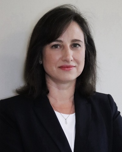 Deborah E. Citrin合作论文数Center for Cancer Research, National Cancer Institute, National Institutes of Health6
 Simon Turcotte合作论文数Département de chirurgie, Faculté de médecine, Universite de Montreal6Daniel J. Powell合作论文数5Laura A Johnson合作论文数5
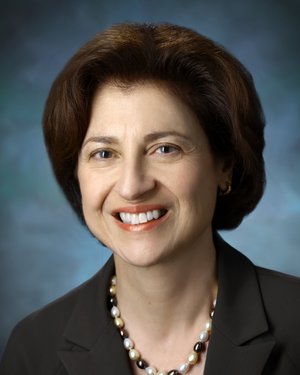 Suzanne Louise Topalian合作论文数Bloomberg-Kimmel Institute for Cancer Immunotherapy, Johns Hopkins University;School of Medicine, Johns Hopkins University5
 Ena Wang合作论文数Nurix Therapeutics, Inc.5Yao Xin合作论文数5




