BACKGROUND:Tissue engineering (TE) offers a potential alternative to fibula-free flap (FFF) reconstruction and may reduce surgical complexity, hospital stay, and health care costs. PURPOSE:The purpose of this study is to estimate and compare the cost-effectiveness of immediate TE and FFF reconstruction. STUDY DESIGN, SETTING, SAMPLE:This study was designed as a retrospective cohort conducted at the University of Texas Health Science Center at Houston from November 2015 to March 2024. Exclusion criteria included subjects with malignant pathologies, those treated with only a fasciocutaneous graft, cases of osteoradionecrosis, and those with incomplete records. PREDICTOR VARIABLE:The primary predictor variable was the type of reconstruction method, either TE bone grafts or FFF reconstruction. MAIN OUTCOME VARIABLES:The main outcome variable was cost-effectiveness which was defined as cost per successful reconstruction. Successful reconstruction was defined as complete union more than 1 year postoperatively without the need for revision surgery and the absence of postoperative complications requiring take-back surgery. COVARIATES:Covariates include age, sex, defect size, and American Society of Anesthesiologists classification. ANALYSES:R statistical software was used for data analysis. Statistical significance was defined as P < .05. RESULTS:The sample was composed of 31 subjects with a mean age of 44.97 ± 18.46 years for TE and 41.93 ± 18.23 years for FFF (P = .7). There were 18 (58%) and 13 (42%) subjects in TE and FFF, respectively. The proportion of successful reconstructions was 95% (n = 18) for TE and 77% (n = 13) for FFF (P = .6). The mean total hospital charges for TE were $247,172 ± $54,080 (P < .001) and $423,008 ± $59,571.75 (P < .001) for FFF. The cost per successful reconstruction was $261,711.64 for TE and $549,910.40 for FFF. CONCLUSIONS AND RELEVANCE:TE may be a more cost-effective alternative to FFF, providing comparable reconstructive success with reduced cost, surgical time, and hospital resource utilization.
PURPOSE:Surgeon-scientists have played a critical role in advancing understanding of human disease and improving patient care. Despite this legacy, the surgeon-scientist pathway in oral and maxillofacial surgery (OMFS) is increasingly vulnerable. For example, among 163 dentist-scientists funded as National Institutes of Health (NIH) principal investigators between 2003 and 2011, only a small fraction were OMFS faculty. This article discusses contemporary challenges facing OMFS surgeon-scientists and proposes a mentorship-based framework to support trainees across the career continuum. APPROACH:Drawing on published literature, workforce data, and the authors' collective experiences as mentors and trainees, we examine barriers encountered by clinician- and surgeon-scientists in the United States. These perspectives are integrated with an illustrative example of an OMFS surgeon-scientist training pathway to highlight practical considerations and potential solutions. KEY OBSERVATIONS:Major challenges include inadequate financial support, limited protected research time, inconsistent access to high-quality mentorship, and increasing trainee disillusionment. These factors contribute to attrition at multiple transition points. In response, we outline a longitudinal mentorship model designed to support OMFS surgeon-scientists from early exposure in dental training through residency, postdoctoral training, and early- to mid-career faculty development. CONCLUSIONS:Financial pressures, structural constraints, and mentorship gaps continue to threaten the sustainability of the OMFS surgeon-scientist workforce. Early recruitment, combined with coordinated and longitudinal mentorship, is essential. Effective mentors must advocate for trainees within their institutions, facilitate access to diverse funding mechanisms, and engage national organizations to help stabilize and grow the OMFS surgeon-scientist pipeline.
ABSTRACTPurposeOsteoradionecrosis of the jaw (ORNJ) is a severe iatrogenic disease characterized by bone death after radiation therapy (RT) to the head and neck. With over 9 published definitions and at least 16 diagnostic/staging systems, the true incidence and severity of ORNJ are obscured by lack of a standard for disease definition and severity assessment, leading to inaccurate estimation of incidence, reporting ambiguity, and likely under-diagnosis worldwide. This study aimed to achieve consensus on an explicit definition and phenotype of ORNJ and related precursor states through data standardization to facilitate effective diagnosis, monitoring, and multidisciplinary management of ORNJ.MethodsThe ORAL Consortium comprised 69 international experts, including representatives from medical, surgical, radiation oncology, and oral/dental disciplines. Using a web-based modified Delphi technique, panelists classified descriptive cases using existing staging systems, reviewed systems for feature extraction and specification, and iteratively classified cases based on clinical/imaging feature combinations.ResultsThe Consortium ORNJ definition was developed in alignment with SNOMED-CT terminology and recent ISOO-MASCC-ASCO guideline recommendations. Case review using existing ORNJ staging systems showed high rates of inability to classify (up to 76%). Ten consensus statements and nine minimum data elements (MDEs) were outlined for prospective collection and classification of precursor/ORNJ stages.ConclusionThis study provides an international, consensus-based definition and MDE foundation for standardized ORNJ reporting in cancer survivors treated with RT. Head and neck surgeons, radiation, surgical, medical oncologists, and dental specialists should adopt MDEs to enable scalable health information exchange and analytics. Work is underway to develop both a human- and machine-readable knowledge representation for ORNJ (i.e., ontology) and multidisciplinary resources for dissemination to improve ORNJ reporting in academic and community practice settings.
Successful dental implant therapy relies on a bone-implant interface that is mechanically strong and capable of dynamic remodeling in response to functional loads. There are a number of medical conditions or therapies that can affect either bone metabolism or the resistance of bone to infection. However, their effects are often mitigated by local factors or individual responses so the impact of these conditions is not clear-cut. This article will review a number of these conditions and therapies and describe existing studies that have studied these conditions to guide practitioners in their implant practice.
The Comprehensive Basic Science Exam (CBSE) has been utilized for nearly a decade ever since the National Board Dental Examination transitioned to a pass/fail grading system. Despite the CBSE being utilized by medical school trainees as a practice test for the United States Medical Licensing Exam (USMLE) Step 1, early studies showed that CBSE scores of oral and maxillofacial surgery (OMS) residency applicants were not correlated with subsequent USMLE Step 1 pass rates. The USMLE Step 1 transitioned to a pass/fail grading system in January 2022. Proponents of this change feel that the transition allows applicant evaluations to refocus on holistic reviews of applications and further promote well-being of medical students and enable them to focus on their pre-clinical curriculum instead of simply preparing for an exam. However, the field of OMS continues to require residency applicants to submit their CBSE score. The primary objective of this study was to determine if OMS residency programs are utilizing a threshold number for the CBSE to evaluate applicants and what that number is and if it changes from year to year. The secondary objectives were to evaluate program directors' perspectives regarding the importance of the CBSE score in evaluating applicants and if they feel it is predictive of resident success. This was a cross-sectional, survey-based research study of PDs of the 88 non-federal OMS programs in the United States. Email solicitations were sent to the PDs listed in the AAOMS directory to participate in the anonymous online survey (Qualtrics). The survey consisted of 9-17 questions with multiple choice, Likert scale, and free response. Descriptive statistics were performed on the survey results. There was a 62.5% (55/88) response rate. Forty-one-point-five percent of responding PDs represented 4-year only programs, 17.0% represented 4/6-year programs, 39.6% represented 6-year only programs, and 1.9% represented 4-year with MD optional programs. Seventy-six-point-four percent of the respondents utilize a threshold number. Eighty-two-point-nine percent of responding programs indicated that the threshold had changed at least 1 time in the past 5 years. Reasons cited include "changes in the scoring system" "applicants with higher scores" and "volume of applicants." The reported threshold numbers included scores on both the 2-digit and 3-digit scales. After separating the responses from the 2 scales, the mode for the 2-digit scale was 70 and the mode for the 3-digit scale was 200. PDs indicated that the CBSE score was more influential in deciding to offer an interview to a candidate than in making the final rank list. We found that 74.1% of PDs agreed or strongly agreed that the CBSE score remains a necessary component in evaluating applicants, however only 29.6% agreed or strongly agreed that the CBSE score is predictive of a successful resident in their specific program. This study found that the majority of OMS residency programs are utilizing the CBSE score as a threshold, however that number has changed for the majority of programs in the past 5 years for institutional and systemic reasons. Additionally, though the majority of PDs feel the CBSE remains an important component of the application, much fewer feel the score is predictive of a successful resident.
Craniofacial defects require a treatment approach that provides both robust tissues to withstand the forces of mastication and high geometric fidelity that allows restoration of facial architecture. When the surrounding soft tissue is compromised either through lack of quantity (insufficient soft tissue to enclose a graft) or quality (insufficient vascularity or inducible cells), a vascularized construct is needed for reconstruction. Tissue engineering using customized 3D printed bioreactors enables the generation of mechanically robust, vascularized bony tissues of the desired geometry. While this approach has been shown to be effective when utilized for reconstruction of non-load bearing ovine angular defects and partial segmental defects, the two-stage approach to mandibular reconstruction requires testing in a large, load-bearing defect. In this study, 5 sheep underwent bioreactor implantation and the creation of a load-bearing mandibular defect. Two bioreactor geometries were tested: a larger complex bioreactor with a central groove, and a smaller rectangular bioreactor that were filled with a mix of xenograft and autograft (initial bone volume/total volume BV/TV of 31.8 ± 1.6%). At transfer, the tissues generated within large and small bioreactors were composed of a mix of lamellar and woven bone and had BV/TV of 55.3 ± 2.6% and 59.2 ± 6.3%, respectively. After transfer of the large bioreactors to the mandibular defect, the bioreactor tissues continued to remodel, reaching a final BV/TV of 64.5 ± 6.2%. Despite recalcitrant infections, viable osteoblasts were seen within the transferred tissues to the mandibular site at the end of the study, suggesting that a vascularized customized bony flap is a potentially effective reconstructive strategy when combined with an optimal stabilization strategy and local antibiotic delivery prior to development of a deep-seated infection.
For maxillofacial surgeons, the reconstruction of mandibular defects that result from the ablation of tumors or traumatic injury poses a formidable, and somewhat controversial challenge. The maxillofacial region is a complex and prominent region with specialized functions. For a patient to maintain a high quality of life, it is important to maintain functions such as speech, deglutination, mastication, facial expression, and airway maintenance. Furthermore, the cosmetics of this region play an equal or greater role in the perceived quality of life of the patient.
The primary purpose of this study is to identify if there is an underlying genetic predisposition for COVID-related macroglossia and if this susceptibility is higher among individuals of African heritage. Secondary objectives include determining if genetic testing of COVID-infected patients who are intubated and prone could identify patients with higher susceptibility to the development of macroglossia. A retrospective chart review was completed for each patient, and prospectively, genetic and histopathologic analyses were completed. Whole-exome sequencing was completed on two patients; immunohistochemistry was completed on the COVID-positive tissue samples. Histopathology of the COVID-positive patient revealed significant peri-lymphocytic infiltrate, which was absent in the COVID-negative patient. Immunohistochemistry confirmed the presence of immune cells. Results from the whole-exome sequencing were inconclusive. The findings of this study are consistent with others that have observed a lymphocytic infiltrate in the organs of patients infected with SARS-CoV-2. On histology, IHC highlighted a CD45 + predominance, indicating that a robust immune response is present in the tissues. The pathobiology of this phenomenon and its role in the development and/or persistence of massive macroglossia requires further study.
Purpose The purpose of this article is to present an interesting, rare case of a patient who experienced avascular necrosis of the maxilla associated with COVID-19 infection. Methods and Results Our team retrospectively evaluated this patient's chart after completion of surgical management. The patient is a 72-year-old male who presented to the University of Texas Health Science Center at Houston for surgical management of his infarcted maxilla, which developed as a sequela of infection with COVID-19. A literature review was completed using PubMed. Twenty-five articles are reviewed and discussed. Conclusions Infection with COVID-19 confers a hypercoagulable state in patients, leading to various complications in the head and neck region. In our case report, we present a patient who developed avascular necrosis of the maxilla secondary to infection with COVID-19. Thromboembolic prophylaxis is imperative in COVID-19 patients due to the high rate of potential systemic complications.
Although the knee joint and temporomandibular joint (TMJ) experience similar incidence of cartilage ailments, the knee orthopedics field has greater funding and more effective end-stage treatment options. Translational research has resulted in the development of tissue-engineered products for knee cartilage repair, but the same is not true for TMJ cartilages. Here, we examine the anatomy and pathology of the joints, compare current treatments and products for cartilage afflictions, and explore ways to accelerate the TMJ field. We examine disparities, such as a 6-fold higher article count and 2,000-fold higher total joint replacement frequency in the knee compared to the TMJ, despite similarities in osteoarthritis incidence. Using knee orthopedics as a template, basic and translational research will drive the development and implementation of clinical products for the TMJ. With more funding opportunities, training programs, and federal guidance, millions of people afflicted with TMJ disorders could benefit from novel, life-changing therapeutics.
Purpose: This article aims to introduce the novel technique of utilization of a custom 3D titanium printed reconstruction combined with in-situ tissue engineering for the reconstruction and dental rehabilitation of an infected severely atrophic mandible. Technique: A 3-staged approach was used to remove the failing implants and debride the mandible, eradicating the infection, followed by reconstruction of the severely atrophic mandible using tissue engineering supported by a customized 3D-printed titanium undercarriage and reconstruction plate for vertical and horizontal augmentation. Finally, a CAD/CAM 3D-printed surgical guide was used for optimal placement of 4 dental implants. Conclusion: A predictable and satisfactory reconstruct of a severely infected mandible with failing hardware to full dental rehabilitation and aesthetics was achieved by utilizing an amalgamation of state-of-the-art technology and tissue engineering.
Osteochondral defects present a unique clinical challenge due to their combination of phenotypically distinct cartilage and bone, which require specific, stratified biochemical cues for tissue regeneration. Furthermore, the articular cartilage exhibits significantly worse regeneration than bone due to its largely acellular and avascular nature, prompting significant demand for regenerative therapies. To address these clinical challenges, we have developed a bilayered, modular hydrogel system that enables the click functionalization of cartilage- and bone-specific biochemical cues to each layer. In this system, the crosslinker poly(glycolic acid)-poly(ethylene glycol)-poly(glycolic acid)-di(but-2-yne-1,4-dithiol) (PdBT) was click conjugated with either a cartilage- or bone-specific peptide sequence of interest, and then mixed with a suspension of thermoresponsive polymer and mesenchymal stem cells (MSCs) to generate tissue-specific, cell-encapsulated hydrogel layers targeting the cartilage or bone. We implanted bilayered hydrogels in rabbit femoral condyle defects and investigated the effects of tissue-specific peptide presentation and cell encapsulation on osteochondral tissue repair. After 12 weeks implantation, hydrogels with a chondrogenic peptide sequence produced higher histological measures of overall defect filling, cartilage surface regularity, glycosaminoglycan (GAG)/cell content of neocartilage and adjacent cartilage, and bone filling and bonding compared to non-chondrogenic hydrogels. Furthermore, MSC encapsulation promoted greater histological measures of overall defect filling, cartilage thickness, GAG/cell content of neocartilage, and bone filling. Our results establish the utility of this click functionalized hydrogel system for in vivo repair of the osteochondral unit.
The basic concepts from the fields of biology and engineering are integrated into tissue engineering to develop constructs for the repair of damaged and/or absent tissues, respectively. The field has grown substantially over the past two decades, with particular interest in bone tissue engineering (BTE). Clinically, there are circumstances in which the quantity of bone that is necessary to restore form and function either exceeds the patient's healing capacity or bone's intrinsic regenerative capabilities. Vascularized osseous or osteocutaneous free flaps are the standard of care with autologous bone remaining the gold standard, but is commonly associated with donor site morbidity, graft resorption, increased operating time, and cost. Regardless of the size of a craniofacial defect, from trauma, pathology, and osteonecrosis, surgeons and engineers involved with reconstruction need to consider the complex three-dimensional (3D) geometry of the defect and its relationship to local structures. Three-dimensional printing has garnered significant attention and presents opportunities to use craniofacial BTE as a technology that offers a personalized approach to bony reconstruction. Clinicians and engineers are able to work together to produce patient-specific space-maintaining scaffolds tailored to site-specific defects, which are osteogenic, osseoconductive, osseoinductive, encourage angiogenesis/vasculogenesis, and mechanically stable upon implantation to prevent immediate failure. In this work, we review biological and engineering principles important in applying 3D printing technology to BTE for craniofacial reconstruction as well as present recent translational advancements in 3D printed bioactive ceramic scaffold technology.
Mandibular reconstruction requires functional and aesthetic repair and is further complicated by contamination from oral and skin flora. Antibiotic-releasing porous space maintainers have been developed for the local release of vancomycin and to promote soft tissue attachment. In this study, mandibular defects in six sheep were inoculated with 106 colony forming units of Staphylococcus aureus; three sheep were implanted with unloaded porous space maintainers and three sheep were implanted with vancomycin-loaded space maintainers within the defect site. During the same surgery, 3D-printed in vivo bioreactors containing autograft or xenograft were implanted adjacent to rib periosteum. After 9 weeks, animals were euthanized, and tissues were analyzed. Antibiotic-loaded space maintainers were able to prevent dehiscence of soft tissue overlying the space maintainer, reduce local inflammatory cells, eliminate the persistence of pathogens, and prevent the increase in mandibular size compared to unloaded space maintainers in this sheep model. Animals with an untreated mandibular infection formed bony tissues with greater density and maturity within the distal bioreactors. Additionally, tissues grown in autograft-filled bioreactors had higher compressive moduli and higher maximum screw pull-out forces than xenograft-filled bioreactors. In summary, we demonstrated that antibiotic-releasing space maintainers are an innovative approach to preserve a robust soft tissue pocket while clearing infection, and that local infections can increase local and remote bone growth.
 Antonios Mikos合作论文数Department of Bioengineering, Rice University54
 Kasper F Kurtis合作论文数Dept Bioengn, Rice Univ28
 John A. Jansen合作论文数Radboud University Nijmegen Medical Centre28
 James C Melville合作论文数Department of Oral and Maxillofacial Surgery, University of Texas Health Science Center at Houston22
 Simon Young合作论文数20
 Nagi demian合作论文数Department of Oral and Maxillofacial Surgery, University of Texas Health Science Center at Houston18Shum Jonathan W合作论文数Department of Oral and Maxillofacial Surgery, University of Texas Health Science Center at Houston16
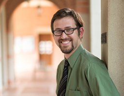 Alexander Tatara合作论文数Dept Bioengn, Rice Univ16
 Jeroen van den Beucken合作论文数Radboud University Nijmegen Medical Center;Radboud University Medical Center16James D. Kretlow合作论文数13
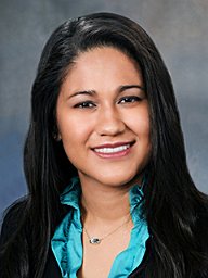 Shah Sarita R合作论文数Department of Bioengineering, Rice University12
 Kyriacos Athanasiou合作论文数Department of Biomedical Engineering, Samueli School of Engineering, University of California, Irvine11Henslee Allan M合作论文数11
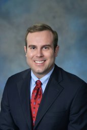 Brandon T Smith合作论文数Department of Bioengineering, Rice University10
 Emma Watson合作论文数Infection, Immunity and Inflammation, University of Leicester10
 Yasuhiko Tabata (田畑泰彦)合作论文数Department of Biomaterials, Institute for Frontier Medical Sciences, Kyoto University9Meng Shi合作论文数8Issa A Hanna合作论文数7
 Lam Johnny合作论文数7Steven Lu合作论文数6
 Victoria Manon合作论文数5Kau Chung H合作论文数5PJ Duke合作论文数4
 Anthony Ratcliffe合作论文数Synthasome, Inc.4L. Scott Baggett合作论文数Department of Statistics, Rice University4
 Zahid Lalani合作论文数Department of Oral and Maxillofacial Surgery, The University of Texas Houston Health Science Center;Private Practice in Oral and Maxillofacial Surgery, Currently4Tang Ho合作论文数4
 Jordan Trachtenberg合作论文数Department of Bioengineering, Rice University3Smoak Mollie合作论文数Department of Biological and Agricultural Engineering, Louisiana State University and Agricultural Center3
 Pearce Hannah合作论文数3
 Jeryl D English合作论文数3Carol Johnston合作论文数3
 Patrick T. Spicer合作论文数School of Chemical Engineering, Faculty of Engineering, The University of New South Wales3
 David Warren Scott合作论文数Department of Statistics, Rice University3
 George N. Bennett合作论文数Microbial Biotechnology Laboratory, Department of Biochemistry and Cell Biology, Rice University3Kyle D. Allen合作论文数2
 Sherif G Kamel合作论文数2Diana M. Yoon合作论文数2T. Ho合作论文数2
 Ramzey Tursun合作论文数2
 T. C. Piepergerdes合作论文数Dept Bioengn, Rice Univ2
 Sergio Barrios合作论文数Department of Bioengineering, Rice University2
 Gerry L Koons合作论文数Med Scientist Training Program, Baylor Coll Med2I. Hanna合作论文数2
 Yu Seon Kim合作论文数2
 Lukasz Witek合作论文数Department of Biomaterials, College of Dentistry, New York University2
 Neeraj Panchal合作论文数Department of Oral and Maxillofacial Surgery, School of Dental Medicine, University of Pennsylvania/Department of Pharmacology, School of Dental Medicine, University of Pennsylvania2
 Boaz Arzi合作论文数Dept Surg & Radiol Sci, Univ Calif Davis2




