Mapping fine axonal projections with high sensitivity remains a major challenge in neuroanatomy. Here, we developed Codon Optimized Membrane Embedded Tracers (COMET), a family of genetically encoded, membrane-anchored fluorescent proteins optimized for adeno-associated virus (AAV) delivery. COMET tracers—based on codon-optimized SuperFolder GFP (gCOMET) and TdTomato (rCOMET) fused to an H-RAS CaaX prenylation motif—enable robust plasma membrane targeting and enhance fluorescent signal localization to fine-caliber axons. In vivo, COMET tracers significantly outperformed conventional soluble fluorophores and chemical tracers in detecting corticospinal tract (CST) projections, revealing previously underappreciated collateralization and fine arborizations. Following spinal cord injury, COMET provided superior sensitivity for detecting regenerating and sprouting axons compared with biotinylated dextran amine (BDA). Importantly, COMET tracers retained bright native fluorescence after tissue clearing by CLARITY, enabling volumetric imaging of long-range projections without antibody amplification. COMET expands the available toolkit for high-resolution connectomics and regeneration studies, offering a versatile platform for sensitive, scalable, and cell-type-specific neuronal tracing in intact and injured mammalian nervous systems.
BDNF is a neurotrophic factor that prevents neuronal death, activates neuronal function, restores synapses and improves learning and memory in animal models of Alzheimer's disease. Beneficial effects of BDNF gene delivery to entorhinal/hippocampal circuits on cognitive performance are observed in APP transgenic mice, aged rats and aged non-human primates. Based on the preceding evidence, we advanced AAV2-BDNF gene delivery to a Phase 1 clinical trial in patients with biomarker-supported mild Alzheimer's disease (AD) and Mild Cognitive Impairment (MCI). 6 patients with mild AD and 6 patients with MCI are being recruited into this clinical trial. Patients undergo MRI-guided injection of AAV2-BDNF into the entorhinal cortex and are monitored over 2 years with serial cognitive testing and FDG PET scans. As of this writing, 6 patients with mild AD have been treated, and we are now beginning treatment of the MCI cohort. 6 patients with mild AD have been treated with followup times ranging from 1 to 18 months. There have been no serious adverse events related to the study procedure. Among three patients that were treated at least 6 months earlier, followup FDG PET scans demonstrate increases in cortical metabolism in entorhinal regions that received AAV2-BDNF, a reversal of the normal pattern of decline typically observed in AD. Cognitive outcomes will be presented at the meeting. Given the safety of treatment to date, we are proceeding to dose the MCI cohort of patients. AAV2-BDNF gene therapy to the entorhinal cortex in mild AD has been safe to date and restores FDG-PET activity in the treated brain region. BDNF gene therapy offers the potential to prevent neuronal loss, activate neuronal function and restore synapses, representing a possible neuro-restorative treatment for AD and MCI.
Depletion or inhibition of core stress granule proteins, G3BP1 in mammals and TIAR-2 in Caenorhabditis elegans, increases the growth of spontaneously regenerating axons. Inhibition of G3BP1 by expression of its acidic or "B-domain" accelerates axon regeneration after nerve injury, bringing a potential therapeutic strategy for peripheral nerve repair. Here, we asked whether G3BP1 inhibition is a viable strategy to promote regeneration in injured mammalian central nervous system (CNS) where axons do not regenerate spontaneously. G3BP1 B-domain expression was found to promote axon regeneration in the transected spinal cord provided with a permissive peripheral nerve graft (PNG) as well as in crushed optic nerve. Moreover, a cell-permeable peptide (CPP) to a subregion of B-domain (rodent G3BP1 amino acids 190 to 208) accelerated axon regeneration after peripheral nerve injury and promoted regrowth of reticulospinal axons into the distal transected spinal cord through a bridging PNG. G3BP1 CPP promoted axon growth from rodent and human neurons cultured on permissive substrates, and this function required alternating Glu/Asp-Pro repeats that impart a unique predicted tertiary structure. The G3BP1 CPP disassembles axonal G3BP1, G3BP2, and FMRP, but not FXR1, granules and selectively increases axonal protein synthesis in cortical neurons. These studies identify G3BP1 granules as a key regulator of axon growth in CNS neurons and demonstrate that disassembly of these granules promotes retinal axon regeneration in injured optic nerve and reticulospinal axon elongation into permissive environments after CNS injury. This work highlights G3BP1 granule disassembly as a potential therapeutic strategy for enhancing axon growth and neural repair.
BACKGROUND:A spinal cord contusion injury is among the most clinically relevant models for studying pathophysiology and for developing potential therapeutic interventions for spinal cord injuries (SCI). METHODS:In this study, we implemented an intra-operative ultrasound (IOU) approach to precisely locate and examine the lesion site at 5 and 10 min post-injury after a cervical hemi-contusion injury in a non-human primate (NHP) model. We assessed acute lesion progression from 5 to 10 min and then compared that to the lesion extent as measured by MRI 3 weeks later. RESULTS:We observed a small increase in the rostrocaudal and mediolateral lesion area (mm2) from 5 to 10 min and a further 26% increase in the mediolateral lesion extent when comparing 5 and 10 min to 3 weeks post-injury. CONCLUSIONS:By enabling high-resolution ultrasound visualization of the hemicontusion lesion in vivo, this approach can provide critical insights into the early progression of SCI. It can help with further refining this preclinical SCI model and provide significant predictive value for the animals' recovery post-injury.
Neuronal growth and regeneration are regulated by local translation of mRNAs in axons. We examined RNA polyadenylation changes upon sensory neuron injury and found upregulation of a subset of polyadenylated B2-SINE repeat elements, hereby termed GI-SINEs (growth-inducing B2-SINEs). GI-SINEs are induced from ATF3 and other AP-1 promoter-associated extragenic loci in injured sensory neurons but are not upregulated in lesioned retinal ganglion neurons. Exogenous GI-SINE expression elicited axonal growth in injured sensory, retinal, and corticospinal tract neurons. GI-SINEs interact with ribosomal proteins and nucleolin, an axon-growth-regulating RNA-binding protein, to regulate translation in neuronal cytoplasm. Finally, antisense oligos against GI-SINEs perturb sensory neuron outgrowth and nucleolin-ribosome interactions. Thus, a specific subfamily of transposable elements is integral to a physiological circuit linking AP-1 transcription with localized RNA translation.
Research on cell therapy for spinal cord injury has yet to achieve sufficient functional recovery. Previous studies in the field grafted oligodendrocyte progenitors, nonspinal neural stem cells or primary spinal neural progenitors. Here we sought to improve functional outcomes by grafting clinically compatible spinal cord neural stem cells derived from human embryonic stem cells (H9-scNSCs). H9-scNSCs significantly improved functional outcomes on a skilled hand task 9.2-fold (P = 2.5 × 10⁻²⁷) in hemisected subjects compared with lesioned controls, achieving a fine object retrieval success of 53.4 ± 19.2
Neural stem cells have exhibited efficacy in pre-clinical models of spinal cord injury (SCI) and are on a translational path to human testing. We recently reported that neural stem cells must be driven to a spinal cord fate to optimize host axonal regeneration into sites of implantation in the injured spinal cord, where they subsequently form neural relays across the lesion that support significant functional improvement. We also reported methods of deriving and culturing human spinal cord neural stem cells derived from embryonic stem cells that can be sustained over serial high passage numbers in vitro, providing a potentially optimized cell source for human clinical trials. We now report further optimization of methods for deriving and sustaining cultures of human spinal cord neural stem cell lines that result in improved karyotypic stability while retaining anatomical efficacy in vivo. This development improves prospects for safe human translation.
Layer 5 neurons of the neocortex receive their principal inputs from layer 2/3 neurons. We seek to identify the nature and extent of the plasticity of these projections with motor learning. Using optogenetic and viral intersectional tools to selectively stimulate distinct neuronal subsets in rat primary motor cortex, we simultaneously record from pairs of corticospinal neurons associated with distinct features of motor output control: distal forelimb vs. proximal forelimb. Activation of Channelrhodopsin2-expressing layer 2/3 afferents onto layer 5 in untrained animals produces greater monosynaptic excitation of neurons controlling the proximal forelimb. Following skilled grasp training, layer 2/3 inputs onto corticospinal neurons controlling the distal forelimb associated with skilled grasping become significantly stronger. Moreover, peak excitatory response amplitude nearly doubles while latency shortens, and excitatory-to-inhibitory latencies become significantly prolonged. These findings demonstrate distinct, highly segregated, and cell-specific plasticity of layer 2/3 projections during skilled grasp motor learning.
Nervous system growth factors are natural proteins of the brain that influence neuronal survival and function throughout life, from embryonic development to old age. In animal models of Alzheimer’s disease (AD), the growth factor brain derived neurotrophic factor (BDNF) prevents neuronal death, activates neuronal function, builds new synapses and improves learning and memory. Accordingly, we are determining whether gene delivery of BDNF in patients with AD will slow disease progression and improve memory. In a previous clinical trial of nerve growth factor (NGF) gene therapy in AD patients (NCT00017940, June 2001), we learned that growth factors can unequivocally elicit classic trophic responses from degenerating neurons in AD. Experience gained from the earlier NGF gene therapy trial is guiding our effort to optimize gene delivery of BDNF in our present clinical program (NCT05040217, June 2021).
Neuroscience dogma avers that astrocytic "scars" inhibit axonal regeneration after spinal cord injury (SCI). A recent report suggested however that astrocytes form "borders" around lesions that are permissive rather than inhibitory to axonal growth. We now provide further evidence supporting a facilitatory role of astrocytes in axonal regeneration after SCI. First, even 6months after SCI, injured axons are retained within regions of densely reactive astrocytes, in direct contact with astrocyte processes without being repelled. Second, 6 month-delayed implants of neural stem cells extend axons into reactive astrocyte borders surrounding lesions, densely contacting astrocyte surfaces. Third, bioengineered hydrogels implanted into sites of SCI re-orient reactive astrocytic processes to align along the rostral-to-caudal spinal cord axis resulting in successful regeneration into the lesion/ scaffold in close association with astrocytic processes. Fourth, corticospinal axons regenerate into neural progenitor cells implanted six months after injury in close association with host astrocytic processes. Thus, astrocytes do not appear to inhibit axonal regeneration, and the close association of newly growing axons with astrocytic processes suggests a facilitatory role in axonal regeneration.
The complex and heterogeneous nature of spinal cord injury has limited translational bench-to-bedside results. The wide variety of data, including injury parameters, biochemical, histological, and behavioral outcome measures represent a ‘big data’ problem, calling for modern data science solutions. There are some instances in which SCI researchers collect sensitive data that needs to remain private, such as datasets designed to meet regulatory approval, sensitive intellectual property, and non-human primate studies. For these types of data, we have developed a Private Data Commons for SCI (PDC-SCI). Our objective is to give an overview of this novel data commons, describing how this type of commons works, how it can benefit the research community, and the cases in which it would be most useful. This private infrastructure is ideal for multi-lab transdisciplinary studies that require a well-organized, scalable data commons for rapid data sharing within a closed, distributed team. As a use-case for the PDC-SCI, we demonstrate the VA Gordon Mansfield SCI Consortium, in which multimodal data from behavior, biomechanics of injury, hospital records, imaging, and histology are integrated, shared, and analyzed to facilitate insights and knowledge discovery.
Introduction The paralysis that occurs after a spinal cord injury, particularly during the early stages of post-lesion recovery (∼6 weeks), appears to be attributable to the inability to activate motor pools well beyond their motor threshold. In the later stages of recovery, however, the inability to perform a motor task effectively can be attributed to abnormal activation patterns among motor pools, resulting in poor coordination. Method We have tested this hypothesis on four adult male Rhesus monkeys ( Macaca mulatta ), ages 6-10 years, by recording the EMG activity levels and patterns of multiple proximal and distal muscles controlling the upper limb of the Rhesus when performing three tasks requiring different levels of skill before and up to 24 weeks after a lateral hemisection at C7. During the recovery period the animals were provided routine daily care, including access to a large exercise cage (5' × 7' × 10') and tested every 3-4 weeks for each of the three motor tasks. Results At approximately 6-8 weeks the animals were able to begin to step on a treadmill, perform a spring-loaded task with the upper limb, and reaching, grasping, and eating a grape placed on a vertical stick. The predominant changes that occurred, beginning at ∼6-8 weeks of the recovery of these tasks was an elevated level of activation of most motor pools well beyond the pre-lesion level. Discussion As the chronic phase progressed there was a slight reduction in the EMG burst amplitudes of some muscles and less incidence of co-contraction of agonists and antagonists, probably contributing to an improved ability to selectively activate motor pools in a more effective temporal pattern. Relative to pre-lesion, however, the EMG patterns even at the initial stages of recovery of successfully performing the different motor tasks, the level of activity of most muscle remained higher. Perhaps the most important concept that emerges from these data is the large combinations of adaptive strategies in the relative level of recruitment and the timing of the peak levels of activation of different motor pools can progressively provide different stages to regain a motor skill.
Neural stem cells (NSCs) implanted into sites of spinal cord injury (SCI) extend very large numbers of new axons over very long distances caudal to the lesion site, and support partial functional recovery. Newly extending graft axons distribute throughout host gray and white matter caudal to the injury. We hypothesized that provision of trophic gradients caudal to the injury would provide neurotrophic guidance to newly extending graft-derived axons to specific intermediate and ventral host gray matter regions, thereby potentially further improving neural relay formation. Immunodeficient rats underwent C5 lateral hemisection lesions, following by implants of human NSC grafts two weeks later. After an additional two weeks, animals received injections of AAV2-BDNF expressing vectors three spinal segments (9 mm) caudal to the lesion in host ventral and intermediate gray matter. After 2 months additional survival, we found a striking, 5.5-fold increase in the density of human axons innervating host ventral gray matter (P < 0.05) and 2.7-fold increase in intermediate gray matter (P < 0.01). Moreover, stem cell-derived axons formed a substantially greater number of putative synaptic connections with host motor neurons (P < 0.01). Thus, trophic guidance is an effective means of enhancing and guiding neural stem cell axon growth after SCI and will be used in future experiments to determine whether neural relay for-mation and functional outcomes can be improved.
One hundred years ago, Ramón y Cajal, considered by many as the founder of modern neuroscience, stated that neurons of the adult central nervous system (CNS) are incapable of regenerating. Yet, recent years have seen a tremendous expansion of knowledge in the molecular control of axon regeneration after CNS injury. We now understand that regeneration in the adult CNS is limited by (1) a failure to form cellular or molecular substrates for axon attachment and elongation through the lesion site; (2) environmental factors, including inhibitors of axon growth associated with myelin and the extracellular matrix; (3) astrocyte responses, which can both limit and support axon growth; and (4) intraneuronal mechanisms controlling the establishment of an active cellular growth programme. We discuss these topics together with newly emerging hypotheses, including the surprising finding from transcriptomic analyses of the corticospinal system in mice that neurons revert to an embryonic state after spinal cord injury, which can be sustained to promote regeneration with neural stem cell transplantation. These gains in knowledge are steadily advancing efforts to develop effective treatment strategies for spinal cord injury in humans.
Abstract Neuroscience dogma avers that astrocytic “scars” inhibit axonal regeneration after spinal cord injury (SCI). A recent report suggested however that astrocytes form “borders” around lesions that are not inhibitory and may be permissive to axonal growth, although direct evidence is lacking. We now provide direct evidence that astrocytes support axonal growth after SCI. First, even 6 months after SCI, injured axons are retained within regions of densely reactive astrocytes, in direct contact with astrocyte processes without being repelled. Second, 6 month-delayed implants of neural stem cells extend axons into reactive astrocyte borders surrounding lesions, densely contacting astrocyte surfaces. Third, bioengineered hydrogels implanted into sites of SCI re-orient reactive astrocytic processes to align along the rostral-to-caudal spinal cord axis resulting in successful regeneration along astrocytic processes. Fourth, corticospinal axons regenerate into neural stem cells implanted six months after injury in close association with host astrocytic processes. Thus, astrocytes support axonal regeneration.
We reported previously that neural progenitor cell (NPC) grafts form neural relays across sites of subacute spinal cord injury (SCI) and support functional recovery. Here, we examine whether NPC grafts after chronic delays also support recovery and whether intensive rehabilitation further enhances recovery. One month after severe bilateral cervical contusion, rats received daily intensive rehabilitation, NPC grafts, or both rehabilitation and grafts. Notably, only the combination of rehabilitation and grafting significantly improved functional recovery. Moreover, improved functional outcomes were associated with a rehabilitation-induced increase in host corticospinal axon regeneration into grafts. These findings identify a critical and synergistic role of rehabilitation and neural stem cell therapy in driving neural plasticity to support functional recovery after chronic and severe SCI.
Neural stem cells (NSCs) expressing GFP were embedded into fibrin matrices containing growth factor cocktails and grafted to sites of severe spinal cord injury. Grafted cells differentiated into multiple cellular phenotypes, including neurons, which extended large numbers of axons over remarkable distances. Extending axons formed abundant synapses with host cells. Axonal growth was partially dependent on mammalian target of rapamycin (mTOR), but not Nogo signaling. Grafted neurons supported formation of electrophysiological relays across sites of complete spinal transection, resulting in functional recovery. Two human stem cell lines (566RSC and HUES7) embedded in growth-factor-containing fibrin exhibited similar growth, and 566RSC cells supported functional recovery. Thus, properties intrinsic to early-stage neurons can overcome the inhibitory milieu of the injured adult spinal cord to mount remarkable axonal growth, resulting in formation of new relay circuits that significantly improve function. These therapeutic properties extend across stem cell sources and species.
We used viral intersectional tools to map the entire projectome of corticospinal neurons associated with fine distal forelimb control in Fischer 344 rats and rhesus macaques. In rats, we found an extraordinarily diverse set of collateral projections from corticospinal neurons to 23 different brain and spinal regions. Remarkably, the vast weighting of this "motor"projection was to sensory systems in both the brain and spinal cord, confirmed by optogenetic and transsynaptic viral intersectional tools. In contrast, rhesus macaques ex-hibited far heavier and narrower weighting of corticospinal outputs toward spinal and brainstem motor systems. Thus, corticospinal systems in macaques primarily constitute a final output system for fine motor control, whereas this projection in rats exerts a multi-modal integrative role that accesses far broader CNS regions. Unique structural-functional correlations can be achieved by mapping and quantifying a single neuronal system's total axonal output and its relative weighting across CNS targets.
 Armin Blesch合作论文数Department of Neurosciences, School of Medicine, University of California, San Diego;School of Medicine, Indiana University69
 Paul Lu合作论文数50
 Rusty Gage合作论文数Laboratory of Genetics, Salk Institute for Biological Studies;Kavli Institute for Brain and Mind, Salk Institute for Biological Studies;Department of Neurosciences, University of California, San Diego37
 James M. Conner合作论文数Department of Biology, University of California34
 Ephron S. Rosenzweig合作论文数32Alan H. Nagahara合作论文数24
 Michael S. Beattie合作论文数Brain and Spinal Injury Center, University of California, San Francisco23
 Jeff Sakamoto合作论文数Department of Mechanical Engineering, University of Michigan22Jacqueline C. Bresnahan合作论文数22
 Adam Ferguson合作论文数Department of Neurological Surgery, School of Medicine, University of California, San Francisco;Brain and Spinal Injury Center, Weill Institute for Neurosciences, University of California, San Francisco;San Francisco VA Medical Center;San Francisco General Hospital;UCSF Medical Center21Lori Graham合作论文数19Leif A. Havton合作论文数16Moseanko Rod合作论文数15
 V. Reggie Edgerton合作论文数Department of Neurobiology, David Geffen School of Medicine, University of California, Los Angeles14
 Jacob Koffler合作论文数Biomedical Engineering and;Interdepartmental Program in Biotechnology, Technion—Israel Institute of Technology;Biomedical Engineering and;Interdepartmental Program in Biotechnology, Technion—Israel Institute of Technology13
 Jessica L Nielson合作论文数Brain and Spinal Injury Center (BASIC),, University of California, San Francisco13Sarah Strand合作论文数12Nout-Lomas Yvette S合作论文数12
 Jeffrey Kordower合作论文数Banner Neurodegenerative Disease Research Center, Arizona State University;Department of Neurological Sciences, Rush Medical College, Rush University12John H. Brock合作论文数11Leonard L Jones合作论文数11
 Grégoire Courtine合作论文数Interschool Institute Neuro-X, School of Life Sciences, Ecole Polytechnique Fédérale de Lausanne;ONWARD;Centre Hospitalier Universitaire Vaudois11Hawbecker Stephanie合作论文数11Salegio Ernesto A合作论文数Dept Neurol Surg, Univ Calif San Francisco11Hiromi Kumamaru合作论文数11Ken Kadoya合作论文数11Heather Mckay合作论文数10
 Roland R. Roy合作论文数Brain Research Institute, University of California, Los Angeles10
 Sparrey Carolyn J合作论文数Department of Mechanical Engineering, University of California9
 Shahriari Dena合作论文数9Biane Jeremy合作论文数Department of Neurosciences, University of California at San Diego9Kovacs Imre合作论文数9John W. Brock合作论文数Centers for Disease Control and Prevention, National Center for Environmental Health9Krystof S. Bankiewicz合作论文数9Julian V. Roberts合作论文数Criminology, University of Ottawa9
 Huie J Russell合作论文数Brain and Spinal Injury Center, University of California San Francisco San Francisco8
 Dulin Jennifer N合作论文数Department of Neurosciences, University of California8
 Raymond T. Bartus合作论文数RTBioconsultants, Inc.8
 Binhai Zheng合作论文数Department of Neurosciences, University of California San Diego8Mehdi Gasmi合作论文数7
 Edmund Hollis合作论文数Neuroscience, Weill Cornell Graduate School of Medical Sciences, Cornell University7
 Castle Michael J合作论文数University of Pennsylvania, University of Pennsylvania7
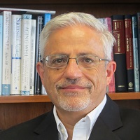 Eliezer Masliah合作论文数Division of Neuroscience, National Institute on Aging, National Institutes of Health;Experimental Neuropathology Laboratory, University of California San Diego7
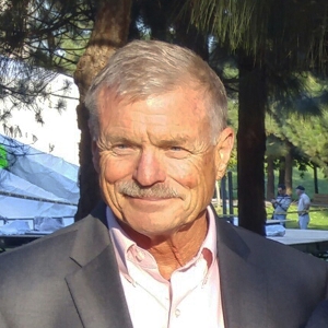 Oswald Steward合作论文数Reeve-Irvine Research Center, Department of Anatomy & Neurobiology, School of Medicine, University of California, Irvine7
 James Fawcett合作论文数King's College, University of Cambridge;John van Geest Centre for Brain Repair, University of Cambridge7
 Yaozhi Wang合作论文数Department of Neurology and Biomedical Research and Integrative NeuroImaging (BRaIN Imaging) Center, University of New Mexico School of Medicine7Daniel Lynam合作论文数College of Engineering, Michigan State University7Yoshio Takashima合作论文数7
 Di Wu合作论文数6




