Myxomatous valve disease (MVD) is the most common form of cardiac valve disease in the developed world. A small fraction of MVD is syndromic and arises in association with matrix protein defects such as those in Marfan syndrome, but most MVD is acquired later in life through an undefined pathogenesis. The KLF2/4 transcription factors mediate endothelial fluid shear responses, including those required to create cardiac valves during embryonic development. Here we test the role of hemodynamic shear forces and downstream endothelial KLF2/4 in mature cardiac valves. We find that loss of hemodynamic forces in heterotopically transplanted hearts or genetic deletion of KLF2/4 in cardiac valve endothelium confers valve cell proliferation and matrix deposition associated with valve thickening, findings also observed in mice expressing the mutant fibrillin-1 protein known to cause human MVD. Transcriptomic and histologic analysis reveals increased monocyte recruitment and TGF-β signaling in both fibrillin-1-mutant valves and valves lacking hemodynamic forces or endothelial KLF2/4 function, but only loss of TGF-β/SMAD signaling rescued myxomatous changes. We observed reduced KLF2/4 expression and augmented SMAD signaling in human MVD. These studies identify hemodynamic activation of endothelial KLF2/4 as an environmental homeostatic regulator of cardiac valves and suggest that non-syndromic MVD may arise in association with disturbed blood flow across the aging valve.
Arterial blood transport into peripheral organs is indispensable for developmental growth, homeostasis and tissue repair. While it is appreciated that defective formation or compromised function of arteries is associated with a range of human diseases, the cellular and molecular mechanisms mediating arterial development remain little understood for most organs. Here, we show with genetic approaches that a small subpopulation of endothelial cells inside the intestinal villi of the embryonic mouse, characterized by the expression of endothelial cell-specific molecule 1 (Esm1/endocan), gives rise to arterial endothelium in the intestinal wall but also in the distant mesenteric vasculature. This involves cell migration but also substantial changes in morphology and gene expression. Immunohistochemistry and single cell RNA-sequencing confirm that intestinal Esm1+ cells have a distinct molecular profile and the capacity to undergo arterial differentiation. Genetic approaches establish that artery formation by the progeny of Esm1+ cells requires integrin β1 and signaling by the growth factor VEGF-C and its receptor VEGFR3. The sum of these findings demonstrates that Esm1+ cells inside the villus capillary network contribute to the formation of intestinal and mesenteric arteries during development.
Definitive hematopoietic stem and progenitor cells (HSPCs) arise from a small number of hemogenic endothelial cells (HECs) within the developing embryo. Understanding the origin and ontogeny of HSPCs is of considerable interest and potential therapeutic value. It has been proposed that the murine placenta contains HECs that differentiate into HSPCs. However, during human gestation HSPCs arise in the aorta considerably earlier than when they can first be detected in the placenta, suggesting that the placenta may primarily serve as a niche. We found that the Runx1 transcription factor, which is required to generate HSPCs from HECs, is not expressed by mouse placental ECs. To definitively determine whether the mouse placenta is a site of HSPC emergence, we performed lineage tracing experiments with a Hoxa13Cre allele that specifically labels ECs in the placenta and umbilical cord (UC), but not in the yolk sac or embryo. Immunostaining revealed Hoxa13Cre lineage-traced HECs and HSPCs in the UC, a known site of HECs, but not the placenta. Consistent with these findings, ECs harvested from the E10.5 aorta and UC, but not the placenta, gave rise to hematopoietic cells ex vivo, while colony forming assays using E14.5 fetal liver revealed only 2% of HSPCs arose from Hoxa13-expressing precursors. In contrast, the pan-EC Cdh5-CreERT2 allele labeled most HSPCs in the mouse placenta. Lastly, we found that RUNX1 and other HEC genes were not expressed in first-trimester human placenta villous ECs, suggesting that human placenta is not hemogenic. Our findings demonstrate that the placenta functions as a site for expansion of HSPCs that arise within the embryo proper and is not a primary site of HSPC emergence.
Brain arteriovenous malformations (AVMs) are vascular lesions characterized by abnormal connections between parenchymal arteries and veins, bypassing a capillary bed, and forming a nidus. Brain AVMs are consequential as they are prone to rupture and associated with significant morbidity. They can broadly be subdivided into hereditary vs. sporadic lesions with sporadic brain AVMs representing the majority of all brain AVMs. However, little had been known about the pathogenesis of sporadic brain AVMs until the landmark discovery in 2018 that the majority of sporadic brain AVMs carry somatic activating mutations of the oncogene, Kirsten rat sarcoma viral oncogene homologue (KRAS), in their endothelial cells. Here, we review the history of brain AVMs, their treatments, and recent advances in uncovering the pathogenesis of sporadic brain AVMs. We specifically focus on the latest studies suggesting that pharmacologically targeting the KRAS/MEK pathway may be a potentially efficacious treatment for sporadic brain AVMs.
Normal placental development and angiogenesis are crucial for fetal growth and maternal health during pregnancy. However, molecular regulation of placental angiogenesis has been difficult to study due to a lack of specific genetic tools that isolate the placenta from the embryo and yolk sac. To address this gap in knowledge we recently developed Hoxa13 Cre mice in which Cre is expressed in allantois-derived cells, including placental endothelial and stromal cells. Mice lacking the transcriptional regulators Yes-associated protein (YAP) and PDZ-binding motif (TAZ) in allantois-derived cells exhibit embryonic lethality at midgestation with compromised placental vasculature. snRNA-seq analysis revealed transcriptional changes in placental stromal cells and endothelial cells. YAP/TAZ mutants exhibited significantly reduced placental stromal cells prior to the endothelial architectural change, highlighting the role of these cells in placental vascular growth. These results reveal a central role for YAP/TAZ signaling during placental vascular growth and implicate Hoxa13 -derived placental stromal cells as a critical component of placental vascularization.
The lymphatic system is a network of vessels, tissues, and organs whose main function is to maintain fluid balance. Lymphatic vessels form a unidirectional conduit system that traffics fluid derived from cell exudate and blood capillary leakage back to the bloodstream via the subclavian veins. This exchange introduces the potential for proteins from the blood to infiltrate the lymphatic system. Accordingly, previous analyses of lymphatic fluid, albeit limited, have established the presence of coagulation factors. Depending on their composition and concentration, these coagulation factors could present a risk for thrombosis in lymphatic fluid, especially when concurrent with other pathological conditions. For instance, although reports of thrombosis within the lymphatic system are rare, disruptions of blood flow, like those in cardiac conditions, can cause lymphedema and contribute to a hypercoagulable state. However, the current body of research lacks a comprehensive evaluation of coagulation within lymphatic fluid, thereby leaving our understanding of lymphatic thrombosis largely undefined. Here we evaluated coagulation in thoracic duct lymphatic fluid collected from pediatric patients at the Children's Hospital of Philadelphia. Coagulation of lymphatic fluid samples was evaluated using a thrombin generation assays (TGAs) as well as prothrombin time (PT) and activated partial thromboplastin time (aPTT) clotting assays. We also characterized antigen concentrations of major clotting factors using ELISAs. In TGAs initiated with low tissue factor (TF; 0.1 pM), most lymphatic fluid from patient samples (n= 52) had thrombin generation (TG) profiles comparable to pooled normal plasma (PNP). Average peak thrombin (IIa) and endogenous thrombin potential (ETP) were around 75% of PNP at 166.2 +/- 17.27 nM and 3185.7 +/- 314.3 nM*min, respectively. Lag times of lymphatic samples were slightly prolonged with an average of 14.1 +/- 1.9 min compared to PNP (10.2 +/- 1.4 min). Consistent with these results, lymphatic fluid samples also demonstrated appreciable clotting activity in PT assays, with most samples having a twofold increase in clot time in comparison to PNP (31.7 +/- 4.3 sec lymph vs. 13.6 +/- 0.6 sec PNP). In contrast to TF-initiated TGAs, initiation via the intrinsic pathway using a dilute aPTT reagent generally produced a weak TG profile (peak IIa: 86.9 +/- 15.4 nM, ETP: 2474.2 +/- 303.8 nM*min, lag time: 22.4 +/- 2.2 min) compared to PNP (peak IIa: 281.2 +/- 20.2 nM, ETP: 4195.58 +/- 279.1 nM*min, lag time: 21.25 +/- 3.75). These lymphatic fluid samples also had markedly prolonged aPTT clotting times compared to both PNP (32.6 +/- 0.4 sec) and FVIII deficient plasmas (96.1 +/- 3.0 sec). These findings suggest concentrations of one or more clotting factors involved in the intrinsic pathway may be low in lymphatic fluid compared to normal plasma. Coagulation assays of lymphatic fluid were supported by measurement of several key coagulation factors. ELISA data for FVIII revealed levels at 30% of PNP concentrations, which is consistent with antigen levels observed in moderate hemophilia A plasma (a FVIII deficiency with 5-40% of PNP antigen levels). Fibrinogen levels were also notably low at 30% of plasma concentrations. In contrast, FV antigen concentrations were just below the normal range of 50-150% antigen level at 43% while lymphatic FIX and FX antigen were above 50%, suggesting that none of these factors is limiting for coagulation in lymphatic fluid. Because lymphatic endothelial cells are known to express thrombomodulin, we also tested lymphatic samples for evidence of a functional Protein C (PC) pathway. The addition of soluble thrombomodulin to lymphatic fluid in TF-initiated TGAs produced a robust anti-thrombogenic effect comparable to a PNP control, confirming the presence of functional PC anticoagulant activity in lymphatic fluid. These data support further investigation of other anticoagulant regulators (e.g. antithrombin, tissue factor pathway inhibitor) in lymphatic fluid. Together these data confirm that human lymphatic fluid supports thrombin generation and clot formation in vitro, and this procoagulant activity is primarily TF-dependent. Further analysis of additional components of the pro- and anticoagulant pathways will provide a more complete understanding of coagulation in the lymphatic system and its potential to contribute to lymphatic thrombosis.
Extra-axial cavernous hemangiomas (ECHs) are complex vascular lesions mainly found in the spine and cavernous sinus. Their removal poses significant risk due to their vascularity and diffuse nature, and their genetic underpinnings remain incompletely understood. Our approach involved genetic analyses on 31 tissue samples of ECHs employing whole-exome sequencing and targeted deep sequencing. We explored downstream signaling pathways, gene expression changes, and resultant phenotypic shifts induced by these mutations, both in vitro and in vivo . In our cohort, 77.4% of samples had somatic missense variants in GNA14 , GNAQ , or GJA4 . Transcriptomic analysis highlighted significant pathway upregulation, with the GNAQ c.626A >G (p.Gln209Arg) mutation elevating PI3K-AKT-mTOR and angiogenesis-related pathways, while GNA14 c.614A >T (p.Gln205Leu) mutation led to MAPK and angiogenesis-related pathway upregulation. Using a mouse xenograft model, we observed enlarged vessels from these mutations. Additionally, we initiated rapamycin treatment in a 14-year-old individual harboring the GNAQ c.626A >G (p.Gln209Arg) variant, resulting in gradual regression of cutaneous cavernous hemangiomas and improved motor strength, with minimal side effects. Understanding these mutations and their pathways provides a foundation for developing therapies for ECHs resistant to current therapies. Indeed, the administration of rapamycin in an individual within this study highlights the promise of targeted treatments in treating these complex lesions.
Cerebral cavernous malformation (CCM) is a hemorrhagic neurovascular disease with no currently available therapeutics. Prior evidence suggests that different cell types may play a role in CCM pathogenesis. The contribution of each cell type to the dysfunctional cellular crosstalk remains unclear. Herein, RNA-seq was performed on fluorescence-activated cell sorted endothelial cells (ECs), pericytes, and neuroglia from CCM lesions and non-lesional brain tissue controls. Differentially Expressed Gene (DEG), pathway and Ligand-Receptor (LR) analyses were performed to characterize the dysfunctional genes of respective cell types within CCMs. Common DEGs among all three cell types were related to inflammation and endothelial-to-mesenchymal transition (EndMT). DEG and pathway analyses supported a role of lesional ECs in dysregulated angiogenesis and increased permeability. VEGFA was particularly upregulated in pericytes. Further pathway and LR analyses identified vascular endothelial growth factor A/ vascular endothelial growth factor receptor 2 signaling in lesional ECs and pericytes that would result in increased angiogenesis. Moreover, lesional pericytes and neuroglia predominantly showed DEGs and pathways mediating the immune response. Further analyses of cell specific gene alterations in CCM endorsed potential contribution to EndMT, coagulation, and a hypoxic microenvironment. Taken together, these findings motivate mechanistic hypotheses regarding non-endothelial contributions to lesion pathobiology and may lead to novel therapeutic targets.
The GPR124/RECK/WNT7 pathway is an essential regulator of CNS angiogenesis and blood-brain barrier (BBB) function. GPR124, a brain endothelial adhesion seven-pass transmembrane protein, associates with RECK, which binds and stabilizes newly synthesized WNT7 that is transferred to frizzled (FZD) to initiate canonical β-catenin signaling. GPR124 remains enigmatic: although its extracellular domain (ECD) is essential, the poorly conserved intracellular domain (ICD) appears to be variably required in mammals versus zebrafish, potentially via adaptor protein bridging of GPR124 and FZD ICDs. GPR124 ICD deletion impairs zebrafish angiogenesis, but paradoxically retains WNT7 signaling upon mammalian transfection. We thus investigated GPR124 ICD function using the mouse deletion mutant Gpr124ΔC. Despite inefficiently expressed GPR124ΔC protein, Gpr124ΔC/ΔC mice could be born with normal cerebral cortex angiogenesis, in comparison with Gpr124-/- embryonic lethality, forebrain avascularity and hemorrhage. Gpr124ΔC/ΔC vascular phenotypes were restricted to sporadic ganglionic eminence angiogenic defects, attributable to impaired GPR124ΔC protein expression. Furthermore, Gpr124ΔC and the recombinant GPR124 ECD rescued WNT7 signaling in culture upon brain endothelial Gpr124 knockdown. Thus, in mice, GPR124-regulated CNS forebrain angiogenesis and BBB function are exerted by ICD-independent functionality, extending the signaling mechanisms used by adhesion seven-pass transmembrane receptors.
Recent studies have described the importance of lymphatics in numerous organ-specific physiological and pathological processes. The role of meningeal lymphatics in various neurological and cerebrovascular diseases has been suggested. It has also been shown that these structures develop postnatally and are altered by aging and that the vascular endothelial growth factor C (VEGFC)/ vascular endothelial growth factor receptor 3 (VEGFR3) signaling plays an essential role in the development and maintenance of them. However, the molecular mechanisms governing the development and maintenance of meningeal lymphatics are still poorly characterized. Recent in vitro cell culture-based experiments, and in vivo studies in zebrafish and mouse skin suggest that collagen and calcium binding EGF domains 1 (CCBE1) is involved in the processing of VEGFC. However, the organ-specific role of CCBE1 in developmental lymphangiogenesis and maintenance of lymphatics remains unclear. Here, we aimed to investigate the organ-specific functions of CCBE1 in developmental lymphangiogenesis and maintenance of meningeal lymphatics during aging. We demonstrate that inducible deletion of CCBE1 leads to impaired postnatal development of the meningeal lymphatics and decreased macromolecule drainage to deep cervical lymph nodes. The structural integrity and density of meningeal lymphatics are gradually altered during aging. Furthermore, the meningeal lymphatic structures in adults showed regression after inducible CCBE1 deletion. Collectively, our results indicate the importance of CCBE1-dependent mechanisms not only in the development, but also in the prevention of the age-related regression of meningeal lymphatics. Therefore, targeting CCBE1 may be a good therapeutic strategy to prevent age-related degeneration of meningeal lymphatics.
Background: Cavernous angiomas (CAs) affect 0.5% of the population, predisposing to serious neurologic sequelae from brain bleeding. A leaky gut epithelium associated with a permissive gut microbiome, was identified in patients who develop CAs, favoring lipid polysaccharide producing bacterial species. Micro-ribonucleic acids along with plasma levels of proteins reflecting angiogenesis and inflammation were also previously correlated with CA and CA with symptomatic hemorrhage. Methods: The plasma metabolome of CA patients and CA patients with symptomatic hemorrhage was assessed using liquid-chromatography mass spectrometry. Differential metabolites were identified using partial least squares-discriminant analysis ( p < 0.05, FDR corrected). Interactions between these metabolites and the previously established CA transcriptome, microbiome, and differential proteins were queried for mechanistic relevance. Differential metabolites in CA patients with symptomatic hemorrhage were then validated in an independent, propensity matched cohort. A machine learning-implemented, Bayesian approach was used to integrate proteins, micro-RNAs and metabolites to develop a diagnostic model for CA patients with symptomatic hemorrhage. Results: Here we identify plasma metabolites, including cholic acid and hypoxanthine distinguishing CA patients, while arachidonic and linoleic acids distinguish those with symptomatic hemorrhage. Plasma metabolites are linked to the permissive microbiome genes, and to previously implicated disease mechanisms. The metabolites distinguishing CA with symptomatic hemorrhage are validated in an independent propensity-matched cohort, and their integration, along with levels of circulating miRNAs, enhance the performance of plasma protein biomarkers (up to 85% sensitivity and 80% specificity). Conclusions: Plasma metabolites reflect CAs and their hemorrhagic activity. A model of their multiomic integration is applicable to other pathologies.
OBJECTIVES Deep venous thrombosis (DVT) causes significant morbidity and mortality after trauma. Recently, we have shown that blood flow patterns at vein valves induce oscillatory stress genes, which maintain an anticoagulant endothelial phenotype that inhibits spontaneous clotting at vein valves and sinuses, is lost in the presence of DVT in human pathological samples, and is dependent on expression of the transcription factor FOXC2. We describe an assay, modifying our mouse multiple injury system, which shows evidence of clinically relevant microthrombosis and hypercoagulability applicable to the study of spontaneous DVT in trauma without requiring direct vascular injury or ligation. Finally, we investigated whether these model findings are relevant to a human model of critical illness by examining gene expression changes by quantitative polymerase chain reaction and immunofluorescence in veins collected from critically ill. METHODS C57/Bl6 mice were subjected to a modified mouse multiple injury model with liver crush injury, crush and pseudofracture of a single lower extremity, and 15% total blood volume hemorrhage. Serum was assayed for d-dimer at 2, 6, 24, and 48 hours after injury by enzyme-linked immunosorbent assay. For the thrombin clotting assay, veins of the leg were exposed, 100 μL of 1 mM rhodamine (6 g) was injected retro-orbitally, and 450 μg/mL thrombin was then applied to the surface of the vein with examination of real-time clot formation via in vivo immunofluorescence microscopy. Images were then examined for percentage area of clot coverage of visible mouse saphenous and common femoral vein. Vein valve specific knockout of FOXC2 was induced with tamoxifen treatment in PROX1 Ert2Cre FOXC2 fl/fl mice as previously described. Animals were then subjected to a modified mouse multiple injury model with liver crush injury, crush and pseudofracture of a single lower extremity, and 15% total blood volume hemorrhage. Twenty-four hours after injury, we examined the valve phenotype in naive versus multiple injury animals, with and without loss of the FOXC2 gene from the vein valve (FOXC2 del ) via the thrombin assay. Images were then examined for proximity of clot formation to the valve present at the junction of the mouse saphenous, tibial, and superficial femoral vein and presence of spontaneous microthrombi present in the veins before exposure to thrombin. Human vein samples were obtained from excess tissue preserved after harvest for elective cardiac surgery and from organ donors after organ procurement. Sections were submitted for paraffin embedding and then assayed by immunofluorescence for PROX1, FOXC2, thrombomodulin, endothelial protein C receptor, and von Willebrand's factor. All animal studies were reviewed and approved by the Institutional Animal Care and Use Committee, and all human studies reviewed and approved by the institutional review board. RESULTS After mouse multiple injuries, enzyme-linked immunosorbent assay for d-dimer showed evidence of products of fibrin breakdown consistent with formation of clot related to injury, fibrinolysis, and/or microthrombosis. The thrombin clotting assay demonstrated higher percentage area of vein covered with clot when exposed to thrombin in the multiple injury animals compared with uninjured (45% vs. 27% p = 0.0002) consistent with a phenotype of hypercoagulable state after trauma in our model system. Unmanipulated FoxC2 knockout mice manifest increased clotting at the vein valve as compared with unmanipulated wild type animals. After multiple injuries, wild type mice manifest increase clotting at the vein after thrombin exposure ( p = 0.0033), and equivalent to that of valvular knockout of FoxC2 (FoxC2del), recapitulating the phenotype seen in FoxC2 knockout animals. The combination of multiple injuries and FoxC2 knockout resulted in spontaneous microthrombi in 50% of the animals, a phenotype not observed with either multiple injuries or FoxC2 deficiency alone (χ 2 , p = 0.017). Finally, human vein samples demonstrated the protective vein valve phenotype of increased FOXC2 and PROX1 and showed decreased expression in the critically ill organ donor population by immunofluorescence imaging in organ donor samples. CONCLUSION We have established a novel model of posttrauma hypercoagulation that does not require direct restriction of venous flow or direct injury to the vessel endothelium to assay for hypercoagulability and can generate spontaneous microthrombosis when combined with valve-specific FOXC2 knockout. We find that multiple injuries induce a procoagulant phenotype that recapitulates the valvular hypercoagulability seen in FOXC2 knockout and, in critically ill human specimens, find evidence for loss of oscillatory shear stress–induced gene expression of FOXC2 and PROX1 in the valvular endothelium consistent with potential loss of DVT-protective valvular phenotype.
The efficacy of immunotherapy is limited by the paucity of T cells delivered and infiltrated into the tumors through aberrant tumor vasculature. Here, we report that phosphoglycerate dehydrogenase (PHGDH)-medi-ated endothelial cell (EC) metabolism fuels the formation of a hypoxic and immune-hostile vascular microen-vironment, driving glioblastoma (GBM) resistance to chimeric antigen receptor (CAR)-T cell immunotherapy. Our metabolome and transcriptome analyses of human and mouse GBM tumors identify that PHGDH expres-sion and serine metabolism are preferentially altered in tumor ECs. Tumor microenvironmental cues induce ATF4-mediated PHGDH expression in ECs, triggering a redox-dependent mechanism that regulates endo-thelial glycolysis and leads to EC overgrowth. Genetic PHGDH ablation in ECs prunes over-sprouting vascu-lature, abrogates intratumoral hypoxia, and improves T cell infiltration into the tumors. PHGDH inhibition ac-tivates anti-tumor T cell immunity and sensitizes GBM to CAR T therapy. Thus, reprogramming endothelial metabolism by targeting PHGDH may offer a unique opportunity to improve T cell-based immunotherapy.
Endothelial damage and vascular pathology have been recognized as major features of COVID-19 since the beginning of the pandemic. Two main theories regarding how severe acute respiratory syndrome coronavirus 2 (SARS-CoV-2) damages endothelial cells and causes vascular pathology have been proposed: direct viral infection of endothelial cells or indirect damage mediated by circulating inflammatory molecules and immune mechanisms. However, these proposed mechanisms remain largely untested in vivo. In the present study, we utilized a set of new mouse genetic tools developed in our lab to test both the necessity and sufficiency of endothelial human angiotensin-converting enzyme 2 (hACE2) in COVID-19 pathogenesis. Our results demonstrate that endothelial ACE2 and direct infection of vascular endothelial cells do not contribute significantly to the diverse vascular pathology associated with COVID-19.
Lymphatic capillaries develop discontinuous cell-cell junctions that permit the absorption of large macromolecules, chylomicrons, and fluid from the interstitium. While excessive vascular endothelial growth factor 2 (VEGFR2) signaling can remodel and seal these junctions, whether and how VEGFR3 can alter lymphatic junctions remains incompletely understood. Here, we use lymphatic-specific Flt4 knockout mice to investigate VEGFR3 signaling in lymphatic junctions. We show that loss of Flt4 prevents specialized button junction formation in multiple tissues and impairs interstitial absorption. Knockdown of FLT4 in human lymphatic endothelial cells results in impaired NOTCH1 expression and activation, and overexpression of the NOTCH1 intracellular domain in Flt4 knockout vessels rescues the formation of button junctions and absorption of interstitial molecules. Together, our data reveal a requirement for VEGFR3 and NOTCH1 signaling in the development of button junctions during postnatal development and may hold clinical relevance to lymphatic diseases with impaired VEGFR3 signaling.
BACKGROUND: Cerebral cavernous malformations (CCMs) are vascular malformations that frequently cause stroke. CCMs arise due to loss of function in one of the genes that encode the CCM complex, a negative regulator of MEKK3-KLF2/4 signaling in vascular endothelial cells. Gain-of-function mutations in PIK3CA (encoding the enzymatic subunit of the PI3K (phosphoinositide 3-kinase) pathway associated with cell growth) synergize with CCM gene loss-of-function to generate rapidly growing lesions. METHODS: We recently developed a model of CCM formation that closely reproduces key events in human CCM formation through inducible CCM loss-of-function and PIK3CA gain-of-function in mature mice. In the present study, we use this model to test the ability of rapamycin, a clinically approved inhibitor of the PI3K effector mTORC1, to treat rapidly growing CCMs. RESULTS: We show that both intraperitoneal and oral administration of rapamycin arrests CCM growth, reduces perilesional iron deposition, and improves vascular perfusion within CCMs. CONCLUSIONS: Our findings further establish this adult CCM model as a valuable preclinical model and support clinical testing of rapamycin to treat rapidly growing human CCMs.
 Issam A. Awad合作论文数Center of Excellence for Cerebral Cavernous Malformations, The University of Chicago Medicine27
 Jisheng Yang合作论文数26Alan T. Tang合作论文数25Girard Romuald合作论文数Department of Surgery, The University of Chicago Medicine and Biological Sciences24Robert Shenkar合作论文数Division of Pulmonary Sciences and Critical Care Medicine and Department of Pathology, University of Colorado Health Sciences Center24Mei Chen合作论文数Univ Penn, Dept Med, Philadelphia, PA 19104 USA21Rhonda Lightle合作论文数20
 Douglas Alan Marchuk合作论文数Department of Molecular Genetics & Microbiology, School of Medicine, Duke University;Duke University Medical Center20
 Hong Chen合作论文数19
 Thomas Lee Moore合作论文数18Paul R. Hess合作论文数17Sharbel Romanos合作论文数Univ Chicago Med & Biol Sci, Dept Surg, Sect Neurosurg, Neurovasc Surg Program, Chicago, IL USA15
 Z. Jakus合作论文数Semmelweis Univ, Sch Med, Dept Physiol, Budapest, Hungary15Zhiying Zou合作论文数15Patricia Mericko-Ishizuka合作论文数14Xiangjian Zheng合作论文数School of Health Sciences, Faculty of Medicine and Health, The University of Sydney14Koskimäki Janne合作论文数Faculty of Medicine, University of Turku;Department of Neurosurgery, Helsinki University Central Hospital;University of Turku, Helsinki University Central Hospital13Ying Cao合作论文数Univ Chicago Med & Biol Sci, Dept Surg, Sect Neurosurg, Neurovasc Surg Program, Chicago, IL USA12
 Polster Sean P合作论文数12Le Shen合作论文数12
 Mark H. Ginsberg合作论文数Division of Rheumatology, Allergy & Immunology, Department of Medicine, School of Medicine, University of California San Diego12
 Lopez-Ramirez Miguel Alejandro合作论文数Yale Cardiovascular Research Center, Yale University11Abhinav Srinath合作论文数10Stadnik Agnieszka合作论文数10Goddard Lauren M合作论文数10
 Shaun R. Coughlin合作论文数Helen Diller Family Comprehensive Cancer Center, University of California San Francisco;Cardiovascular Research Institute, University of California San Francisco;School of Medicine, University of California San Francisco10
 Julián Carrión-Penagos合作论文数Univ Chicago Med & Biol Sci, Dept Surg, Sect Neurosurg, Neurovasc Surg Program, Chicago, IL USA9Patricia Mericko合作论文数Univ Penn, Dept Med, Philadelphia, PA 19104 USA8Dongdong Zhang合作论文数Univ Chicago Med & Biol Sci, Sect Neurosurg, Neurovasc Surg Program, Chicago, IL USA8
 Guillermo Oliver合作论文数Department of Medicine, Feinberg School of Medicine, Northwestern University;Center for Vascular and Developmental Biology, Feinberg Cardiovascular and Renal Research Institute, Feinberg School of Medicine, Northwestern University8
 Lawrence F. Brass合作论文数Department of Medicine, Perelman School of Medicine, University of Pennsylvania8
 Ren Aileen合作论文数Diabetic Cardiovascular Disease Center and Department of Medicine, Washington University School of Medicine8Zeineddine Hussein A合作论文数8
 Eric Sebzda合作论文数8
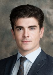 Seán B Lyne合作论文数Univ Chicago Med & Biol Sci, Dept Surg, Sect Neurosurg, Neurovasc Surg Program, Chicago, IL USA7
 Daniel A Snellings合作论文数Duke Univ, Sch Med, Mol Genet & Microbiol Dept, Durham, NC USA7Courtney C Hong合作论文数7
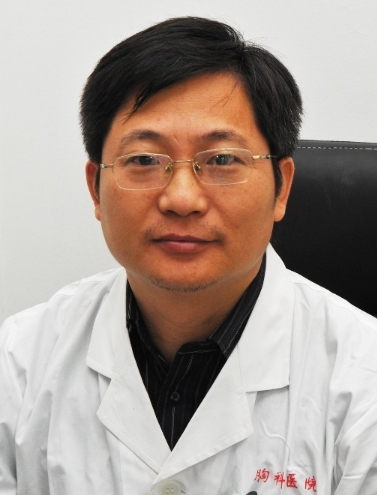 Yiqing Yang (杨奕清)合作论文数Shanghai Chest Hospital7Amy Akers合作论文数7Lindsay Sceats合作论文数6
 Anneli Peters合作论文数6A Young Kim合作论文数6
 Nicholas Hobson合作论文数6MinMin Lu合作论文数6
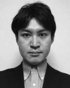 Takashi Morita合作论文数6
 Lan Cheng合作论文数6Stephanie Sterling合作论文数6
 Siqi Gao合作论文数6
 Elizabeth Gardiner合作论文数John Curtin School of Medical Research, College of Health and Medicine, The Australian National University6




