X-ray and neutron tomography are applied as a bi-modal approach for the 3D characterisation of a Monturaqui impactite formed by shock metamorphism during the impact of an iron meteorite with the target rocks in the Monturaqui crater (Chile). The particular impactite exhibits structural heterogeneities on many length scales: its composition is dominated by silicate-based glassy and crystalline materials with voids and Fe/Ni-metal and oxihydroxides particles generally smaller than 1 mm in diameter. The non-destructive investigation allowed us to apply a novel bi-modal imaging approach that provides a more detailed and quantitative understanding of the structural and chemical composition compared to standard single mode imaging methods, as X-ray and neutron interaction with matter results in different attenuation coefficients with a non-linear relation. The X-ray and neutron data sets have been registered, and used for material segmentation, porosity and metallic content characterization. The bimodal data enabled the segmentation of a large number of different materials, their morphology as well as distribution in the specimen including the quantification of volume fractions. The 3D data revealed an evaporite type of material in the impactite not noticed in previous studies. The present study is exemplary in demonstrating the potential for non-destructive characterisation of key features of complex multi-phase objects such as impactites.
Recent developments in multispectral X-ray detectors allow for an efficient identification of materials based on their chemical composition. This has a range of applications including security inspection, which is our motivation. In this paper, we analyze data from a tomographic setup employing the MultiX detector, that records projection data in 128 energy bins covering the range from 20 to 160 keV. Obtaining all information from this data requires reconstructing 128 tomograms, which is computationally expensive. Instead, we propose to reduce the dimensionality of projection data prior to reconstruction and reconstruct from the reduced data. We analyze three linear methods for dimensionality reduction using a dataset with 37 equally-spaced projection angles. Four bottles with different materials are recorded for which we are able to obtain similar discrimination of their content using a very reduced subset of tomograms compared to the 128 tomograms that would otherwise be needed without dimensionality reduction.
Nowadays, the use of MULTIX detectors which provide imaging of many channels once has been so popular in some applications such as security. On the other hand, the reconstruction of energy channels can be time consuming. In this paper, we will try to reduce the number of energy beams so that we do not lose a lot of information. The results shows that we can get nearly the same result with dimensional reduction as without. It can speed up the process of reconstruction to great extend.
Energy dispersive X-ray diffraction (EDXRD) can be applied for identification of liquid threats in luggage scanning in security applications. To define the instrumental design, the framework for data reduction and analysis and test the performance of the threat detection in various scenarios, a flexible laboratory EDXRD test setup was build. A data set of overall 570 EDXRD spectra has been acquired for training and testing of threat identification algorithms. The EDXRD data was acquired with limited count statistics and at multiple detector angles and merged after correction and normalization. Initial testing of the threat detection algorithms with this data set indicate the feasibility of detection levels of > 95 % true positive with < 6 % false positive alarms.
Active Appearance Models (AAM) are used for annotating or segmenting shapes in biomedical images. Performance relies heavily on the image data used to train the AAM. In this paper we improve the generalization properties of the model by making it robust to slowly varying spatial intensity inhomogeneities which are often seen in Light Sheet Fluorescence Microscopy (LSFM) images. This robustness is achieved by modelling the appearance of an image as a regularized Normalized Gradient Field (rNGF). We perform two experiments to challenge the model. First it is tested using a repeated leave-one-out approach on images with minimal imperfections where the left out images are corrupted by a simulated bias field and segmented using the AAM. Secondly we test the model on LSFM images with common acquisition problems. In both experiments the proposed approach outperforms the often used AAM implementation based on Sum of Squared Differences.
Accurate tumor segmentation plays an important role in radiosurgery planning and the assessment of radiotherapy treatment efficacy. In this paper we propose a method combining an ensemble of 2D convolutional neural networks for doing a volumetric segmentation of magnetic resonance images. The segmentation is done in three steps; first the full tumor region, is segmented from the background by a voxel-wise merging of the decisions of three networks learned from three orthogonal planes, next the segmentation is refined using a cellular automaton-based seed growing method known as growcut. Finally, within-tumor sub-regions are segmented using an additional ensemble of networks trained for the task. We demonstrate the method on the MICCAI Brain Tumor Segmentation Challenge dataset of 2014, and show improved segmentation accuracy compared to an axially trained 2D network and an ensemble segmentation without growcut. We further obtain competitive Dice scores compared with the most recent tumor segmentation challenge.
Background: There is a large unmet need for treatments for patients with progressive multiple sclerosis (MS). Phase 2 studies with cerebrospinal fluid (CSF) biomarker outcomes may be well suited for the initial evaluation of efficacious treatments.Objective: To evaluate the effect of monthly oral methylprednisolone pulse treatment on intrathecal inflammation in progressive MS.Methods: In this open-label phase 2A study, 15 primary progressive and 15 secondary progressive MS patients received oral methylprednisolone pulse treatment for 60 weeks. Primary outcome was changes in CSF concentrations of osteopontin. Secondary outcomes were other CSF biomarkers of inflammation, axonal damage and demyelination; clinical scores; magnetic resonance imaging measures of disease activity, magnetization transfer ratio (MTR) and diffusion tensor imaging (DTI); motor evoked potentials; and bone density scans.Results: We found no change in the CSF concentration of osteopontin, but we observed significant improvement in clinical scores, MTR, DTI and some secondary CSF outcome measures. Adverse events were well-known side effects to methylprednisolone.Conclusion: Monthly methylprednisolone pulse treatment was safe, but had no effect on the primary outcome. However, improvements in secondary clinical and MRI outcome measures suggest that this treatment regimen may have a beneficial effect in progressive MS.
Hippocampal volumetry derived from structural magnetic resonance imaging (MRI) has been endorsed by the Alzheimer's disease (AD) diagnostic guidelines as a radiological marker of disease progression. Among the top performing automated hippocampal segmentation methods are multi-atlas segmentation methods, which rely on manual annotations. In this study, we investigate a combination of such method with annotations from a new Harmonized Hippocampal Protocol (HHP). We compare its capabilities to a FreeSurfer method and verify its impact on segmentation and diagnostic group separation capabilities. 40 manual HHP hippocampal annotations (12 normal control (NC), 11 mild cognitive impairment (MCI), 17 AD) were transformed to a common segmentation space. The corresponding 1.5T MRIs were preprocessed using FreeSurfer. An automated Non-Local Patch-based segmentation technique (N-L Patch) was used to segment the left- and right hippocampus, separately. All 40 HHP annotations were used as atlases during pre-selection, but only the 9 most similar contributed to the final segmentation. Leave-one-out cross-validation was performed on the 40 atlases, and the corresponding DICE-scores with the manual annotations were calculated. A standardized ADNI dataset containing 1.5T MRIs from 504 subjects (169 NC, 234 MCI, 101 AD) at baseline and month 12 was segmented using the method described above and atrophy rate calculated as percentage volume change was estimated. Mean (±sd) cross-validation DICE-scores of the 40 atlases segmented using N-L Patch and cross-sectional FreeSurfer were 0.868 (±0.019) and 0.781 (±0.031), respectively. A paired t-test between N-L Patch and FreeSurfer DICE-scores showed significance (p<0.001).Statistics in terms of AUC and Cohens' D were used to evaluate differences in atrophy rates between diagnostic groups for N-L Patch and FreeSurfer segmentations. N-L Patch performed significantly better in separating AD from NC and AD from MCI, Table 1. Including the HHP labels in a multi-atlas segmentation method resulted in better segmentation consensuses with the new hippocampal label standard than a state-of-the-art method, FreeSurfer. Furthermore, N-L Patch yielded significantly better group separation than FreeSurfer in separating AD from NC and AD from MCI. This illustrates the longitudinal robustness of segmentations when annotations from the new hippocampal label standard are included in automated segmentation methods.
Multiple sclerosis (MS) damages central white matter pathways which has considerable impact on disease-related disability. To identify disease-related alterations in anatomical connectivity, 34 patients (19 with relapsing remitting MS (RR-MS), 15 with secondary progressive MS (SP-MS) and 20 healthy subjects underwent diffusion magnetic resonance imaging (dMRI) of the brain. Based on the dMRI, anatomical connectivity mapping (ACM) yielded a voxel-based metric reflecting the connectivity shared between each individual voxel and all other brain voxels. To avoid biases caused by inter-individual brain-shape differences, they were estimated in a spatially normalized space. Voxel-based statistical analyses using ACM were compared with analyses based on the localized microstructural indices of fractional anisotropy (FA). In both RR-MS and SP-MS patients, considerable portions of the motor-related white matter revealed decreases in ACM and FA when compared with healthy subjects. Patients with SP-MS exhibited reduced ACM values relative to RR-MS in the motor-related tracts, whereas there were no consistent decreases in FA between SP-MS and RR-MS patients. Regional ACM statistics exhibited moderate correlation with clinical disability as reflected by the expanded disability status scale (EDSS). The correlation between these statistics and EDSS was either similar to or stronger than the correlation between FA statistics and the EDSS. Together, the results reveal an improved relationship between ACM, the clinical phenotype, and impairment. This highlights the potential of the ACM connectivity indices to be used as a marker which can identify disease related-alterations due to MS which may not be seen using localized microstructural indices.
Natalizumab inhibits the migration of systemic immune cells to the CNS and may be beneficial in progressive multiple sclerosis (MS). The objective of the study was to examine the effects of natalizumab in progressive MS.In an open-label phase 2A study, 24 patients with progressive MS were included to receive natalizumab treatment for 60 weeks. Response to natalizumab was assessed in CSF and MRI studies. The primary endpoint was change in CSF osteopontin, a biomarker of intrathecal inflammation, from baseline to week 60.Seventeen patients completed the study. No new safety issues were encountered. CSF osteopontin decreased by 65 ng/mL (95% confidence interval 34-96 ng/mL; p = 0.0004) from baseline to week 60 in conjunction with decreases in other CSF biomarkers of inflammation, axonal damage, and demyelination. Magnetization transfer ratio increased in both cortical gray and normal-appearing white matter and correlated with decreases in CSF neurofilament light chain.Natalizumab treatment of progressive MS reduces intrathecal inflammation and tissue damage, supporting a beneficial effect of natalizumab treatment in progressive MS and suggesting that systemic inflammation contributes to the pathogenesis. Moreover, the study establishes the feasibility of using CSF biomarkers in proof-of-concept trials, allowing a low number of participants and short study duration.This study provides Class IV evidence that in patients with progressive MS, natalizumab reduces biomarkers of intrathecal inflammation.
Objective:Natalizumab inhibits the migration of systemic immune cells to the CNS and may be beneficial in progressive multiple sclerosis (MS). The objective of the study was to examine the effects of natalizumab in progressive MS.Methods:In an open-label phase 2A study, 24 patients with progressive MS were included to receive natalizumab treatment for 60 weeks. Response to natalizumab was assessed in CSF and MRI studies. The primary endpoint was change in CSF osteopontin, a biomarker of intrathecal inflammation, from baseline to week 60.Results:Seventeen patients completed the study. No new safety issues were encountered. CSF osteopontin decreased by 65 ng/mL (95% confidence interval 34-96 ng/mL; p = 0.0004) from baseline to week 60 in conjunction with decreases in other CSF biomarkers of inflammation, axonal damage, and demyelination. Magnetization transfer ratio increased in both cortical gray and normal-appearing white matter and correlated with decreases in CSF neurofilament light chain.Conclusions:Natalizumab treatment of progressive MS reduces intrathecal inflammation and tissue damage, supporting a beneficial effect of natalizumab treatment in progressive MS and suggesting that systemic inflammation contributes to the pathogenesis. Moreover, the study establishes the feasibility of using CSF biomarkers in proof-of-concept trials, allowing a low number of participants and short study duration.Classification of evidence:This study provides Class IV evidence that in patients with progressive MS, natalizumab reduces biomarkers of intrathecal inflammation.
Identifying specific structures of the brain where pathology differs between groups of subjects may aid to develop imaging-based markers for disease diagnosis. We propose a new technique for doing multivariate statistical analysis on white matter tracts with sheet like shapes. Previous works assume tube-like shapes, not always suitable for modelling the white matter tracts of the brain. The tract-oriented technique aimed at group studies, integrates the usage of multivariate features and outputs a single value of significance indicating tract-specific differences. This is in contrast to voxel based analysis techniques which outputs a significance per voxel basis, and requires multiple comparison correction. We demonstrate our technique by comparing a group of controls with a group of Multiple Sclerosis subjects obtaining significant differences on 11 different fascicle structures.
Introduction Anatomical Connectivity Mapping (ACM) based on diffusion MRI generates a scalar map that reflects the connectivity of each voxel with the rest of the brain [1, 2]. The value of an ACM voxel reflects white mater integrity both locally and globally and the ACM is therefore able to reveal effects not seen with traditional diffusion maps such as Mean Diffusivity (MD) or Fractional Anisotropy (FA), specifically with a disease that results in a varying degree of local and global pathology changes along white matter axons. Multiple Sclerosis (MS) is a disease that fits this profile with its heterogeneous disease pattern where lesions along a given tract may have varying impact on the global conductivity/connectivity of the underlying axons. The ACM map is estimated using probabilistic tractography performed within a brain mask which is usually made by manual delineation by a clinician or obtained from automatic brain segmentation tools. We suggest a variation of ACM particularly suited for group studies that avoid brain masks defined in individual subject space as done in previously [2] and demonstrate it on a group of patients with MS. It is known that MS patients with higher Expanded Disability Status Scale (EDSS) have an increased motor handicap and our aim is to show that this effect is measurable using ACM by testing the hypothesis that a group of patients with high motor impairment (High EDSS) has a significant decreased connectivity profile along the corticospinal tract (CST) compared to a group of patients with low motor impairment (Low EDSS). The results demonstrate that ACM is able to reveal group wise differences not seen in FA.
We propose a method for the segmentation of Multiple Sclerosis lesions. The method is based on probability maps derived from a K-Nearest Neighbours classification. These are used as a non parametric likelihood in a Bayesian formulation with a prior that assumes connectivity of neighbouring voxels. The formulation is solved using the method of Iterated Conditional Modes (ICM). The parameters of the method are found through leave-one-out cross validation on training data after which it is evaluated on previously unseen test data. The multi modal features investigated are 3 structural MRI modalities, the diffusion MRI measures of Fractional Anisotropy (FA), Mean Diffusivity (MD) and several spatial features. Results show a benefit from the inclusion of diffusion primarily to the most difficult cases. Results shows that combining probabilistic K-Nearest Neighbour with a Markov Random Field formulation leads to a slight improvement of segmentations.
Introduction Echo Planar Imaging (EPI) sequences are subject to imaging artifacts, caused by subject motion, eddy currents effects and field inhomogeneity distortions (susceptibility) causing a geometric displacement of voxel intensities along the phase encode direction. Inhomogeneity correction is important to obtain an anatomical correct image which can be aligned with structural MR images. To estimate the displacement field that allows correction, we extend an existing technique, based on acquiring a full EPI sequence and one additional EPI image acquired with reversed gradient polarity along the phase encoding direction [2, 6]. The EPI with reversed gradient polarity contains the same intensity information as a corresponding image of the EPI sequence but with distortions causing voxel shifts in the reverse directions. To find the displacement field between two reversely distorted EPI’s, an image registration problem is solved as in [2]. We propose to use the simpler, more efficient Thirion's demons algorithm [3, 4] and suggest a different registration pipeline for obtaining the displacement fields and name it phase reversed demons (PRD). The PRD is compared to two other correction methods that require additional MRI sequences, leading to increased scan time. These are the gradient field map (FM) [1] and the point spread function (PSF) [5]. We compare the three methods applied to five subjects. The results are compared visually and quantitatively by estimating the statistical dependence with a structural T1-weighted image. The quantitative results indicate that the (PRD) approach is competitive by being more similar with the structural image but inspection of regions in subjects also demonstrates individual cases where the other methods are favorable.
This paper investigates and demonstrates a 4D lung CT reconstruction/registration method which results in a complete volumetric model of the lung that deforms according to a respiratory motion field. The motion field is estimated iteratively between all available slice samples and a reference volume which is updated on the fly. The method is two part and the second part of the method aims to correct wrong phase information by employing another iterative optimizer. This two part iterative optimization allows for complete reconstruction at any phase and it will be demonstrated that it is better than using an optimization which does not correct for phase errors. Knowing how the lung and any tumors located within the lung deforms is relevant in planning the treatment of lung cancer.
 Rasmus Larsen合作论文数Technical University of Denmark12
 Hartwig R. Siebner合作论文数Department of Clinical Medicine, University of Copenhagen;Center for Functional and Diagnostic Imaging and Research, Danish Research Center for Magnetic Resonance, Copenhagen University Hospital Hvidovre;Faculty of Health and Medical Sciences, Institute of Clinical Medicine, University of Copenhagen9
 Per Soelberg Sørensen合作论文数Department of Clinical Medicine, University of Copenhagen8
 Ellen Garde合作论文数7
 Tim Dyrby合作论文数Department of Applied Mathematics and Computer Science, Technical University of Denmark;Danish Research Centre for Magnetic Resonance, Copenhagen University Hospital6
 Finn Sellebjerg合作论文数Institute of Clinical Medicine, University of Copenhagen;Danish Multiple Sclerosis Center, Copenhagen University Hospital Rigshospitalet4
 R. Ratzer合作论文数4
 Anders Bjorholm Dahl合作论文数Department of Applied Mathematics and Computer Science, Technical University of Denmark4
 Morten Blinkenberg合作论文数Danish Multiple Sclerosis Center;Department of Neurology, Rigshospitalet;Copenhagen University Hospital4
 Mads Fogtmann合作论文数3
 Timo Erkinjuntti合作论文数Department of Neurology, Helsinki University Central Hospital3
 Lars Bornsen合作论文数3Ulrik Lund Olsen合作论文数Center for Fundamental Research: Metal Structures in Four Dimensions, the Technical University of Denmark3
 Knut Conradsen合作论文数3J. Romme Christensen合作论文数2Mina Kheirabadi合作论文数2Cecilie Anker合作论文数2Dogonowski Anne-Marie合作论文数2
 Jeppe Romme Christensen合作论文数2
 Akshay Pai合作论文数2
 Mads Nielsen合作论文数Department of Computer Science, Faculty of Science, University of Copenhagen2
 Geoff Parker合作论文数Department of Med Phys & Biomedical Eng, University College London;Bioxydyn Limited2
 Lauge Sørensen合作论文数Department of Computer Science, University of Copenhagen2
 Lars Kai Hansen合作论文数Department of Applied Mathematics and Computer Science, Technical University of Denmark1
 Sune Darkner合作论文数Department of Computer Science, University of Copenhagen1Louise B Johansen合作论文数1
 Wesley Thompson合作论文数Division of Biostatistics & Bioinformatics, Department of Family Medicine and Public Health, School of Medicine, University of California, San Diego1
 Cecilie Ammitzbøll合作论文数1
 Anna Fedrigo合作论文数Rutherford Appleton Lab, ISIS Facil1Kasper Marstal合作论文数1Jensen, C.B.合作论文数1Mikael Agn合作论文数1
 Vedrana Andersen Dahl合作论文数Department of Applied Mathematics and Computer Science, Technical University of Denmark1
 Wail Mustafa合作论文数1Reislev Nina L合作论文数1Madsen Cg合作论文数1
 Peter Iversen合作论文数Copenhagen Prostate Cancer Center, Rigshospitalet;Institut for Klinisk Medicin, Københavns Universitet1Kehres Jan合作论文数1
 Anna L Secher合作论文数1
 Gary Zhang合作论文数Department of Computer Science, Faculty of Engineering Science, University College London1
 Terry Jernigan合作论文数Center for Human Development, University of California, San Diego;Department of Cognitive Science, Psychiatry, and Radiology, University of California, San Diego1Hartwig R Siebner合作论文数1
 Daniel Alexander合作论文数Department of Computer Science, Faculty of Engineering Sciences, University College London1
 Henrik Pedersen合作论文数Department of Physics, Chemistry and Biology, Linköping University1
 Carsten Gundlach合作论文数School of Mathematical Sciences, University of Southampton1
 Kristoffer Hougaard Madsen合作论文数Informatics and Mathematical Modelling;Technical University of Denmark;Informatics and Mathematical Modelling, Technical University of Denmark1
 Tue Herlau合作论文数Department of Informatics, Technical University of Denmark1
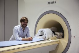 Henrik Lundell合作论文数1
 Martin Lillholm合作论文数Department of Computer Science, University of Copenhagen;CEREBRIU1




