The inferior olive (IO) supports motor learning by supplying the cerebellum with critical sensory and motor input. In adult rats, that input includes externally generated limb stimulation. In contrast, the IO of Postnatal Day 8 (P8) rats does not exhibit responses to external stimuli. Instead, IO activity primarily reflects corollary discharges associated with the production of self-generated limb twitches during active (REM) sleep. Because corollary discharges are necessary for the computation of internal models, we tested the hypothesis that IO-related corollary discharge is necessary for the expression of cerebellar-dependent feed-forward activity during development. First, by recording from the IO of P12 and P20 rats of both sexes, we confirmed the presence of twitch-related corollary discharge at both ages; however, whereas the IO at P20 responded to limb stimulation, the IO at P12 did not. Next, using a protocol for selectively lesioning the climbing fibers that connect the IO to the cerebellum, including the interpositus nucleus (IP), we confirmed that lesioning at P12 prevents the IP's expression of corollary discharge at P13. Finally, we assessed the necessity of IO input to the cerebellum for the typical development of an internal model by lesioning climbing fibers at P12 or P19 and testing for the model's expression in the thalamus at P20. Only when the lesions occurred at P12 was the expression of the internal model severely disrupted. These findings provide the most direct evidence to date linking twitch-related corollary discharge to the developmental emergence of a cerebellar-dependent internal model.
Abstract Introduction Though sleep is crucial for motor memory consolidation, the brain mechanisms underlying its role in real-world, whole-body movement, such as dance, remain poorly understood. Here, we examine how daytime naps and specific features of sleep, such as twitches during REM sleep, impact improvement across a three-day dance-learning protocol. Methods Nineteen adults (12 females, 7 males) completed three days of dance classes, learning a standardized choreography. Each participant took one baseline nap before any training and one experimental nap that occurred on either Day 1, Day 2, or Day 3, followed by a post-nap dance test to assess nap-dependent improvement. We developed a method to systematically evaluate dance performances from video, based on four error types: detail (incorrect movement execution), order (sequence mistakes), omission (missing moves), and addition (extra unintended movements). For each nap, we calculated the rate of twitching (twitches/min) and the proportion of twitching for each body part. We used repeated-measures ANOVA, linear mixed-effects models, and Tukey-adjusted comparisons to evaluate learning, nap timing, and twitch characteristics. Results In preliminary analyses, we observed robust learning across days, with a significant linear reduction in error scores across the three classes. Given the strong linear effect of learning across classes, we examined the nap effect while controlling for this linear learning effect. Nap day significantly predicted post-nap performance change, with naps on Day 1 showing significant improvement, and naps on Days 2 and 3 having minimal effects. We compared twitching between the baseline nap (pre-dance class) and the experimental naps for each body part. Twitching differed by region, with hand twitches decreasing and foot twitches increasing from baseline to experimental naps. Conclusion Though data collection is ongoing, these early findings indicate that earlier naps yield larger performance benefits during whole-body motor learning, and the body regions most engaged during dancing (feet) showed more twitching after learning. Our findings suggest a systematic approach to studying whole-body motor learning and sleep. Support (if any)
Limb twitching is among the earliest observable behaviors in human development and a hallmark of active (rapid eye movement, REM) sleep. Systematic assessments in full-term infants reveal a spatiotemporal organization of twitching that informal observation cannot. Because preterm infants spend even more time asleep and are at heightened risk for neurodevelopmental disorders, we provide the first systematic characterization of twitching at 34-35 weeks postmenstrual age. Preterm infants exhibit an immense quantity of twitching across the body, underscoring its potential functional significance. The spatiotemporal structure of twitching also changes with age, including a selective increase in finger and toe twitching. Unexpectedly, during periods of tracé alternant, a precursor to quiet sleep, twitching appears in brief bouts that are almost exclusively restricted to the legs. These findings show how this abundant but overlooked sleep behavior provides a sensitive assay of the developing neural control of movement, with implications for understanding typical and atypical development.
The emergence of the cortical delta rhythm (1-4 Hz) during quiet sleep (QS) is a major milestone in brain development. In rats, this milestone is achieved between 8 and 12 days of postnatal (P) age. We previously reported an age-dependent increase in PZ delta-rhythmic activity that is synchronized with cortical delta and entrained by breathing. Here, we ask whether this long-distance synchrony persists in response to perturbations to sleep homeostasis or respiration. First, using male and female P12 rats, we investigated the coupling strength between frontal cortex and PZ in response to a short but intense period of sleep deprivation. During recovery sleep, we observed a rebound in delta power in both PZ and cortex, even in the absence of increased QS duration, indicating that PZ and cortical delta power are equivalent markers of homeostatic sleep regulation. Analyses of phase-locking and lagged cross-correlation revealed persistent temporal coupling between the two rhythms such that cortical delta reliably lagged PZ delta regardless of changes in sleep pressure. Curiously, we also observed an increase in breathing depth during recovery sleep, which we confirmed in a separate cohort of pups. Next, using mild hypercapnia (5% CO2) to alter breathing frequency and depth, we produced decreases in cortical and PZ delta power along with decreases in the depth of breathing. These findings provide additional support for the notion that PZ and cortical delta rhythms function as distantly interconnected components within a developmentally emerging sleep-homeostatic system that is also intimately tied with the brainstem respiratory network.
The methods used for assessing infants' sleep vary widely, and the insights provided by each method are not necessarily interchangeable. Direct physiological measurements are often performed in hospital environments with extensive and controllable instrumentation, whereas at-home assessments have been based on parental questionnaires and diaries or actigraphy measurements. There is a rising need for the establishment of objective at-home methods to improve ecological validity in assessing infant sleep. Here, we review how specific research questions can be aligned with at-home assessment methods, ranging from indirect parental interviews to direct modern measures with unobtrusive sensors (wearables, in-bed sensors, video), and their integrated machine learning -based analytics. We discuss the nature of information available from each method in relation to the physiological or behavioural sleep phenomena. The currently available methods may support disruptive science, including globally harmonized and scaled-up studies across cultures and geographic locations, including fully decentralized developmental research and sleep-focused health care. IMPACT: The paper raises the practical and scientific need to study infant sleep using methods that are both objective and ecologically valid, thereby supporting the use of direct at-home measurements. The paper emphasizes the multifaceted nature of infant sleep, which precludes its assessment as a solitary entity and calls for clear research questions to identify measurable phenomena in sleep-related physiology or behavior Presently available research methods for at-home studies are described and compared in relation to their information content, practical utility and caveats Various analytic results and sleep metrics are described in relation to the underlying sleep physiology or behavior. The overall message emphasizes the need to establish a solid end-to-end waterfall framework in infant sleep research, spanning study questions, sleep phenomena, recording methods, and data analytics.
Twitches are discrete movements that characterize rapid eye movement (REM) sleep. However, recent work showed that twitches also occur during non-REM (NREM) sleep in human infants beginning around 3 months of age, a time when sleep spindles and the cortical delta rhythm are also emerging. Further, NREM twitches are coupled with sleep spindles, suggesting a unique contribution to sensorimotor development. Given that NREM sleep is composed of distinct substages, we investigated whether twitching and twitch-spindle coupling are differentially expressed during N2 and N3 sleep. In 6-month-old human infants (n = 21; 7 females), we recorded electroencephalogram, respiration, and video during daytime sleep. We found high-intensity twitching during N2 and REM but not N3 sleep. In contrast, sleep spindles exhibited similar temporal characteristics during N2 and N3. Also, despite differences in the intensity of twitching during N2 and N3, significant twitch-spindle coupling occurred in both stages. Finally, the rate of twitching was inversely related to delta power across NREM periods. These findings suggest that although twitching occurs during REM, N2, and N3 sleep at this age, its expression is compatible with some sleep components (e.g., rapid eye movements, sleep spindles) but not others (e.g., cortical delta), highlighting the continuing need to better understand the dynamic organization of sleep and its individual components in early development.
Beginning around postnatal day 12 (P12) in rats, quiet sleep (QS, or NREM sleep) increases at the expense of active sleep (AS, or REM sleep). The developmental increase in QS is accompanied by the sudden emergence of cortical delta (0.5-4 Hz). We recently discovered a novel delta rhythm in a medullary structure, the parafacial zone (PZ), a region implicated in regulating QS in adults. At P12, PZ delta was generated locally and was highly coherent with cortical delta. We hypothesized that if PZ delta is a homolog of cortical delta, then the two rhythms should exhibit parallel homeostatic responses to sleep deprivation. P12 rats (n=8) were deprived of sleep for 30 minutes while recording from the PZ and frontal cortex. Cold stimuli were applied to the snout to induce arousal whenever cortical delta was observed. After deprivation, pups were allowed to sleep undisturbed for an additional 60 minutes for recovery. Control pups (n = 8) were matched for sex and age and were left undisturbed for 90 min. PZ and cortical LFP, PZ unit activity, respiration, and behavioral measures (EMG, video) were obtained for each pup. Sleep deprivation produced intense sleep pressure, as operationalized by the need to increase the rate of arousing stimulations over the 30-minute deprivation period. Over the recovery period, PZ and cortical delta showed parallel homeostatic responses consisting of initially increased delta power that decreased in lock-step toward baseline over time. The durations of QS and AS showed a similar pattern. Brief arousals (< 10 s) occurred more often during the recovery period; these arousals elicited simultaneous decreases in delta power in PZ and cortex. Surprisingly, pups breathed more deeply at the start of the recovery period, providing additional evidence of a link between respiration and delta power at this age. These results support the notion that PZ and cortical delta are closely linked components within the sleep-regulatory system. Also, the findings suggest intimate connections between delta- and respiration-generating structures in the brainstem, a possibility that is currently being investigated. NIH grant R37-HD081168 to M.S.B.
The nativist-empiricist debate and the nativist commitment to the idea of core knowledge and endowments that exist without relevant postnatal experience continue to distract attention from the reality of developmental systems. The developmental systems approach embraces the concept of epigenesis, that is, the view that development emerges via cascades of interactions across multiple levels of causation, from genes to environments. This view is rooted in a broader interpretation of experience and an appreciation for the nonobvious nature of development. We illustrate this systems approach with examples from studies of imprinting, spatial cognition, and language development, revealing the inadequacies of the nativist-empiricist debate and the inconvenient truths of development. Developmental scientists should no longer abide the nativist-empiricist debate and nativists’ ungrounded focus on origins. Rather, the future lies in grounding our science in contemporary theory and developmental process.
Twitches are discrete movements that contribute to the self-organization of the sensorimotor system. At 3 months of age, infants begin to twitch during both NREM and REM sleep, and twitches during NREM occur in synchrony with sleep spindles. In the present study, we examine in 6-month-olds how the specific stage of NREM sleep (i.e., N2 vs. N3) modulates the characteristics of twitches and their coupling with spindles. EEG and video were recorded during daytime naps in 21 infants (7F, 6.19 ± 0.10 months). Twitches of the arms, legs, and face, and rapid eye movements, were visually identified by two independent raters. EEG was scored in 30-s epochs using AASM scoring criteria. Spindles were identified in the C3 electrode during artifact-free periods using an automated detection algorithm. Differences in twitch rate (twitches/min) between sleep stages (i.e., REM vs. NREM) and substages (i.e., N2 vs N3) were analyzed. Paired t tests were used to assess differences in spindle rate (spindles/min) and twitch-spindle co-occurrences between NREM substages. The rate of twitching in NREM was not significantly different from that during REM (NREM: 9.13 ± 1.16, REM: 13.70 ± 2.68; N = 18 infants; Z = -1.24). During NREM, infants twitched significantly more in N2 than in N3 (N2: 13.45 ± 1.95; N3: 5.68 ± 0.77; N = 20 infants; Z = - 3.21, p = 0.001), but the spindle rate was the same across the substages (N2: 3.28 ± 0.27; N3: 3.03 ± 0.33; N=20 infants; t(19)=.904). Regardless of substage, there was a high probability that a spindle occurred at the onset of a twitch. Moreover, the proportion of twitches with co-occurring spindles was not different between substages (N2: 0.26 ± 0.03; N3: 0.24 ± 0.03; N=17 infants; t(16) = 0.80). These results indicate that 6-month-old infants twitch more during N2 than N3, but exhibit similar rates of spindle production. Regardless of NREM substage twitches are equally likely to co-occur with spindles. Thus, it may be that it is the presence of slow waves during N3 that is incompatible with the production of twitches. This possibility is currently being examined. R01-HD104616 to MSB
By recording brain activity in seven lizard species, humans, rats and pigeons, we demonstrate an infraslow brain rhythm during sleep in all species. This rhythm is tightly coupled with eye movements, muscle tone, heart and breathing rate in lizards, with skin brightness in chameleons and with pulsatile changes in cerebrovascular volume throughout sleep in bearded dragons and during non-rapid eye movement sleep in mice. These findings indicate that the infraslow rhythm is conserved across amniotes, questioning the evolution of sleep states.
In early development, active sleep is the predominant sleep state before it is supplanted by quiet sleep. In rats, the developmental increase in quiet sleep is accompanied by the sudden emergence of the cortical delta rhythm (0.5-4 Hz) around postnatal day 12 (P12). We sought to explain the emergence of the cortical delta by assessing developmental changes in the activity of the parafacial zone (PZ), a medullary structure thought to regulate quiet sleep in adults. We recorded from the PZ in P10 and P12 rats and predicted an age -related increase in neural activity during increasing periods of delta -rich cortical activity. Instead, during quiet sleep, we discovered sleep -dependent rhythmic spiking activity-with intervening periods of total silence-phase locked to a local delta rhythm. Moreover, PZ and cortical delta were coherent at P12 but not at P10. PZ delta was also phase locked to respiration, suggesting sleep -dependent modulation of PZ activity by respiratory pacemakers in the ventral medulla. Disconnecting the main olfactory bulbs from the cortex did not diminish cortical delta, indicating that the influence of respiration on delta at this age is not mediated indirectly through nasal breathing. Finally, we observed an increase in parvalbumin-expressing terminals in the PZ across these ages, supporting a role for local GABAergic inhibition in the PZ's rhythmicity. The unexpected discovery of delta -rhythmic neural activity in the medulla-when cortical delta is also emerging-provides a new perspective on the brainstem's role in regulating sleep and promoting long-range functional connectivity in early development.
Abstract Introduction An important function of sleep is neuroplasticity, which is essential to functional recovery after stroke. However, inpatient acute care settings may not be conducive to restful and consolidated sleep. The goals of the present study were to test the feasibility of using wearable technology to objectively quantify sleep during inpatient stay in the first few days of hospitalization post stroke and examine relations of sleep quality and architecture with stroke severity. Methods Data collection is ongoing, preliminary analyses are based on 8 patients (5F/3M, 41-78 yrs, mean age=61.7 yrs). A headband style EEG acquisition device (Sleep Profiler) was used for one night (0-8 days following a first-ever stroke, mean=3 days) either in ICU or neurology inpatient floors. Sleep was scored based on standard AASM criteria. The National Institute of Health Stroke Scale (NIHSS) was administered to assess stroke/disability severity. Results We observed great variability in sleep duration (2.1-6.2 hrs, mean=4.1 hrs), sleep efficiency (29-81%, mean=51%) and increased time spent in N3 (compared to standard sleep architecture; 21.2-72.8%, mean=41.7% of total sleep time). Stroke severity correlated with WASO (r=.82, p=.02) and time spent in supine position after sleep onset (r=.83, p=.02). Conclusion While preliminary, these findings identify profound sleep restriction and fragmentation experienced by stroke survivors during the first few days of hospitalization. Further, stroke severity predicts increased sleep fragmentation, possibly due to more intensive medical care required. Future plans involve examining long-term cognitive and functional outcomes in patients to test the hypothesis that better sleep during the acute phase of stroke enhances recovery. Support (if any) Iowa Neuroscience Institute Research Program of Excellence
Animals must distinguish the sensory consequences of self-generated movements (reafference) from those of other-generated movements (exafference). Only self-generated movements entail the production of motor copies (i.e., corollary discharges), which are compared with reafference in the cerebellum to compute predictive or internal models of movement. Internal models emerge gradually over the first three postnatal weeks in rats through a process that is not yet fully understood. Previously, we demonstrated in postnatal day (P) 8 and P12 rats that precerebellar nuclei convey corollary discharge and reafference to the cerebellum during active (REM) sleep when pups produce limb twitches. Here, recording from a deep cerebellar nucleus (interpositus, IP) in P12 rats of both sexes, we compared reafferent and exafferent responses with twitches and limb stimulations, respectively. As expected, most IP units showed robust responses to twitches. However, in contrast with other sensory structures throughout the brain, relatively few IP units showed exafferent responses. Upon finding that exafferent responses occurred in pups under urethane anesthesia, we hypothesized that urethane inhibits cerebellar cortical cells, thereby disinhibiting exafferent responses in IP. In support of this hypothesis, ablating cortical tissue dorsal to IP mimicked the effects of urethane on exafference. Finally, the results suggest that twitch-related corollary discharge and reafference are conveyed simultaneously and in parallel to cerebellar cortex and IP. Based on these results, we propose that twitches provide opportunities for the nascent cerebellum to integrate somatotopically organized corollary discharge and reafference, thereby enabling the development of closed-loop circuits and, subsequently, internal models.
In early development, active sleep is the predominant sleep state before it is supplanted by quiet sleep. In rats, the developmental increase in quiet sleep is accompanied by the sudden emergence of the cortical delta rhythm (0.5-4 Hz) around postnatal day 12 (P12). We sought to explain the emergence of cortical delta by assessing developmental changes in the activity of the parafacial zone (PZ), a medullary structure thought to regulate quiet sleep in adults. We recorded from PZ in P10 and P12 rats and predicted an age-related increase in neural activity during increasing periods of delta-rich cortical activity. Instead, during quiet sleep we discovered sleep-dependent rhythmic spiking activity-with intervening periods of total silence-phase-locked to a local delta rhythm. Moreover, PZ and cortical delta were coherent at P12, but not at P10. PZ delta was also phase-locked to respiration, suggesting sleep-dependent modulation of PZ activity by respiratory pacemakers in the ventral medulla. Disconnecting the main olfactory bulbs from the cortex did not diminish cortical delta, indicating that the influence of respiration on delta at this age is not mediated indirectly through nasal breathing. Finally, we observed an increase in parvalbumin-expressing terminals in PZ across these ages, supporting a role for GABAergic inhibition in PZ's rhythmicity. The discovery of delta-rhythmic neural activity in the medulla-when cortical delta is also emerging-opens a new path to understanding the brainstem's role in regulating sleep and synchronizing rhythmic activity throughout the brain.
Cognition in preverbal human infants must be inferred from overt motor behaviors such as gaze shifts, head turns, or reaching for objects. However, infant mammals - including human infants - show protracted postnatal development of cortical motor outflow. Cortical control of eye, face, head, and limb movements is absent at birth and slowly emerges over the first postnatal year and beyond. Accordingly, the neonatal cortex in humans cannot generate the motor behaviors routinely used to support inferences about infants' cognitive abilities, and thus claims of developmental continuity between infant and adult cognition are suspect. Recognition of the protracted development of motor cortex should temper rich interpretations of infant cognition and motivate more serious consideration of the role of subcortical mechanisms in early cognitive development.
Humans and other mammals never sleep more than when they are young. However, as any new parent is aware, infant sleep differs from that of adults in that it accumulates in short bouts across the day and night. It is only with age that sleep bouts consolidate and occur preferentially during the night. These basic developmental processes of consolidation and emergence of circadian rhythmicity are typical of mammals, whether they occur prenatally (in precocial species such as sheep) or postnatally (in altricial species such as rats). Another feature of sleep development is that some of the components that are used to define it in adults are initially absent in infants. Close inspection has revealed that sleep–wake cycles are expressed initially as relatively rapid fluctuations of high and low muscle tone coupled with wake-related behaviors (e.g., kicking, stretching) or sleep-related movements called myoclonic twitches. It is upon this foundation that other components of sleep, including the delta activity that characterizes quiet sleep, are added. Although brainstem mechanisms are sufficient to support the foundational components of sleep–wake cycling, forebrain components are necessary for consolidation, circadian rhythmicity, and sleep rebound after periods of sleep deprivation. Finally, although it is not yet fully understood why infants exhibit such high quantities of sleep – especially active sleep – in early infancy, or why infants exhibit such high rates of twitching, recent work is shedding new light on these important issues by focusing on the perinatal period as a time of increased brain plasticity.
In developing rats, behavioral state exerts a profound modulatory influence on neural activity throughout the sensorimotor system, including primary motor cortex (M1). We hypothesized that similar state-dependent modulation occurs in prefrontal cortical areas with which M1 forms functional connections. Here, using 8- and 12-day-old rats cycling freely between sleep and wake, we record neural activity in M1, secondary motor cortex (M2), and medial prefrontal cortex (mPFC). At both ages in all three areas, neural activity increased during active sleep (AS) compared with wake. Also, regardless of behavioral state, neural activity in all three areas increased during periods when limbs were moving. The movement-related activity in M2 and mPFC, like that in M1, is driven by sensory feedback. Our results, which diverge from those of previous studies using anesthetized pups, demonstrate that AS-dependent modulation and sensory responsivity extend to prefrontal cortex. These findings expand the range of possible factors shaping the activity-dependent development of higher-order cortical areas.
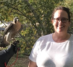 Greta Sokoloff合作论文数Department of Psychology;University of Iowa;Department of Psychology, University of Iowa71
 John H Freeman合作论文数31
 Scott R. Robinson合作论文数Laboratory for Psychobiological Research, Oregon State University29
 James Clinton Dooley合作论文数13
 Ethan J Mohns合作论文数10
 Scott K. Robinson (Scott Robinson)合作论文数Florida Museum of Natural History, University of Florida;Center for African Studies, College of Liberal Arts and Sciences, University of Florida10
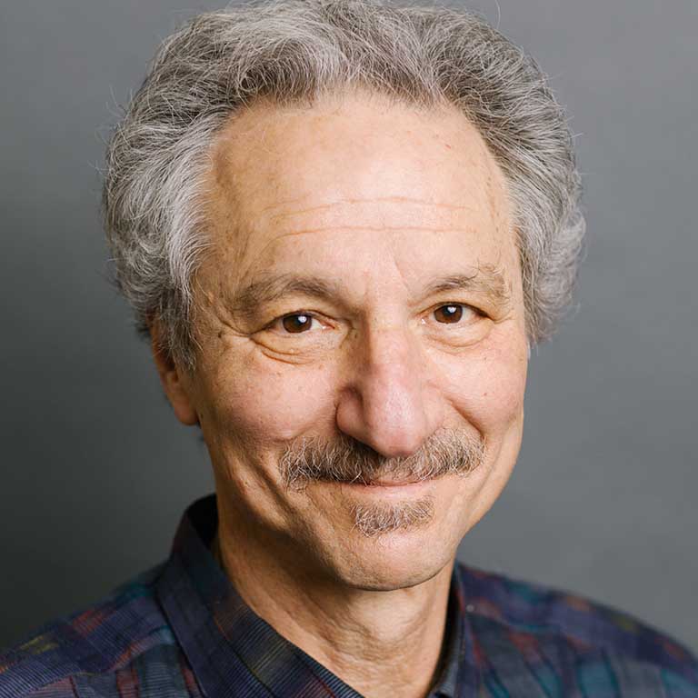 Jeffrey Alberts合作论文数Department of Psychological and Brain Sciences, The College of Arts & Sciences, Indiana University Bloomington9
 Alexandre Tiriac合作论文数Department of Biological Sciences, Vanderbilt University8
 Del Rio-Bermudez Carlos合作论文数Department of Psychology, University of Iowa8
 Andrew J Gall合作论文数8Rf Kirby合作论文数7Eric D. Johnson合作论文数7Kae Karlsson合作论文数6
 William D Todd合作论文数6
 Bob Mcmurray合作论文数Department of Psychological and Brain Sciences , College of liberal Arts and Sciences, University of Iowa6
 Joy Kreider合作论文数6A. J. Gall合作论文数5Karl Æ Karlsson合作论文数5
 Ryan M Glanz合作论文数Dept Psychol & Brain Sci, Univ Iowa5
 Adele Seelke合作论文数Department of Psychology, Physiology, and Behavior, University of California5Middlemis-Brown Jessica E合作论文数5Kent K J合作论文数5
 Howard Moltz合作论文数University of Chicago5AM Seelke合作论文数4
 Edward Wasserman合作论文数Department of Psychological and Brain Sciences, College of Liberal Arts and Sciences, The University of Iowa4Coleman Cassandra M合作论文数4Kim Jangjin合作论文数Department of Psychology, University of Iowa4
 Didhiti Mukherjee合作论文数All India Institute of Medical Sciences4
 John P Spencer合作论文数Department of Psychology;University of Iowa;Department of Psychology, University of Iowa4KyoungBin Im合作论文数3
 Alan Plumeau合作论文数3Mark Eric Dyken合作论文数3
 Julie A. Mennella合作论文数Monell Chemical Senses Center3Lucas D E合作论文数3Glenn Christine L合作论文数3
 James Bruce Tomblin合作论文数Department of Communication Sciences and Disorders, College of Liberal Arts and Sciences, University of Iowa3
 M. Bridget Zimmerman合作论文数Department of Biostatistics, College of Public Health, University of Iowa3
 Larissa K Samuelson合作论文数Department of Psychology;University of Iowa;Department of Psychology, University of Iowa3
 Robert Rodnitzky合作论文数3
 Deborah C. Lin-Dyken合作论文数3
 Sandra Lewis合作论文数Portland Cardiovascular Institute3
 Joshua Weiner合作论文数College of Liberal Arts and Sciences University of Iowa3Connie L. Grobe合作论文数2K.Æ. Karlsson合作论文数2Rebecca Y Wen合作论文数2Meredith M Hickerson合作论文数2
 Lex J. Gomez合作论文数Interdisciplinary Graduate Program in Neuroscience, University of Iowa2
 Brandt D Uitermarkt合作论文数University of Iowa2
 Markus Schmidt合作论文数2




