Computed tomography (CT) images are frequently degraded by acquisition artifacts, including noise, blur, streaking, aliasing, and metal artifacts. Yet CT enhancement is still largely evaluated using image quality metrics with limited perceptual and clinical validity, while existing datasets remain focused on isolated restoration tasks, hindering unified benchmarking across diverse degradation types. We present CT-DegradBench, a dataset and benchmark for CT degradation detection and severity estimation under controlled single- and mixed-artifact settings. CT-DegradBench enables systematic evaluation across multiple degradation families and severity levels within a common experimental framework. We further propose SeSpeCT (Semantic-Spectral CT degradation estimation), a framework that combines semantic priors from medical vision-language models with complementary frequency-domain cues for artifact analysis. SeSpeCT constructs a training-free semantic quality axis in the multimodal embedding space using radiology-informed text prompts, without task-specific fine-tuning, and combines it with spectral features that capture degradation-specific frequency patterns. The resulting representation enables joint prediction of artifact type and severity. Experimental results show that SeSpeCT consistently outperforms the evaluated baselines under both single- and mixed-degradation settings. The framework is available at https://github.com/yousranb/CT-DEGRADBENCH.
Medical image segmentation remains difficult to scale because high-performing methods typically rely on dense expert annotations and task-specific training. We introduce GazeRefine, a training-free framework that uses gaze as an inference-time prompt for zero-shot medical image segmentation. Sparse, duration-weighted fixations are converted into foreground and background priors that initialize semantic prototypes in frozen DINOv3 feature space. These prototypes are iteratively refined through foreground-background discrimination, feature-space affinity propagation, and anchoring to the initial gaze guidance, allowing segmentation to extend beyond directly fixated regions while limiting semantic drift. GazeRefine requires no segmentation masks, fine-tuning, adapters, prompt encoders, or gradient updates. We evaluate the method on gaze-annotated polyp segmentation and prostate MRI segmentation. The results show strong performance on colonoscopy images and competitive performance on prostate MRI, supporting gaze-guided prototype refinement as a promising approach for segmentation-label-efficient, human-in-the-loop medical image segmentation. Our tools and code can be found in the following repository: https://github.com/MohammedOussamaBEN/GazeRefine.git
Scanpath similarity metrics are central to eye-movement research, yet existing methods predominantly evaluate spatial and temporal alignment while neglecting semantic equivalence between attended image regions. We present a semantic scanpath similarity framework that integrates vision-language models (VLMs) into eye-tracking analysis. Each fixation is encoded under controlled visual context (patch-based and marker-based strategies) and transformed into concise textual descriptions, which are aggregated into scanpath-level representations. Semantic similarity is then computed using embedding-based and lexical NLP metrics and compared against established spatial measures, including MultiMatch and DTW. Experiments on free-viewing eye-tracking data demonstrate that semantic similarity captures partially independent variance from geometric alignment, revealing cases of high content agreement despite spatial divergence. We further analyze the impact of contextual encoding on description fidelity and metric stability. Our findings suggest that multimodal foundation models enable interpretable, content-aware extensions of classical scanpath analysis, providing a complementary dimension for gaze research within the ETRA community.
We introduce GazeVaLM, a public eye-tracking dataset for studying clinical perception during chest radiograph authenticity assessment. The dataset comprises 960 gaze recordings from 16 expert radiologists interpreting 30 real and 30 synthetic chest X-rays (generated by diffusion based generative AI) under two conditions: diagnostic assessment and real-fake classification (Visual Turing test). For each image-observer pair, we provide raw gaze samples, fixation maps, scanpaths, saliency density maps, structured diagnostic labels, and authenticity judgments. We extend the protocol to 6 state-of-the-art multimodal LLMs, releasing their predicted diagnoses, authenticity labels, and confidence scores under matched conditions - enabling direct human-AI comparison at both decision and uncertainty levels. We further provide analyses of gaze agreement, inter-observer consistency, and benchmarking of radiologists versus LLMs in diagnostic accuracy and authenticity detection. GazeVaLM supports research in gaze modeling, clinical decision-making, human-AI comparison, generative image realism assessment, and uncertainty quantification. By jointly releasing visual attention data, clinical labels, and model predictions, we aim to facilitate reproducible research on how experts and AI systems perceive, interpret, and evaluate medical images. The dataset is available at https://huggingface.co/datasets/davidcwong/GazeVaLM.
Understanding human visual attention is key to preserving cultural heritage We introduce SPGen a novel deep learning model to predict scanpaths the sequence of eye movementswhen viewers observe paintings. Our architecture uses a Fully Convolutional Neural Network FCNN with differentiable fixation selection and learnable Gaussian priors to simulate natural viewing biases To address the domain gap between photographs and artworks we employ unsupervised domain adaptation via a gradient reversal layer allowing the model to transfer knowledge from natural scenes to paintings Furthermore a random noise sampler models the inherent stochasticity of eyetracking data. Extensive testing shows SPGen outperforms existing methods offering a powerful tool to analyze gaze behavior and advance the preservation and appreciation of artistic treasures.
Accurate assessment of spheno-occipital synchondrosis (SOS) maturation is a key indicator of craniofacial growth and a critical determinant for orthodontic and surgical timing. However, SOS staging from cone-beam CT (CBCT) relies on subtle, continuously evolving morphological cues, leading to high inter-observer variability and poor reproducibility, especially at transitional fusion stages. We frame SOS assessment as a fine-grained visual recognition problem and propose a progressive representation-learning framework that explicitly mirrors how expert clinicians reason about synchondral fusion: from coarse anatomical structure to increasingly subtle patterns of closure. Rather than training a full-capacity network end-to-end, we sequentially grow the model by activating deeper blocks over time, allowing early layers to first encode stable cranial base morphology before higher-level layers specialize in discriminating adjacent maturation stages. This yields a curriculum over network depth that aligns deep feature learning with the biological continuum of SOS fusion. Extensive experiments across convolutional and transformer-based architectures show that this expert-inspired training strategy produces more stable optimization and consistently higher accuracy than standard training, particularly for ambiguous intermediate stages. Importantly, these gains are achieved without changing network architectures or loss functions, demonstrating that training dynamics alone can substantially improve anatomical representation learning. The proposed framework establishes a principled link between expert dental intuition and deep visual representations, enabling robust, data-efficient SOS staging from CBCT and offering a general strategy for modeling other continuous biological processes in medical imaging.
Human visual search is serial: the fovea must land on a candidate to confirm it, and those landings form a scanpath. Whether multimodal large language models (MLLMs), given the same foveated input, search as humans do bears on their use as models of human vision and on attention-alignment scores. We compare three general-purpose MLLMs with human eye-movement scanpaths on goal-directed search (COCO-Search18), driving each model fixation by fixation through an identical, human-matched foveated view and assessing it along three axes: the decision of target presence, the efficiency of reaching the target, and the gaze process itself. The axes dissociate. On the decision and on target acquisition the models match or exceed humans, detecting present targets near ceiling and reaching them on the first saccade more often than people do. The gaze process is not human. Under the human-matched condition, all three share one signature: low-entropy, large-amplitude, self-consistent scanpaths that agree with themselves far more closely than two humans agree with each other. That is consistent with a single-pass, non-serial architecture rather than a limit of acuity. Matched retinal input reproduces where humans look but not how the looking unfolds in time, and no degradation regime recovers human-like search at human-like success. The gap sits on a process axis that answer-alignment and saliency metrics do not measure. Because they miss it, such metrics cannot certify human-like vision, and zero-shot models suit outcome and spatial questions but not temporal, process-level ones.
How different populations visually explore artworks bears on cognitive science and on accessibility design, yet most eye-tracking work in autism has used social scenes rather than art, and has analysed where the eyes land while ignoring when and in what order. We present a comparative free-viewing study across three groups, autistic adults (ASD), trained artists, and neurotypical observers, who each viewed 30 paintings for 15s. We introduce a directed, metric-grounded framework that compares groups along two complementary axes: a spatial axis, in which one group's fixation-density map predicts another's fixations under six saliency metrics (AUC-Judd, NSS, CC, SIM, KL, Information Gain); and a temporal axis, in which individual scanpaths are compared with MultiMatch, ScanMatch, a foveal-disc IoU score (FDISS), and dynamic time warping (DTW). Fixations are extracted uniformly for all groups with a dispersion-threshold algorithm. Three results converge. (i)Artists and neurotypicals are almost indistinguishable in both space (density-map correlation CC=0.96) and time (they form the most alignable scanpath pair), whereas ASD gaze diverges from both. (ii)ASD attention is dissociated: it matches artists' wide spatial exploration (dispersion, explored area) but carries a distinct temporal signature, shorter fixations, less dwell, and the most idiosyncratic (least self-consistent) scanpaths of any group. (iii)ASD gaze is not selectively artist-like on any metric; if anything it is marginally closer to neurotypical. Together these findings indicate that autistic viewing of art is a distinct, group-specific attentional profile in both space and time, and they motivate population-conditioned models of aesthetic attention. We release all analysis code and per-stimulus results.
Knee osteoarthritis (KOA) diagnosis from radiographs remains challenging due to the subtle morphological details that standard deep learning models struggle to capture effectively. We propose a novel multimodal framework that combines anatomical structure with radiographic features by integrating a morphological graph representation - derived from Segment Anything Model (SAM) segmentations - with a vision encoder. Our approach enforces alignment between geometry-informed graph embeddings and radiographic features through mutual information maximization, significantly improving KOA classification accuracy. By constructing graphs from anatomical features, we introduce explicit morphological priors that mirror clinical assessment criteria, enriching the feature space and enhancing the model's inductive bias. Experiments on the Osteoarthritis Initiative dataset demonstrate that our approach surpasses single-modality baselines by up to 10% in accuracy (reaching nearly 80%), while outperforming existing state-of-the-art methods by 8% in accuracy and 11% in F1 score. These results underscore the critical importance of incorporating anatomical structure into radiographic analysis for accurate KOA severity grading.
Dubai's rapid urban development and its architectural uniqueness pose a challenge in tracking urbanization due to poor delineation in DubaiSat2 dataset labels, where multiple buildings are often grouped in a single block. This study investigates the feasibility of correcting these inaccuracies through Unsupervised Domain Adaptation (UDA). We introduce our model, the Gradient Reversal UNet (GRUNet), which combines a UNet structure with a ResNet34 backbone and a Gradient Reversal Layer (GRL). This approach leverages accurately labeled remote sensing datasets as a source domain to improve the delineation of buildings in the target domain; DubaiSat2. This enhances generalization and offers a potential solution for more accurate building segmentation labeling with minimal human intervention. Our findings, supported by quantitative and qualitative evaluation, indicate the effectiveness of our approach. The code is available on the following repository: https://github.com/NourO93/UDA_Labels.
Eye-tracking analysis plays a vital role in medical imaging, providing key insights into how radiologists visually interpret and diagnose clinical cases. In this work, we first analyze radiologists' attention and agreement by measuring the distribution of various eye-movement patterns, including saccades direction, amplitude, and their joint distribution. These metrics help uncover patterns in attention allocation and diagnostic strategies. Furthermore, we investigate whether and how doctors' gaze behavior shifts when viewing authentic (Real) versus deep-learning-generated (Fake) images. To achieve this, we examine fixation bias maps, focusing on first, last, short, and longest fixations independently, along with detailed saccades patterns, to quantify differences in gaze distribution and visual saliency between authentic and synthetic images.
This position paper argues that Mean Opinion Score (MOS), while historically foundational, is no longer sufficient as the sole supervisory signal for multimedia quality assessment models. MOS reduces rich, context-sensitive human judgments to a single scalar, obscuring semantic failures, user intent, and the rationale behind quality decisions. We contend that modern quality assessment models must integrate three interdependent capabilities: (1) context-awareness, to adapt evaluations to task-specific goals and viewing conditions; (2) reasoning, to produce interpretable, evidence-grounded justifications for quality judgments; and (3) multimodality, to align perceptual and semantic cues using vision-language models. We critique the limitations of current MOS-centric benchmarks and propose a roadmap for reform: richer datasets with contextual metadata and expert rationales, and new evaluation metrics that assess semantic alignment, reasoning fidelity, and contextual sensitivity. By reframing quality assessment as a contextual, explainable, and multimodal modeling task, we aim to catalyze a shift toward more robust, human-aligned, and trustworthy evaluation systems.
Knee Osteoarthritis (KOA) is a prevalent degenerative joint disease, with diagnosis and management predominantly reliant on radiographic analysis, particularly the Kellgren-Lawrence (KL) scale. Current deep learning methodologies for the automatic diagnosis of KOA primarily utilize raw X-ray images, which are often compromised by noisy texture artifacts. These artifacts can obscure critical shape cues that clinicians prioritize to determine the KL grading system. In this study, we present a novel morphological approach that emphasizes the geometric structure of the knee joint, thereby simplifying the model's inductive bias and directing training towards meaningful bone shapes. Our method employs the segmentation of input images to automatically extract precise anatomical landmarks on the femur and tibia, utilizing the Segment Anything Model (SAM). These landmarks are represented as nodes within a graph, with edges defined by geometric relationships, such as distances, thereby encoding both local and global structural information of the joint. An EdgeConv-based Graph Neural Network (GNN) classifier is subsequently employed to process this graph representation, effectively capturing interand intra-bone relationships to predict the KL grades of KOA severity. By adopting a graph-centric framework, our approach exhibits inherent invariance to translation, rotation, and scaling, while robustly representing subtle morphological changes that are critical for accurate diagnosis. To the best of our knowledge, this represents the first deep learning method that leverages morphological features for KOA assessment. Validation experiments conducted across various KL grade configurations (including all classes, $0-1$, and $0-2$) consistently demonstrate state-of-the-art classification performance.
Deep learning models have great potential in medical imaging, including orthodontics and skeletal maturity assessment. However, using a model on data different from its training set can lead to unreliable predictions that may impact patient care. To address this, we introduce a Gradient Attention Map (GAM)-based framework that evaluates a model's suitability for new data by examining its attention patterns. Using Grad-CAM, we generate attention maps and compare them with metrics such as IoU, Dice Similarity, SSIM, Cosine Similarity, Pearson Correlation, KL Divergence, and Wasserstein Distance. A Random Forest classifier then distinguishes between models that are well-suited and those that are misapplied. Experimental results show that our method effectively filters out unsuitable models, promoting safer and more reliable use of deep learning in medical imaging.
In this paper, we introduce a benchmark dataset for the diagnosis and treatment planning of Class III malocclusion, a condition that requires precise evaluation to determine the necessity of surgical intervention. Our dataset comprises paired lateral cephalometric X-rays and profile photographs, each annotated with a treatment plan mainly indicating whether surgery is required or not. We assess state-of-the-art deep learning models on both imaging modalities to explore their potential for automating diagnosis and treatment decisions. Notably, the dataset facilitates research into non-radiographic diagnostic approaches, potentially enabling treatment planning based solely on profile photographs. We release this dataset as a resource to advance automated applications in orthodontics and maxillofacial surgery.
The demand for high-quality synthetic data for model training and augmentation has never been greater in medical imaging. However, current evaluations predominantly rely on computational metrics that fail to align with human expert recognition. This leads to synthetic images that may appear realistic numerically but lack clinical authenticity, posing significant challenges in ensuring the reliability and effectiveness of AI-driven medical tools. To address this gap, we introduce GazeVal, a practical framework that synergizes expert eye-tracking data with direct radiological evaluations to assess the quality of synthetic medical images. GazeVal leverages gaze patterns of radiologists as they provide a deeper understanding of how experts perceive and interact with synthetic data in different tasks (i.e., diagnostic or Turing tests). Experiments with sixteen radiologists revealed that 96.6% of the generated images (by the most recent state-of-the-art AI algorithm) were identified as fake, demonstrating the limitations of generative AI in producing clinically accurate images.
The advancement of image restoration, especially in reconstructing missing or damaged image areas, has benefited significantly from self-supervised learning techniques, notably through the recent Masked Auto Encoder (MAE) strategy. In this project, we leverage eye-tracking data to enhance image reconstruction quality, and more specifically, with fixation-based saliency combined with the MAE strategy. By examining the emergent properties of representation learning and drawing parallels to human perceptual observation, we focus on how eye-tracking data informs the selection of image patches for reconstruction, aligning computational methods with human visual perception. Our findings reveal the potential of integrating eye-tracking insights to improve the accuracy and perceptual relevance of self-supervised learning models in computer vision. This study thus underscores the synergy between computational image restoration methods and human perception, facilitated by eye-tracking technology, opening new directions and insights for both fields. Our experiments are available for reproducibility in this GitHub Repository.
Preserving cultural heritage is paramount for societal and historical identity. Paintings, spanning ancient to modern eras, are pivotal subjects under constant scrutiny. As art reflects human creativity, studying human visual behavior toward paintings becomes increasingly vital. Thus, we introduce the AVAtt dataset, providing eye movement data for a diverse collection of painting styles across various ages and geographical origins. This dataset aims to facilitate the development and evaluation of computational saliency and scanpath prediction methods in the unique domain of painting (Dataset available at Github).
Conventional imaging diagnostics frequently encounter bottlenecks due to manual inspection, which can lead to delays and inconsistencies. Although deep learning offers a pathway to automation and enhanced accuracy, foundational models in computer vision often emphasize global context at the expense of local details, which are vital for medical imaging diagnostics. To address this, we harness the Swin Transformer's capacity to discern extended spatial dependencies within images through the hierarchical framework. Our novel contribution lies in refining local feature representations, orienting them specifically toward the final distribution of the classifier. This method ensures that local features are not only preserved but are also enriched with task-specific information, enhancing their relevance and detail at every hierarchical level. By implementing this strategy, our model demonstrates significant robustness and precision, as evidenced by extensive validation of two established benchmarks for Knee OsteoArthritis (KOA) grade classification. These results highlight our approach's effectiveness and its promising implications for the future of medical imaging diagnostics. Our implementation is available on Github.
 Aladine Chetouani合作论文数Institute of Galilee, University Paris XIII36
 Alessandro Bruno合作论文数19
 Mohamed Amine Kerkouri合作论文数Université d'Orléans11
 Giuseppe Valenzise合作论文数Politecnico di Milano8
 Frédéric Dufaux合作论文数Division of Telecom and Networking, Laboratoire des Signaux et Systèmes, Centre National de la Recherche Scientifique;Université Paris-Saclay8
 Rachid Jennane合作论文数PRISME Laboratory, University of Orléans5
 Ulas Bagci合作论文数ISTP Research Fellow
NATIONAL INSTITUTES OF HEALTH (NIH)5Aymen Sekhri合作论文数4
Ulas Bagci合作论文数ISTP Research Fellow
NATIONAL INSTITUTES OF HEALTH (NIH)5Aymen Sekhri合作论文数4 Arzu Coltekin合作论文数Dept Geog, Univ Zurich4
 Mohammed H Elnagar合作论文数Dept Orthodont, Univ Illinois3
 Ayis Pyrros合作论文数Dupage Med Grp, Glen Ellyn, IL USA3
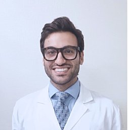 Nicolo Gennaro合作论文数Dept Radiol, Netherlands Canc Inst3
 Ahmet Enis Çetin合作论文数Department of Electrical and Computer Engineering, College of Engineering, University of Illinois at Chicago3
 Elizabeth Krupinski合作论文数Department of Radiology and Imaging Sciences, School of Medicine, Emory University;Winship Cancer Institute, Emory University3Eric Hart合作论文数2Yassine Nasser合作论文数2
 Mohamed Sayah合作论文数Lab LITIO, Univ Oran12
 Camila Lopes Vendrami合作论文数Northwestern Mem Hosp, Northwestern Univ2
 Frank H Miller合作论文数Department of Radiology, Feinberg School of Medicine, Northwestern University2
 Keleş Elif合作论文数Department of Biochemistry and Molecular Biology, Poznań University of Medical Sciences2
 Hussain Al-Ahmad合作论文数2
 Amir Ali Rahsepar合作论文数Clinical and Translational Sciences Institute, Northwestern University;Feinberg School of Medicine, Northwestern University2
 Ulaş Bağcı合作论文数ISTP Research Fellow
NATIONAL INSTITUTES OF HEALTH (NIH)2
Ulaş Bağcı合作论文数ISTP Research Fellow
NATIONAL INSTITUTES OF HEALTH (NIH)2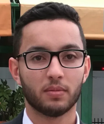 Yasser Dahou合作论文数Technology Innovation Institute1Nadia Aguillon-Hernandez合作论文数1Mina Al-Saad合作论文数Collage Engn & IT, Univ Dubai1
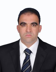 Bashir Ghariba合作论文数Dept Elect & Comp Engn, Elmergib Univ1
 Yasser Abdealziz Dahou Djilali合作论文数Insight Ctr Data Analyt, Dublin City Univ1
 Hatice Savas合作论文数Dept Radiol, Univ Michigan1
 Nour Aburaed合作论文数Collage Engn & IT, Univ Dubai1
 Saeed Al Mansoori合作论文数Mohammed Bin Rashid Space Ctr1
 Mohamed Shehata合作论文数Department of Computer Science, Mathematics, Physics and Statistics, Irving K. Barber Faculty of Science, The University of British Columbia1
 Amir A Borhani合作论文数Department of Radiology, Feinberg School of Medicine, Northwestern University1
 Evangelos Maltezos合作论文数1
 M. Sami Zitouni合作论文数1
 Faouzi Alaya Cheikh合作论文数tampere university of technology1
 Akshay Chaudhari合作论文数Department of Biomedical Data Science, School of Medicine, Stanford University1
 Noel O'Connor合作论文数School of Electronic Engineering, Dublin City University;Insight SFI Research Centre for Data Analytics1
 Pier Luigi Mazzeo合作论文数Institute of Intelligent Systems for Automation, Bari, Italy1Claire Wardak合作论文数1
 Kevin McGuinness合作论文数School of Electronic Engineering, Dublin City University1
 Rachid Harba合作论文数1
 Cosimo Distante合作论文数Università del Salento1
 Veerasathpurush Allareddy合作论文数Boston1
 Mohammed El Hassouni合作论文数FLSHR, Mohammed V University1
 Habib Zaidi合作论文数Department of Radiology and Medical Informatics, University of Geneva;Division of Nuclear Medicine Molecular Imaging, Geneva University Hospital;University of Groningen;University of Southern Denmark;Tehran University of Medical Sciences1
 Jayaram K. Udupa合作论文数Medical Imaging Section, Department of Radiology, Perelman School of Medicine, University of Pennsylvania1
 Marie Luong合作论文数Centre de Recherche en Automatique de Nancy - CNRS UA1Emmanuelle Houy-Durand合作论文数1




