The Integrated Neurological Change Score (INCS) combines changes in sensory and motor subscores from the International Standards for Spinal Cord Injury Classification (ISNCSCI) into a composite measure. We evaluated the INCS sensitivity to therapeutic outcomes, robustness against ceiling effects, and association to functional improvement in patients with acute cervical spinal cord injury (SCI). To this end, we conducted a retrospective analysis of data from the Nogo Inhibition in Spinal Cord Injury (NISCI) clinical trial alongside a matched cohort from the European Multicenter Study about Spinal Cord Injury (EMSCI). The NISCI trial assessed the safety and efficacy of the anti-Nogo-A antibody (NG-101) in acute cervical SCI, reporting a potential therapeutic effect in motor incomplete patients. Our findings show that the sensitivity of INCS to the effect of NG-101 is comparable to that obtained employing the changes in the Upper Extremity Motor Score (the primary outcome in the NISCI trial). Moreover, the INCS is less susceptible to ceiling effects compared to measures of upper and lower extremity or total motor scores, as observed in the NISCI trial and in the matched EMSCI cohort. This robustness may facilitate the design of more inclusive clinical trials without compromising statistical power. Finally, INCS correlates strongly with functional outcomes in self-care and walking ability, outperforming ISNCSCI motor scores. In conclusion, the INCS represents a sensitive measure of neurological change corroborating the value of ISNCSCI standards for use in SCI trials.
Abstract NG101 is a recombinant antibody that neutralizes the nerve growth inhibitor Nogo-A, promoting neural repair and improving upper extremity motor function in spinal cord injury (SCI). This study evaluated spinal cord MRI biomarkers to detect treatment-related structural changes and enhance patient stratification using data from 106 participants with acute cervical SCI in the phase 2b NISCI trial. We assessed lesion volume, tissue bridges, and remote changes in cross-sectional cord area (CSA), and tract-specific myelin-sensitive magnetization transfer saturation (MTsat) over six months. Compared to placebo, NG101-treated participants exhibited faster lesion volume reduction and a slower decline of CSA and MTsat in the corticospinal tracts and dorsal columns. Crucially, multimodal stratification incorporating MRI and electrophysiological measures substantially enhanced the detection of clinical treatment effects. These findings suggest NG101 slows trauma-induced progressive macro- and microstructural degeneration or promotes fiber sprouting. Combining MRI with electrophysiology enables sensitive detection of treatment effects and efficient trial designs. ClinicalTrials.gov identifier: NCT03935321.
There are no approved interventional therapies, aside from neurorehabilitation, that enhance neurological recovery after acute traumatic spinal cord injury. A key challenge is the lack of biomarkers surpassing clinical standards for optimal stratification. We evaluated electrophysiological markers of preserved neuronal function to improve enrichment strategies over clinical measures. We hypothesized that participants with preserved electrophysiological markers would achieve greater neurological and functional recovery in response to a plasticity-inducing intervention. We conducted a retrospective stratification analysis of data from the recently completed randomized, placebo-controlled, phase 2b Nogo Inhibition in spinal cord injury (NISCI) trial (NCT03935321) investigating the efficacy of NG101, a recombinant human antibody that neutralizes the neurite outgrowth-inhibiting protein Nogo-A. Participants aged 18-70 years with acute (4-28 days) cervical spinal cord injury were eligible. At screening, all participants underwent clinical neurological examination and electrophysiological recordings (i.e. somatosensory evoked potentials). Treatment effect sizes for the recovery of upper extremity motor scores and spinal cord independence measure of self-care (6-month change) between NG101 and placebo groups were compared for stratification based on clinical versus electrophysiological criteria. Power analyses were conducted to estimate the required sample sizes needed for each method. The cohort included 116 participants (45.5 ± 16.8 years old, 74 NG101 and 41 placebo). Clinical stratification showed greater functional recovery in motor-incomplete participants treated with NG101 versus placebo [estimate 0.02 (95% confidence interval: 0.006-0.038), P = 0.007]. Electrophysiological stratification revealed greater functional recovery in participants with preserved somatosensory evoked potentials treated with NG101 versus placebo [0.04 (0.015-0.054), P < 0.001]. Effect sizes were large for electrophysiological stratification (Cohen's d = 0.94) but small for clinical stratification (Cohen's d = 0.46). Power analyses demonstrated smaller required sample sizes for electrophysiological stratification (required n = 32) versus clinical stratification (required n = 120). This study shows the value of electrophysiology in comparison to clinical measures for biomarker-driven enrichment and improved power in acute spinal cord injury trials. We emphasize the importance of functionally spared neuronal pathways in promoting recovery in response to plasticity-inducing interventions, such as anti-Nogo-A antibodies.
A straightforward path to successful scientific translation remains uncharted, particularly in a complex progressive condition such as spinal cord injury (SCI), which affects multiple body functions simultaneously. Evolving regulatory requirements add to the complexity and expense of attaining a treatment that is both safe and efficacious. Although rare, there are examples of SCI scientists who have successfully navigated the "valley of death" from discovery science to completed clinical trials. This article reflects the translational journey of five SCI scientists who have encountered similar and different scenarios while striving to launch or complete a clinical trial. Learning from these experiences has identified lessons learned and gaps, particularly with respect to funding and support for SCI translation.
Background Spinal cord injury results in permanent neurological impairment and disability due to the absence of spontaneous regeneration. NG101, a recombinant human antibody, neutralises the neurite growth-inhibiting protein Nogo-A, promoting neural repair and motor recovery in animal models of spinal cord injury. We aimed to evaluate the efficacy of intrathecal NG101 on recovery in patients with acute cervical traumatic spinal cord injury. Methods This randomised, double-blind, placebo-controlled phase 2b clinical trial was done at 13 hospitals in the Czech Republic, Germany, Spain, and Switzerland. Patients aged 18-70 years with acute, complete or incomplete cervical spinal cord injury (neurological level of injury C1-C8) within 4-28 days of injury were eligible for inclusion. Participants were initially randomly assigned 1:1 to intrathecal treatment with 45 mg NG101 or placebo (phosphate- buffered saline); 18 months into the study, the ratio was adjusted to 3:1 to achieve a final distribution of 2:1 to improve enrolment and drug exposure. Randomisation was done using a centralised, computer-based randomisation system and was stratified according to nine distinct outcome categories with a validated upper extremity motor score (UEMS) prediction model based on clinical parameters at screening. Six intrathecal injections were administered every 5 days over 4 weeks, starting within 28 days of injury. Investigators, study personnel, and study participants were masked to treatment allocation. The primary outcome was change in UEMS at 6 months, analysed alongside safety in the full analysis set. The completed trial was registered at ClinicalTrials.gov, NCT03935321. Findings From May 20, 2019, to July 20, 2022, 463 patients with acute traumatic cervical spinal cord injury were screened, 334 were deemed ineligible and excluded, and 129 were randomly assigned to an intervention (80 patients in the NG101 group and 49 in the placebo group). The full analysis set comprised 78 patients from the NG101 group and 48 patients from the placebo group. 107 (85%) patients were male and 19 (15%) patients were female, with a median age of 515 years (IQR 300-600). Across all patients, the primary endpoint showed no significant difference between groups (with UEMS change at 6 months 137 [95% CI -144 to 418]; placebo group mean 1920 [SD 1178] at baseline and 3091 [SD 1549] at day 168; NG101 group mean 1823 [SD 1514] at baseline and 3131 [1954] at day 168). Treatment-related adverse events were similar between groups (nine in the NG101 group and six in the placebo group). 25 severe adverse events were reported: 18 in 11 (14%) patients in the NG101 group and seven in six (13%) patients in the placebo group. Although no treatment-related fatalities were reported in the NG101 group, one fatality not related to treatment occurred in the placebo group. Infections were the most common adverse event affecting 44 (92%) patients in the placebo group and 65 (83%) patients in the NG101 group. Interpretation NG101 did not improve UEMS in patients with acute spinal cord injury. Post-hoc subgroup analyses assessing UEMS and Spinal Cord Independence Measure of self-care in patients with motor-incomplete injury indicated potential beneficial effects that require investigation in future studies.
Stroke is one of the leading causes of disability worldwide. Although preclinical studies have shown promising results of pharmacotherapies to enhance stroke recovery, no drug has been approved for stroke recovery in patients. In this article, we review the preclinical data of one promising treatment, inhibition of NgR1 (Nogo receptor 1) signaling, for stroke recovery. Our scoping review followed the Preferred Reporting Items for Systematic Reviews and Meta-Analysis Extension for Scoping Reviews and surveyed the peer-reviewed literature on PubMed and citation searching. Studies were eligible if they evaluated the pharmacological inhibition of Nogo/NgR1 signaling in nonhuman models of stroke assessing sensorimotor or functional outcomes. There were no restrictions in years considered. An assessment of the risk of bias was performed using the Systematic Review center for Laboratory Animal Experimentation tool. Three hundred seventeen articles were screened based on the inclusion criteria. Thirty preclinical studies were included. Most studies (90%) were conducted in rodents, with only 3 studies (10%) completed in nonhuman primates. Anti-Nogo-A antibody was the most common pharmacological intervention, used in 21 (70%) studies. Most preclinical studies delivered the study drug directly into the central nervous system (intrathecal, intraventricular, or intraparenchymal), with far fewer studies (n=5) testing subcutaneous, intravenous, or intranasal administration. Among the 30 studies included, 27 (90%) report a beneficial effect. In conclusion, there is a large body of preclinical evidence to support Nogo/NgR1 inhibition in promoting stroke recovery. A clinical trial to assess the safety, feasibility, and efficacy among patients with stroke is warranted.
OBJECTIVE:Impaired ability to induce stepping after incomplete spinal cord injury (SCI) can limit the efficacy of locomotor training, often leaving patients wheelchair-bound. The cuneiform nucleus (CNF), a key mesencephalic locomotor control center, modulates the activity of spinal locomotor centers via the reticulospinal tract. Even with severe corticospinal damage, the widely distributed reticulospinal fibers frequently cross the lesion, and lumbosacral spinal locomotor centers remain responsive. Unilateral deep brain stimulation (DBS) of the CNF (CNF-DBS) can increase modulatory input to sublesional locomotor centers and was shown to induce stepping and promote locomotor recovery in rodent models of severe incomplete SCI. Given the evolutionarily conserved CNF-reticulospinal system, we hypothesize that CNF-DBS can augment training and improve gait in humans with incomplete SCI above the lumbosacral levels. METHODS:Aiming at bench-to-bedside translation, we investigate CNF-DBS in non-ambulatory patients (clinicaltrials.gov, NCT03053791). Here, we present the first 2 individuals with chronic tetraplegia who underwent 6 months of locomotor training supported by unilateral CNF-DBS, with regular follow-up assessments of adverse and therapeutic effects performed without and with stimulation. RESULTS:The walking distance covered during the 6-Minute Walking Test (6MWT) after 6 months compared to baseline served as the primary study end point, which was reached by patient 1 in the off-condition and by patient 2 in the off- and the on-condition. No serious adverse events occurred. INTERPRETATION:We show that the CNF-DBS was well tolerated and had therapeutic potential in the first 2 patients, and discuss the lessons learnt with resulting implementations for the next patients. ANN NEUROL 2026;99:161-177.
Neurite outgrowth inhibitor A (Nogo-A) is a major player in neural development and regeneration and the target of clinical trials aiming at promoting the regeneration of the central nervous system upon traumatic and ischemic injury. In this work, we investigated the functions of Nogo-A during tooth development to determine its role in dental physiology and pathology. Using immunohistochemistry and in situ hybridization techniques, we showed that Nogo-A is highly expressed in the developing mouse teeth and, most specifically, in the ameloblasts that are responsible for the formation of enamel. Using both Nogo-A knockout and K14-Cre;Nogo-A fl/fl transgenic mice, we showed that Nogo-A deletion in the dental epithelium leads to the formation of defective enamel. This phenotype is associated with overexpression of a set of specific genes involved in ameloblast differentiation and enamel matrix production, such as amelogenin, ameloblastin and enamelin. By characterising the interactome of Nogo-A in the dental epithelium of wild-type and mutant animals, we found that Nogo-A directly interacts with molecules important for regulating gene expression, and its deletion disturbs their cellular localisation. Furthermore, we demonstrated that inhibition of the intracellular, but not cell-surface, Nogo-A is responsible for gene expression modulation in ameloblasts. Taken together, these results reveal an unexpected function for Nogo-A in tooth enamel formation by regulating gene expression and cytodifferentiation events.
Neurite outgrowth inhibitor A (Nogo-A) is a major player in neural development and regeneration and the target of clinical trials aiming at promoting the regeneration of the central nervous system upon traumatic and ischemic injury. In this work, we investigated the functions of Nogo-A during tooth development to determine its role in dental physiology and pathology. Using immunohistochemistry and in situ hybridization techniques, we showed that Nogo-A is highly expressed in the developing mouse teeth and, most specifically, in the ameloblasts that are responsible for the formation of enamel. Using both Nogo-A knockout and K14-Cre;Nogo-A fl/fl transgenic mice, we showed that Nogo-A deletion in the dental epithelium leads to the formation of defective enamel. This phenotype is associated with overexpression of a set of specific genes involved in ameloblast differentiation and enamel matrix production, such as amelogenin, ameloblastin and enamelin. By characterising the interactome of Nogo-A in the dental epithelium of wild-type and mutant animals, we found that Nogo-A directly interacts with molecules important for regulating gene expression, and its deletion disturbs their cellular localisation. Furthermore, we demonstrated that inhibition of the intracellular, but not cell-surface, Nogo-A is responsible for gene expression modulation in ameloblasts. Taken together, these results reveal an unexpected function for Nogo-A in tooth enamel formation by regulating gene expression and cytodifferentiation events.
Most human spinal cord injuries are anatomically incomplete, leaving some fibers still connecting the brain with the sublesional spinal cord. Spared descending fibers of the brainstem motor control system can be activated by deep brain stimulation (DBS) of the cuneiform nucleus (CnF), a subnucleus of the mesencephalic locomotor region (MLR). The MLR is an evolutionarily highly conserved structure which initiates and controls locomotion in all vertebrates. Acute electrical stimulation experiments in female adult rats with incomplete spinal cord injury conducted in our lab showed that CnF-DBS was able to re-establish a high degree of locomotion five weeks after injury, even in animals with initially very severe functional deficits and white matter lesions up to 80–95%. Here, we analyzed whether CnF-DBS can be used to support medium-intensity locomotor training and long-term recovery in rats with large but incomplete spinal cord injuries. Rats underwent rehabilitative training sessions three times per week in an enriched environment, either with or without CnF-DBS supported hindlimb stepping. After 4 weeks, animals that trained under CnF-DBS showed a higher level of locomotor performance than rats that trained comparable distances under non-stimulated conditions. The MLR does not project to the spinal cord directly; one of its main output targets is the gigantocellular reticular nucleus in the medulla oblongata. Long-term electrical stimulation of spared reticulospinal fibers after incomplete spinal cord injury via the CnF could enhance reticulospinal anatomical rearrangement and in this way lead to persistent improvement of motor function. By analyzing the spared, BDA-labeled giganto-spinal fibers we found that their gray matter arborization density after discontinuation of CnF-DBS enhanced training was lower in the lumbar L2 and L5 spinal cord in stimulated as compared to unstimulated animals, suggesting improved pruning with stimulation-enhanced training. An on-going clinical study in chronic paraplegic patients investigates the effects of CnF-DBS on locomotor capacity.
Nogo-A is a transmembrane protein with multiple functions in the central nervous system (CNS), including restriction of neurite growth and synaptic plasticity. Thus far, Nogo-A has been predominantly considered a cell contact-dependent ligand signaling via cell surface receptors. Here, we show that Nogo-A can be secreted by cultured cells of neuronal and glial origin in association with extracellular vesicles (EVs). Neuron- and oligodendrocyte-derived Nogo-A containing EVs inhibited fibroblast spreading, and this effect was partially reversed by Nogo-A receptor S1PR2 blockage. EVs purified from HEK cells only inhibited fibroblast spreading upon Nogo-A over-expression. Nogo-A-containing EVs were found in vivo in the blood of healthy mice and rats, as well as in human plasma. Blood Nogo-A concentrations were elevated after acute stroke lesions in mice and rats. Nogo-A active peptides decreased barrier integrity in an in vitro blood-brain barrier model. Stroked mice showed increased dye permeability in peripheral organs when tested 2 weeks after injury. In the Miles assay, an in vivo test to assess leakage of the skin vasculature, a Nogo-A active peptide increased dye permeability. These findings suggest that blood borne, possibly EV-associated Nogo-A could exert long-range regulatory actions on vascular permeability.
Intrathecal drug administration represents a promising method to deliver biologics effectively to the central nervous system (CNS). However, little is known about the tolerability and pharmacokinetics of intrathecally applied antibodies. Hence, the focus of this study was to evaluate the toxicity, pharmacokinetic, and pharmacodynamic properties of an intrathecally administered human monoclonal antibody against the growth inhibitory CNS membrane protein Nogo-A in the non-human primate (NHP). The antibody was repeatedly injected into the lumbar cerebrospinal fluid (CSF) sack of NHPs, Macaca fascicularis (N = 18), at three dose levels (placebo, 75 and 150 mg antibody/injection, n = 6/group). CSF and serum samples were collected for pharmacokinetic analysis. The health status was constantly monitored to detect any treatment-related abnormalities. After sacrifice, the CNS tissues were evaluated by immunohistochemistry and biochemistry to study the antibody distribution and target interaction in the spinal cord and brain. No treatment-related side effects were observed, and the treatment was well tolerated by NHPs. After administration, the antibody was rapidly cleared from the CSF with a half-life of 6.4 h and accumulated in the serum where it showed a half-life of 13.7 days. The antibody distributed over the spinal cord and brain, penetrated into the CNS parenchyma where it bound to Nogo-A expressing neurons and oligodendrocytes, and induced significant (P < 0.05) downregulation of the target antigen Nogo-A. Collectively, these results support the direct administration of therapeutic antibodies into the CSF and are of relevance for the antibody-based therapeutics currently in development for different CNS diseases.
Nogo-A is a major regulator of neural development and regeneration, but its role in tooth innervation remains largely unknown. Neurons from trigeminal ganglia support teeth homeostasis and regeneration, and disorders of their function could have significant pathophysiological consequences. In this study, we show that Nogo-A is expressed in the trigeminal ganglia and in the neurons innervating the teeth, and that its deletion affects both the number and patterning of neurons in teeth. In organotypic cultures, Nogo-A blocking antibodies affect the trigeminal ganglia-derived neuronal outgrowths and allow premature innervation of tooth germs. RNA sequencing analysis revealed that Nogo-A deletion induces alterations linked to functions at synapses and interference with neurotrophin signalling during the differentiation and maturation of trigeminal neurons. Taken together, these results reveal for the first time the importance of Nogo-A as a major regulator of tooth innervation and point to its potential as a clinical therapeutic target. ### Competing Interest Statement The authors have declared no competing interest.
The Barnes maze is a task used to assess spatial learning and memory in rodents. It requires animals to learn the position of a hole that can be used as an escape from a bright and open arena. The often-used parameters of latency and path length to measure learning and memory do not reflect the different navigation strategies chosen by the animals. Here, we propose an 11-point scoring scheme to classify the search strategies developed by the animals during the initial training as well as after the change of the escape target to a new position. Strategy scores add an important dimension to time and path length to assess the behavior in this popular maze.
Objective: Functional tests are required to assess the efficacy of therapeutic approaches augmenting peripheral nerve regeneration. Currently available experimental tools are complex, expensive, lack reliability and are difficult to perform. Here we evaluated a swim test for monitoring functional recovery following peripheral nerve lesion in rats.
Recombinant Abs are gaining increasing importance for the treatment of certain cancers or immunological or neurologic disorders. The ELISA is one of the most used analytical tools for detecting and quantifying Abs of interest. However, the performance of ELISAs often varies because of nonstandard experimental procedures as well as inadequate data analysis. In our study, we standardized a procedure and statistical analysis for a highly sensitive ELISA of a mouse Ab in mouse (C57BL/6J) CNS tissue. The following steps are of crucial importance: 1) calculation of the limit of detection based on control tissue lysate samples in the same testing buffer as the testing samples; 2) calculation of the limit of quantification as measured with acceptable accuracy and precision; and 3) a five-parameter logistic regression model to interpolate the symmetric and asymmetric standard curves. We also show that three amplification Abs can significantly increase the sensitivity of the ELISA compared with a two amplification Ab setup. This standardized procedure may be a valuable tool to increase the sensitivity, reproducibility, and precision of ELISA studies in basic science and translational research.
Antibody delivery to the CNS remains a huge hurdle for the clinical application of antibodies targeting a CNS antigen. The blood-brain barrier and blood-CSF barrier restrict access of therapeutic antibodies to their CNS targets in a major way. The very high amounts of therapeutic antibodies that are administered systemically in recent clinical trials to reach CNS targets are barely viable cost-wise for broad, routine applications. Though global CNS delivery of antibodies can be achieved by intrathecal application, these procedures are invasive. A non-invasive method to bring antibodies into the CNS reliably and reproducibly remains an important unmet need in neurology. In the present study, we show that intranasal application of a mouse monoclonal antibody against the neurite growth-inhibiting and plasticity-restricting membrane protein Nogo-A leads to a rapid transfer of significant amounts of antibody to the brain and spinal cord in intact adult rats. Daily intranasal application for 2 wk of anti-Nogo-A antibody enhanced growth and compensatory sprouting of corticofugal projections and functional recovery in rats after large unilateral cortical strokes. These findings are a starting point for clinical translation for a less invasive route of application of therapeutic antibodies to CNS targets for many neurological indications.
The molecular mechanisms of angiogenesis have been intensely studied, but many genes that control endothelial behavior and fate still need to be described. Here, we characterize the role of Apold1 (Apolipoprotein L domain containing 1) in angiogenesis in vivo and in vitro. Single-cell analyses reveal that - across tissues - the expression of Apold1 is restricted to the vasculature, and that Apold1 expression in endothelial cells (ECs) is highly sensitive to environmental factors. Using Apold1-/- mice, we find that Apold1 is dispensable for development and does not affect postnatal retinal angiogenesis nor alters the vascular network in adult brain and muscle. However, when exposed to ischemic conditions following photothrombotic stroke as well as femoral artery ligation, Apold1-/- mice display dramatic impairments in recovery and revascularization. We also find that human tumor endothelial cells express strikingly higher levels of Apold1, and that Apold1 deletion in mice stunts the growth of subcutaneous B16 melanoma tumors, which have smaller and poorly perfused vessels. Mechanistically, Apold1 is activated in ECs upon growth factor stimulation as well as in hypoxia, and Apold1 intrinsically controls EC proliferation but not migration. Our data demonstrate that Apold1 is a key regulator of angiogenesis in pathological settings, whereas it does not affect developmental angiogenesis, thus making it a promising candidate for clinical investigation.
Background The Rotarod test with commercial apparatus is widely used to assess locomotor performance, balance and motor learning as well as the deficits resulting from diverse neurological disorders in laboratory rodents due to its simplicity and objectivity. Traditionally, the test ends when rodents drop from the accelerating, turning rod, and the only parameter used commonly is “latency to fall”. The values of individual animals can often vary greatly. Results In the present study, we established a procedure for mice with 4 consecutive days of training with 4 trials per day and modified the testing procedure by placing the mice back on the rod repeatedly after each fall until the trial ends (5 min). Data from the fourth training day as baseline results showed that the second, third and fourth trial were more consistent than the first, probably due to habituation or learning. There was no difference between the second, third and fourth trial, two trials may be sufficient in testing. We also introduced 3 additional read-outs: Longest duration on the rod (s), Maximal distance covered (cm), and Number of falls to better evaluate the motor capacity over the 5 min of testing. We then used this 4-parameter analysis to capture the motor deficits of mice with mild to moderate traumatic brain injuries (by a weight dropping on the skull (Marmarou model)). We found that normalization of data to individual baseline performance was needed to reduce individual differences, and 4 trials were more sensitive than two to show motor deficits. The parameter of Maximal distance was the best in detecting statistically significant long-term motor deficits. Conclusions These results show that by making adjustments to the protocol and employing a more refined analysis, it is possible to expand a widely used routine behavioral test with additional accessible parameters that detect relevant deficits in a model of mild to moderate traumatic brain injury. The modified Rotarod test maybe a valuable tool for better preclinical evaluations of drugs and therapies.
Despite the fact that a majority of patients with an injury to the spinal cord develop lower urinary tract dysfunction, only few treatment options are available currently once the dysfunction arises. Tibial nerve stimulation has been used in pilot clinical trials, with some promising results. Hence, we investigated whether the early application of transcutaneous tibial nerve stimulation in the animal model of spinal cord injured rats can prevent the development of detrusor overactivity and/or detrusor-sphincter-dyssynergia. Rats were implanted with a bladder catheter and external urethral sphincter electromyography electrodes. A dorsal over-hemisection, resulting in an incomplete spinal cord injury at the T8/9 spinal level, induced immediate bladder paralysis. One week later, the animals received daily tibial nerve or sham stimulation for 15 days. Effects of stimulation on the lower urinary tract function were assessed by urodynamic investigation. Measurements showed improvements of several key parameters of lower urinary tract function-in particular, non-voiding bladder contractions and intravesical pressure-immediately after the completion of the stimulation period in the stimulated animals. These differences extinguished one week later, however. In the dorsal horn of the lumbosacral spinal cord, a small significant increase of the density of C-fiber afferents layers I-II was found in the stimulated animals at four weeks after spinal cord injury. Tibial nerve stimulation applied acutely after spinal cord injury in rats had an immediate beneficial effect on lower urinary tract dysfunction; however, the effect was transitory and did not last over time. To achieve more sustainable, longer lasting effects, further studies are needed looking into different stimulation protocols using optimized stimulation parameters, timing, and treatment schedules.
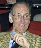 Hans Thoenen合作论文数Max Planck Institute for Neurobiology57
 Lisa Schnell合作论文数Brain Research Institute University of Zürich August Forelstrasse 1 CH-8092 Zürich Switzerland47Oliver Weinmann合作论文数31Anis K. Mir合作论文数28Miriam Gullo合作论文数19Rouiller Eric M合作论文数18Thomas Oertle合作论文数16
 Ruslan Rust合作论文数Zilkha Neurogenetic Institute, Keck School of Medicine, University of Southern California15
 Montani Laura合作论文数Instituto de Biologia Molecular e Celular, 4150-180 Porto, Portugal.15Vincent Pernet合作论文数14
 Karim Fouad合作论文数Rehabilitation Medicine, University of Alberta14
 Patrick Freund合作论文数13
 Benjamin Victor Ineichen合作论文数Brain Research Institute, University of Zurich13
 Michaela Thallmair合作论文数13CE Bandtlow合作论文数12Anna-Sophie Hofer合作论文数12Marta Zagrebelsky合作论文数12
 Jocelyne Bloch合作论文数Department of Neurosurgery, University Hospital of Lausanne12Franziska Christ合作论文数12Leda Dimou合作论文数Inst Physiol, Univ Munich12Thomas M Kessler合作论文数11GL Kartje合作论文数11
 Andrea Sartori合作论文数11Björn Tews合作论文数11Sandrine Joly合作论文数Département de Sciences biologiques, Université de Montréal11
 Florence M Bareyre合作论文数11Brösamle C合作论文数10
 Linard Filli合作论文数Balgrist University Hospital, University of Zurich10
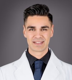 Zemmar Ajmal合作论文数Brain Research Institute, University of Zurich10Armin Curt合作论文数10Pico Caroni合作论文数10
 Hansjoerg Kasper合作论文数10Gwendolyn L. Kartje合作论文数10Eric Schmidlin合作论文数10GAS Metz合作论文数9Bjoern Zoerner合作论文数9carmen barske合作论文数9Anissa Kempf合作论文数9Lisa Grönnert合作论文数9
 Michelle Starkey合作论文数9
 Anna-Sophia Wahl合作论文数Department of Health Sciences and Technology, ETH Zurich;Brain Research Institute, University of Zurich;Brain Research Institute, University of Zurich9
 Bjorn Zorner合作论文数9Schmandke Andre合作论文数Swiss Federal Institute of Technology, University of Zurich9
 Arne Skerra合作论文数School of Life Sciences, Technical University of Munich9
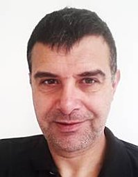 Paolo Paganetti合作论文数Novartis Pharma AG, Novartis Institutes for BioMedical Research9Thierry Wannier合作论文数9Uwe Pott合作论文数9
 Olivier Raineteau合作论文数Inserm9Michael A. Maurer合作论文数8




