Budesonide is recommended for mild-to-moderate ulcerative colitis (UC); however, cytomegalovirus (CMV) colitis may still emerge during therapy. We report the case of a 69-year-old woman with left-sided UC who developed worsening diarrhea and hematochezia while receiving budesonide rectal foam and MMX. Colonoscopy revealed longitudinal, undermined, and "double-tracked" ulcers in the rectosigmoid. CMV pp65 antigenemia was positive, and immunohistochemical staining of a biopsy specimen revealed CMV-infected cells. Oral valganciclovir was initiated, leading to rapid improvement, with antigenemia converting to negative; follow-up endoscopy showed shallower ulcers. This case illustrates that locally acting budesonide MMX does not eliminate the risk of CMV colitis, and viral reactivation can occur even during its use. Accordingly, when clinical worsening occurs during budesonide MMX therapy, early CMV testing should be considered to avoid unnecessary escalation to systemic corticosteroids or advanced agents.
Objectives: Advanced-stage colorectal cancers (CRCs) are occasionally detected on routine abdominal CT scans performed in daily clinical practice. This study aimed to evaluate the diagnostic performance of routine CT scans for detecting advanced CRCs. Methods: We studied patients who underwent routine CT examinations for gastrointestinal symptoms or abnormalities between March 2015 and April 2025, and who subsequently underwent colonoscopy within six months. CT images were interpreted by two radiologists as part of routine clinical practice. Colonoscopic and pathological findings were used as reference standards for advanced CRC (p-T2 or deeper). Diagnostic performance was expressed as percentages with 95% confidence intervals. Results: A total of 350 patients were reviewed (female 44.6%; mean age 64.6 ± 17.2 years; intravenous contrast administration 44.6%). Colonoscopy identified 46 advanced CRCs (median size 56 mm; range 16-105 mm; T2 n=1, T3 n=16, T4 n=18, unknown n=11; proximal colon n=13, distal colon n=16, rectum n=17). Except for one CRC obscured by a large gastric gastrointestinal stromal tumor, all remaining 45 CRCs were suspected on CT (sensitivity 97.8% [88.5-99.9]). Among 304 patients without advanced CRC, 292 were correctly classified as negative (specificity 96.1% [93.2-97.9]). The positive predictive value, negative predictive value, and overall accuracy were 78.9% [66.1-88.6], 99.7% [98.1-100], and 96.3% [93.7-98.0], respectively. In subgroup analysis stratified by contrast enhancement, diagnostic accuracy was 96.8% [92.7-99.0] with contrast and 95.9% [92.0-98.2] without contrast (p=0.65). Conclusions: Routine abdominal CT without colonographic technique demonstrated excellent diagnostic performance for detecting advanced CRC.
ABSTRACT Subepithelial lesions (SELs) of the head and neck have a low diagnostic yield with mucosal biopsy and carry a bleeding risk. Among endoscopic ultrasound–tissue acquisition (EUS‐TA) techniques, fine‐needle biopsy (FNB) provides higher specimen adequacy and diagnostic accuracy than fine‐needle aspiration (FNA). A forward‐viewing curved linear‐array echoendoscope (FV‐EUS) is useful for mobile lesions and those in narrow spaces that are difficult to puncture with conventional oblique‐viewing EUS (OV‐EUS), but FV‐EUS–guided EUS‐FNB has not been reported for head and neck lesions. We report a 72‐year‐old man in whom a lesion at the esophageal inlet was not apparent on initial upper gastrointestinal endoscopy performed with a small‐caliber endoscope. Stenosis was subsequently noted at the time of endoscopic submucosal dissection for early gastric cancer. Contrast‐enhanced computed tomography showed an approximately 20‐mm solid mass on the posterior hypopharyngeal wall. Under general anesthesia with laryngoscopic exposure, an elevated subepithelial lesion without mucosal exposure was observed on the posterior pharyngeal wall. Using FV‐EUS with a cap device attached to the scope tip, a 22 × 18 mm hypoechoic subepithelial mass was clearly visualized, and consecutive EUS‐FNB was performed with a 22‐gauge needle. No complications, including bleeding, occurred. Histopathology and immunohistochemistry demonstrated moderately differentiated squamous cell carcinoma, and the patient was referred for treatment as primary hypopharyngeal cancer. This case illustrates the feasibility and safety of FV‐EUS–guided FNB for pharyngeal SELs and suggests a wider role for FV‐EUS in head and neck disorders.
AbstractRectal neuroendocrine tumor (NET) G1 is a docile-looking tumor. We herein report a 77-year-old male whose rectal NET G1 measuring 10 mm recurred eight years after endoscopic mucosal resection. Tumor recurrence was detected by his mild general fatigue. Abdominal computed tomography revealed widespread metastasis, including the liver, lymph nodes around the rectum, and cervical spines. Subsequent somatostatin receptor scintigraphy, as well as histological reassessment of previously resected specimen, proved that these metastatic diseases originated from the rectal NET G1 with vascular invasion and possible positive vertical margin. First, octreotide long-acting repeatable 30 mg i.m. was administered every four weeks. Second, transcatheter arterial chemoembolization (TACE) for multiple liver metastases and irradiation therapy for spinal metastasis were carried out. One year after these treatments started, everolimus (mTOR inhibitor) 10 mg p.o. was administered every day. For the last five years, he received two rounds of TACE treatments and one round of irradiation. Although recurrent diseases were temporally controlled by these multidisciplinary treatments for the last month, he died of multiple metastases six years later, after tumor recurrence was detected. This case underscores the potential for late recurrence even in rectal NET G1 with unfavorable histological features, highlighting the importance of long-term surveillance.
The patient was a 40-year-old woman who initially presented with ascites in September 2016. She was observed for suspected fatty liver disease and IgA nephropathy. In April 2023, she was admitted to the hospital after sustaining a self-inflicted injury. She presented with ascites and impaired liver and kidney function. A liver biopsy revealed no significant fibrosis, and she was diagnosed with idiopathic portal hypertension (IPH). The ascites did not improve with diuretics or paracentesis. A colonoscopy revealed multiple hyperplastic polyps, and a protein leakage scintigraphy identified protein leakage from the sigmoid colon. This led to a diagnosis of Cronkhite-Canada syndrome (CCS). She presented with characteristic skin symptoms of CCS, including nail atrophy and taste disturbances. Prior to the onset of symptoms, she reported experiencing psychological stress. Steroid therapy improved the ascites and pleural effusion, and the liver dysfunction gradually improved. This case suggests that an autoimmune background may have triggered or exacerbated the IPH and that CCS should be considered in the differential diagnosis of refractory ascites.
OBJECTIVES:In inflammatory bowel disease therapy, thiopurines have been essential. However, several reports have investigated factors affecting thiopurine metabolism to date. This study investigated factors affecting intracellular concentrations of 6-thioguanine nucleotide (6-TGN) and 6-methylmercaptopurine (6-MMP) in a real-world setting. METHODS:Between May 2013 and October 2021 in one institution, 44 patients (median age 44 years;male 35, female 9;ulcerative colitis 32, Crohn's disease 12) receiving thiopurines were reviewed. Intracellular 6-TGN/6-MMP concentrations were measured by high-performance liquid chromatography, and the initial measurement in each patient was used for the study. RESULTS:The 6-TGN level was significantly higher in females, with mild disease activity, absence of NUDT15 polymorphism, and allopurinol administration. A higher trend was observed with high thiopurine dosage (>50 mg). 6-MMP levels were significantly lower with concomitant use of time-dependent 5-aminosalicylic acid (5-ASA) and allopurinol, and higher with high thiopurine dosage. On multivariate analysis of variance, logarithmically transformed 6-TGN levels were significantly higher in females, with high thiopurine dosage, and allopurinol administration. Similarly, logarithmically transformed 6-MMP levels were significantly higher with time-dependent 5-ASA administration and high thiopurine dosage. CONCLUSIONS:Patients who received allopurinol, a high dose of thiopurine, or were female showed higher 6-TGN levels.
Colorectal cancer remains a leading cause of mortality worldwide, and early detection is essential for improving outcomes. CT colonography (CTC) has emerged as a promising alternative to optical colonoscopy for colorectal cancer screening. This article explores the potential of CTC in Japan, focusing on quality control, patient acceptability, complications, and its role in screening programs. CTC has demonstrated high sensitivity and specificity for detecting colorectal polyps, with its diagnostic performance comparable to colonoscopy for lesions ≥ 10 mm. Techniques such as fecal tagging and dual-position imaging significantly enhance diagnostic accuracy. However, the variability in diagnostic outcomes underscores the need for rigorous interpretation training and quality control. The American College of Radiology recommends training with at least 50 cases verified by colonoscopy. Despite its advantages, the adoption of CTC in Japan remains limited due to low awareness among medical professionals, a shortage of trained radiologists, and the absence of specific guidelines endorsing its use. Patient acceptability for CTC is high due to its non-invasive nature, shorter examination time, and reduced bowel preparation requirements compared to colonoscopy. Nonetheless, complications such as bowel perforation, albeit rare, necessitate careful risk assessment. While CTC has been recognized in the U.S. and Europe for screening and diagnostic follow-up, its integration into Japan’s colorectal cancer screening guidelines is crucial to expand its utilization. To maximize the benefits of CTC, efforts must focus on standardizing methodologies, establishing quality indicators, and generating robust evidence on mortality reduction and cost-effectiveness.
ABSTRACT A 55‐year‐old woman presented with postprandial abdominal pain and diarrhea. Contrast‐enhanced abdominal computed tomography revealed a large tumor in the ileocecal region. Colonoscopy demonstrated a pedunculated polyp originating from the terminal ileum, intermittently prolapsing into the cecum with a stalk‐like base. Biopsy specimens showed nonspecific inflammatory changes. Initial hot snare polypectomy was unsuccessful due to the polyp's large size and mobility. Therefore, endoscopic submucosal dissection using the underwater pocket‐creation method was performed, with the polyp stabilized using a traction device anchored to its apex and the opposite side of the ileocecal valve. This technique enabled safe resection of the lesion from its broad stalk. Although marked submucosal fibrosis was observed beneath the lesion, en bloc resection was successfully completed without perforation in 63 min. Retrieval of the resected specimen via conventional endoscopic methods was unsuccessful due to difficulty passing through the hepatic flexure. Instead, the specimen was retrieved following natural elimination the next day. The resected specimen was a prolate spheroid measuring 62 × 40 × 22 mm. Histopathological examination confirmed an inflammatory fibroid polyp (IFP), consisting of edematous stroma with dense inflammatory cell infiltration. The patient resumed oral intake on postoperative day 2 and had an uneventful recovery. Follow‐up colonoscopy at 6 months revealed no residual or recurrent lesion. To our knowledge, this case represents the largest IFP of the small intestine ever resected endoscopically. For a giant, mobile lesion in the terminal ileum, the combination of the pocket‐creation method, underwater technique, and lesion anchoring was an effective strategy.
This case illustrates the complex interactions of the immune responses after vaccination and highlights their potential connections to various autoimmune conditions. A 22-year-old man with quiescent ulcerative colitis (UC) presented with abdominal pain, rectal bleeding, and thrombocytopenia 7 days after receiving the third coronavirus disease 2019 mRNA vaccination. Laboratory data confirmed the diagnosis of immune thrombocytopenia. High-dose intravenous immunoglobulin administration boosted the patient's platelet count. Simultaneously, colonoscopy revealed that his UC had relapsed. Although salazosulfapyridine briefly improved his symptoms, his stool frequency worsened one week later. The patient also developed pyoderma gangrenosum. Subsequent treatment with infliximab notably improved both pyoderma gangrenosum and UC.
Fukushima Kenritsu Ika Daigaku Aizu Iryo Center, Japan; Aizu Daigaku, Japan.
Objectives: Endoscopic submucosal dissection (ESD) of colorectal lesions was invented in Japan, but postoperative management including hospital stay has not been reconsidered due to the Japanese insurance system. To explore appropriate postoperative management after colorectal ESD, we reviewed short-term outcomes after ESD in non-selected consecutive patients. Methods: Patients who underwent colorectal ESD from April 2013 to September 2020 in one institution were reviewed. The primary outcome measure was the occurrence of adverse events stratified by the Clavien-Dindo classification with five grades. A logistic regression model with the Firth procedure was applied to investigate predictors of severe (grade III or greater) adverse events. Results: A total of 330 patients (female 40%, male 60%; median 72 years; IQR 65-80 years) with colorectal lesions (median 30 mm, IQR 23-40 mm; colon 77%, rectum 23%; serrated lesion 4%, adenoma 47%, mucosal cancer 30%, invasive cancer 18%) was evaluated. The en bloc resection rate was 97%. The median dissection time was 58 min (IQR: 38-86). Intraprocedural perforation occurred in 3%, all successfully treated by endoscopic clipping. No delayed perforations occurred. Postprocedural bleeding occurred in 3% on days 1-10 (median day 2); all were controlled endoscopically. Severe adverse events included only delayed bleeding. In analyzing severe adverse events in a multivariate logistic regression model with the Firth procedure, antithrombotic agent use (p = 0.016) and rectal lesions (p = 0.0010) were both significant predictors. Conclusions: No serious adverse events occurred in this series. Four days of hospitalization may be too long for the majority of patients after ESD.
 Kazutomo Togashi合作论文数Department of Surgery 3311-1, Jichi Medical School59Nemoto Daiki合作论文数46Kenichi Utano合作论文数43
 Shungo Endo合作论文数37Noriyuki Isohata合作论文数36
 Alan Kawarai Lefor合作论文数Jichi Medical University22Yuki Nakajima合作论文数20Daisuke Takayanagi合作论文数16Tetsutaro Nemoto合作论文数15Kohei Suzuki合作论文数14Kumamoto Kensuke合作论文数12Goro Shibukawa合作论文数12
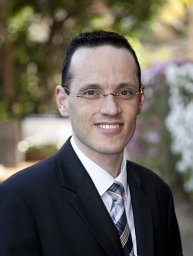 David G. Hewett合作论文数Skills Development Centre, Queensland Health10
 Xin Zhu合作论文数The University of Aizu8Zhe Guo (郭哲)合作论文数Biomedical Information Engineering Lab, The University of Aizu;The Second Xiangya Hospital, Central South University;The University of Aizu, Central South University6Yoshikazu Hayashi合作论文数6Kenta Ueda合作论文数5Hiroshi Hojo合作论文数5
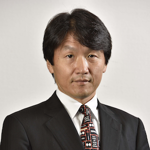 Hironori Yamamoto合作论文数Department of Medicine, Division of Gastroenterology, Jichi Medical University5Shinichi Katsuki合作论文数5Qin Li合作论文数4Takahito Takezawa合作论文数4Yosuke Takahata合作论文数3Yuichi Sagara合作论文数3
 Tadayuki Takagi合作论文数3Yasuyuki Miyakura合作论文数3
 Takuro Saito合作论文数3Nobutoshi Soeta合作论文数3
 Toshio Uraoka合作论文数2Tomoyuki Iwaki合作论文数2Takuya Tsunoda合作论文数2Yu Wang合作论文数2Osamu Suzuki合作论文数2
 Ruiyao Zhang合作论文数2Takashi Kato合作论文数2Oshibe Ikuro合作论文数2Suzuki Sho合作论文数Department of Gastroenterology and Hepatology, Tokyo Medical University2Matsuda Tomoki合作论文数2Tanaka Noriko合作论文数2Yamashina Takeshi合作论文数2Fujita Tomoki合作论文数2
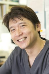 Yoji Takeuchi (竹内洋司)合作论文数Department of Endoscopy and Endoscopic Surgery, Gunma University;Gunma University Hospital2Hideki Mori合作论文数2
 Hidetaka Eguchi合作论文数2Jun Wada合作论文数2Mitsuru Saka合作论文数2
 Hirotsugu Sakamoto合作论文数Kitaibaraki City General Hospital2Osamu Ichii合作论文数2




