Amid the challenges of teacher recruitment, the retention of early career teachers has become even more crucial for schools. Matt Walker and Suzanne Straw consider how the teaching profession can effectively support and retain early career teachers
Transmission electron microscopy (EM) is a versatile technique that can be used to image biological specimens ranging from intact eukaryotic cells to individual proteins >150kDa. There are several strategies for preparing samples for imaging by EM, including negative staining and cryogenic freezing. In the last few years, cryo-EM has undergone a ‘resolution revolution’, owing to both advances in imaging hardware, image processing software, and improvements in sample preparation, leading to growing number of researchers using cryo-EM as a research tool. However, cryo-EM is still a rapidly growing field, with unique challenges. Here, we summarise considerations for imaging of a range of specimens from macromolecular complexes to cells using EM.
Cordon-Bleu (Cobl) is a regulator of actin dynamics in neural development and ciliogenesis. Its function is associated with three adjacent actin binding WASP Homology 2 (WH2) domains. We showed that these WH2 repeats confer multifunctional regulation of actin dynamics, which makes Cobl a « dynamizer » of actin assembly, inducing fast turnover of actin filaments and oscillatory polymerization regime via nucleation, severing, and rapid depolymerization activities. Cobl is the most efficient severer of actin filaments characterized so far. To understand which primary sequence elements determine the filament severing activity of the WH2 repeats, here we combine a mutagenetic/domain swapping approach of the minimal fully active Cobl-KAB construct, which comprises the lysine rich region K preceding the two first WH2 domains A and B. The mutated Cobl constructs display variable loss of the original filament nucleating activities of native Cobl-KAB, without any strict correlation with a loss in actin binding, which emphasizes the functional importance of the electrostatic environment of WH2 domains. Filament severing displayed the greatest stringency and was abolished in all mutated forms of Cobl-KAB. Filament severing and re-annealing by Cobl-KAB, which is key in its rapid remodeling of a population of actin filaments, and most likely responsible for its function in ciliogenesis, was analyzed by electron microscopy in comparison with Spire and ADF.
techniques for the genetic manipulation of this model are well established. Our major goal is to establish an isolation method and further refine it to examine the structure and functioning of both native zebrafish cardiac myosin thick filaments and filaments with mutations in the thick filament associated proteins. In the previous isolation techniques for mammalian cardiac thick filaments, the use of potentially damaging proteolytic enzymes such as elastase and calpain has normally been required. We have successfully isolated thick filaments from zebrafish cardiac muscle, using a procedure similar to those for mammalian heart, only this time without the use of any proteolytic enzyme, and have analyzed their structure by negative staining, transmission electron microscopy and SDS-electrophoresis gels. These results could help to improve the 3D reconstruction of the zebrafish cardiac thick filament for the study of the changes in the cardiac thick filament associated with disease processes. Supported by an NIH grant SC1HL096017 to RWK.
The giant protein titin (chain weight ∼3 MDa) has important roles in assembly and contractile function in vertebrate striated muscle sarcomeres. The molecule consists principally of ∼300 Ig and Fn3 domains in a chain of more than 1 μm long spanning half the sarcomere. The A-band part is attached to the thick (myosin) filament with six molecules in each half filament. The I-band part makes an elastic connection between the tip of the filament and the Z-disc. The central region of A-band titin (C-zone, ∼0.5 μm long) contains 11 copies of what is called the large super-repeat, Ig-Fn3-Fn3-Ig-Fn3-Fn3-Fn3-Ig-Fn3-Fn3-Fn3. The lengths of the large super-repeat (∼43 nm) and of its sub-periods (∼14 nm) correspond to the two main periodicities of the thick filament. C-zone titin is thought to make at least three distinct types of interactions: with itself, with myosin, and with myosin binding protein-C. We studied three recombinant 2-domain fragments from the three sub-periods of a large super-repeat. At physiological ionic strength all three constructs were soluble and mostly monomeric, whereas reduced salt resulted in self-association. Self-association was strongest in the case of the third sub-period construct leading to its complete sedimentation. Electron microscopy revealed large net-like oligomers suggesting regularity in the domains interactions. In the constructs from the first two sub-periods, side-by-side dimers were observed and oligomers were seen only rarely. Co-sedimentation and solid phase assays showed binding of the constructs to the light meromyosin part of myosin but did not reveal any significant differences in the interactions. Modelling of the proteins folds and electrostatics was consistent with the observed interactions being mainly electrostatic.
Dynein ATPases are the largest known cytoskeletal motors and perform critical functions in cells: carrying cargo along microtubules in the cytoplasm and powering flagellar beating. Dyneins are members of the AAA+ superfamily of ring-shaped enzymes, but how they harness this architecture to produce movement is poorly understood. Here, we have used cryo-EM to determine 3D maps of native flagellar dynein-c and a cytoplasmic dynein motor domain in different nucleotide states. The structures show key sites of conformational change within the AAA+ ring and a large rearrangement of the "linker" domain, involving a hinge near its middle. Analysis of a mutant in which the linker "undocks" from the ring indicates that linker remodeling requires energy that is supplied by interactions with the AAA+ modules. Fitting the dynein-c structures into flagellar tomograms suggests how this mechanism could drive sliding between microtubules, and also has implications for cytoplasmic cargo transport.
The giant protein titin has important roles in the assembly, signalling and passive mechanical properties of muscle sarcomeres. Titin is formed by a single polypeptide with isoforms ranging between 3 and 4 MDa. This folds into ∼300 immunoglobulin (Ig) and fibronectin (Fn3) domains in a beads-on-a-string-like chain more than 1 μm long. The N-terminal half of the molecule forms an elastic connection between the end of the thick filament and the Z-line. The C-terminal half is bound to the thick (myosin) filament. Through most of the thick filament region, the Ig and Fn3 domains are arranged in a distinctive eleven domain 'large super-repeat', Ig-Fn-Fn-Ig-Fn-Fn-Fn-Ig-Fn-Fn-Fn. Eleven copies of the large super-repeat make up ∼0.5 μm of the titin molecule length. In an attempt to reconstruct the structure of this region, we have studied a set of two- and three-domain recombinant fragments forming a large super-repeat using electron microscopy, synchrotron X-ray solution scattering and analytical ultracentrifugation. The data illustrate different average conformations in different domain pairs, correlating with differences in lengths of the inter-domain linkers. They also illustrate a level of flexibility between domains in all pairs around average states. Overall, the results suggest the large super-repeat forms an irregular helix, and is also likely to be dimerized in situ.
Titin is a giant protein of striated muscle with important roles in the assembly, intracellular signalling and passive mechanical properties of sarcomeres. The molecule consists principally of similar to 300 immunoglobulin and fibronectin domains arranged in a chain more than 1 mu M long. The isoform-dependent N-terminal part of the molecule forms an elastic connection between the end of the thick filament and the Z-line. The larger, constitutively expressed C-terminal part is bound to the thick filament. Through most of the thick filament part, the immunoglobulin and fibronectin domains are arranged in a repeating pattern of 11 domains termed the 'large super-repeat'. There are 11 contiguous copies of the large super-repeat making up a segment of the molecule nearly 0.5 mu m long. We have studied a set of two-domain and three-domain recombinant fragments from the large super-repeat region by electron microscopy, synchrotron Xray solution scattering and analytical ultracentrifugation, with the goal of reconstructing the overall structure of this part of titin. The data illustrate different average conformations in different domain pairs, which correlate with differences in interdomain linker lengths. They also illustrate interdomain bending and flexibility around average conformations. Overall, the data favour a helical conformation in the super-repeat. They also suggest that this region of titin is dimerised when bound to the thick filament. (C) 2010 Elsevier Ltd. All rights reserved.
 John Trinick合作论文数22
 Peter J Knight合作论文数15
 Stan A Burgess合作论文数13
 Kazuhiro Oiwa (大岩和弘)合作论文数Advanced ICT Research Institute, National Institute of Information and Communications Technology6
 Hitoshi Sakakibara (榊原均)合作论文数Department of Applied Biosciences, Graduate School of Bioagricultural Sciences, Nagoya University6
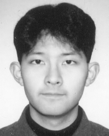 Takahide Kon合作论文数6Sa Burgess合作论文数5H White合作论文数5Numata Naoki合作论文数4
 Caroline Sharp合作论文数4
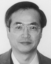 Kazuo Sutoh合作论文数4Reiko Ohkura合作论文数4Bara Malkova合作论文数4Suzanne Straw合作论文数4Hd White合作论文数3S Schmitz合作论文数3
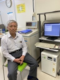 Fumio Arisaka合作论文数Department of Molecular Bioprocessing, Tokyo Institute of Technology3Larissa Tskhovrebova合作论文数3David Teeman合作论文数3Emma Scott合作论文数3Ghulam N. Khan合作论文数2Paula Smith合作论文数2adel varnai合作论文数2F Wang合作论文数2
 J. Gunter Grossmann合作论文数2
 James Sellers合作论文数Laboratory of Molecular Physiology, National Heart, Lung, and Blood Institute, National Institutes of Health2Roberts Anthony J合作论文数2
 Yusuke Kato合作论文数Graduate School of Agricultural and Life Sciences Bunkyo-ku, University of Tokyo2
 Kavitha Thirumurugan合作论文数Centre for Biomedical Research, VIT University2John C. Sparrow合作论文数2Matt Barnes合作论文数University of Bath and the, University of Warwick2Claire Easton合作论文数2Iain W. Manfield合作论文数2
 Gae Spedalieri合作论文数University of York2
 Thomas A Edwards合作论文数1hilary grayson合作论文数1Andy Baron合作论文数1Ryan Conder合作论文数1Helen M Kent合作论文数1H. E. McCreath合作论文数1J Holt合作论文数1G Nasir Khan合作论文数1
 Neil Ranson合作论文数1ellie mundy合作论文数1Xz Zhang合作论文数1




