ObjectiveTo determine whether human milk feeding in children with cleft palate (CP ± L) reduces tympanostomy tubes and improves hearing, speech, and language outcomes.DesignRetrospective cohort study.SettingTertiary children's hospital at a Cleft Craniofacial Specialty Clinic.Patients, Participants319 children with CP ± L who were born between April 2005 and April 2015 were included.InterventionsImpacts on otological, hearing, and speech/language outcomes were analyzed in children fed human milk for any duration, ≥3 months, and ≥6 months.Main Outcome Measure(s)Tympanostomy tube placement history, hearing status before and after tube placement, and speech/language development at 2 and 5 years of age.ResultsChildren with CP ± L fed human milk for ≥3 months (143/319, 44.8%) had decreased odds of abnormal hearing tests (odds ratio [OR]: 0.174, 95% confidence interval [CI]: 0.0266-0.845) or tympanograms (OR: 0.059, 95% CI: 0.008-0.936) before tube placement and decreased odds of bilateral effusion at tube placement (OR: 0.236, 95% CI: 0.089-0.628), even when accounting for Robin sequence and insurance type. There was no significant association between human milk feeding and hearing after tube placement. However, language delays (OR: 0.279, 95% CI: 0.084-0.930) and speech sound production disorder (OR: 0.488, 95% CI: 0.246-0.967) at 5 years of age were less common in those who received human milk of any duration.ConclusionsFeeding human milk to children with CP ± L was associated with improved otological, hearing, speech, and language outcomes. Despite the known challenges of feeding infants with CP ± L, this study demonstrates the added benefits of human milk in this population.
Objective Explore barriers and prioritize supports that could increase breast milk feeding (BMF) rates. Design Survey study. Setting Tertiary children's hospital. Patients, Participants 204 mothers [102 with child with cleft palate (CP), 102 with child with intact palate], ages 0-3 years old. Interventions Survey questions about older sibling feeding history, maternal education, and breastfeeding/breast milk pumping experience. Main Outcome Measure(s) Differences in survey responses between groups; associations between socioeconomic (SES) proxies and BMF. Results Median child age at survey completion was 17.7 months (range 9 days-3.9 years). Direct breastfeeding was less common in the group with CP (46.1% vs. 73.5%, P < .001), who were also more frequently advised against breastfeeding (34.0% vs. 10.9%, P < .001) and more often formula fed (94.1% vs. 85.3%, P = .04). Despite this, the control and CP groups had equal initiation of BMF (77.5%), with no significant difference in median duration (1.0 vs. 1.5 months). Sustained BMF to 6 months was seen in 23.1% of CP group and 28.9% of controls. In the CP group, postnatal counseling (odds ratio [OR] 21.8, P < .001), receiving a breast pump (OR 40.8, P < .001), family support (OR 7.44, P < .001), prior experience with BMF (OR 11.4, P < .001), and maternal education (OR 4.30, P = .006) increased the odds of BMF. Proxies of higher SES were associated with longer BMF in the CP group but not controls (all P < .02). Conclusions Targeted supports for mothers of children with CP such as integrating early feeding specialists and education on pump retrieval are vital to decrease the barriers to sustained BMF.
LEARNING OBJECTIVES:After studying this article, the participant should be able to: 1. Have a clear understanding of the evolution of concepts of velopharyngeal dysfunction, especially as it relates to patients with a cleft palate. 2. Explain the subjective and objective evaluation of speech in children with velopharyngeal dysfunction. 3. On the basis of these diagnostic findings, be able to classify types of velopharyngeal dysfunction. 4. Develop a safe, evidence-based, patient-customized treatment plan for velopharyngeal dysfunction founded on objective considerations.SUMMARY:Velopharyngeal dysfunction is improper function of the dynamic structures that work to control the velopharyngeal sphincter. Approximately 30 percent of patients having undergone cleft palate repair require secondary surgery for velopharyngeal dysfunction. A multidisciplinary team using multimodal instruments to evaluate velopharyngeal function and speech should manage these patients. Instruments may include perceptual speech analysis, video nasopharyngeal endoscopy, multiview speech videofluoroscopy, nasometry, pressure-flow, and magnetic resonance imaging. Velopharyngeal dysfunction may be amenable to surgical or nonsurgical treatment methods or a combination of each. Nonsurgical management may include speech therapy or prosthetic devices. Surgical interventions could include palatal re-repair with repositioning of levator veli palatini muscles, posterior pharyngeal flap, sphincter pharyngoplasty, or soft palate or posterior wall augmentation. Treatment interventions should be based on objective assessment and rating of the movement of lateral and posterior pharyngeal walls and the palate to optimize speech outcomes. Treatment should be tailored to specific anatomical and physiologic findings and the overall needs of the patient.
Background:. The International Consortium of Health Outcome Measurements (ICHOM) standard set for cleft care appraisal recommends clinicians assess articulation with percentage consonants correct (PCC) and velopharyngeal function with velopharyngeal competency rating (VPC-R). This study explores the utility and limitations of these generic measures in detecting cleft speech sound disorders by comparing them with two cleft-specific speech-rating systems, cleft audit protocol of speech–augmented Americleft modification (CAPS-A-AM) and Pittsburgh weighted speech scale (PWSS). Methods:. Consecutive children with repaired, nonsyndromic cleft lip/palate, aged 5 years or older (n = 27) underwent prospective speech evaluations conducted at a single academic institution. These evaluations were conducted, recorded, and evaluated by blinded speech-language pathologists experienced with all tools. Results:. When comparing measures of articulation, PCC scores correlated better with scores for relevant subcomponents of CAPS-A-AM than PWSS. When comparing measures of velopharyngeal function, VPC-R scores correlated well with relevant components of both scales. Using a “screening test versus diagnostic test” analogy, VPC-R ratings were 87.5% sensitive and 73.7% specific for detecting velopharyngeal dysfunction according to subcomponents of CAPS-A-AM, and 70.6% sensitive and 100% specific according to subcomponents of PWSS. Conclusions:. This exploratory study demonstrates that PCC and VPC-R perform moderately well in detecting articulatory and velopharyngeal dysfunction in patients with cleft lip/palate; however, these tools cannot describe nuances of cleft speech sound disorder. Thus, although PCC and VPC-R adequately track basic minimum outcomes, we encourage teams to consider extending the standard set by adopting a cleft-specific measurement system for further evaluation of the tools.
Objective To characterize the prevalence and presentation of laryngomalacia and efficacy of supraglottoplasty (SGP) in a cohort of patients with Pierre Robin Sequence (PRS). Design Retrospective cohort study. Setting Tertiary-care children's hospital. Patients, Participants Consecutive patients with PRS born between January 2010 and June 2018. Main Outcome Measures Chart review included demographics, comorbid airway obstruction including laryngomalacia, timing of surgical interventions, clinical symptoms, sleep study data, and modified barium swallow study data. Results 126 patients with PRS were included; 54% had an associated syndrome, 64% had an overt cleft palate, and 22% had a submucous cleft palate. 64/126 were noted to have laryngomalacia (51%). Patients with concurrent PRS and laryngomalacia were significantly more likely to have submucous cleft palate (P = .005) and present with aspiration with cough (P = .01) compared to patients with PRS without laryngomalacia. Patients with concurrent laryngomalacia and PRS showed a significant decrease in apnea–hypopnea index (AHI) and obstructive AHI (OAHI) after mandibular distraction, with a median AHI and OAHI improvement of 22.3 (P = .001) and 19.8 (P = .002), respectively. Patients who underwent only SGP did not show significant improvement in these parameters (P = .112 for AHI, P = .064 for OAHI). Conclusions The prevalence of laryngomalacia in our PRS cohort was 51%. Patients with PRS and laryngomalacia are more likely to present with overt aspiration compared to patients with PRS without laryngomalacia. These data support that laryngomalacia does not appear to be a contraindication to pursuing MDO.
OBJECTIVE Investigate associations between socioeconomic indicators of healthcare access with family compliance with cleft-related otologic and audiologic care within an interdisciplinary model. DESIGN Retrospective case series. SUBJECTS AND SETTING Children born 2005-2015 who presented to the Cleft-Craniofacial Clinic (CCC) at a quaternary care children's hospital. INTERVENTIONS Associations between main outcome measures and Area Deprivation Index (ADI), median household income for zip code, distance from hospital, and insurance status were evaluated. MAIN OUTCOME MEASURES Cleft types, ages at presentation to outpatient clinic (cleft, otolaryngology, and audiology), and ages at procedures (first tympanostomy tube insertion (TTI), lip repair, and palatoplasty) were measured. RESULTS Most patients were male (147/230, 64%) with cleft lip and palate (157/230, 68%). Median age at first cleft, otolaryngology, and audiology visits were 7 days, 86 days, and 5.9 months, respectively. Private insurance predicted lower no-show rates (p = .04). Age at first CCC visit was younger for patients with private insurance (p = .04) and older for those who lived further from the hospital (p = .002). Age at lip repair was positively correlated with national ADI (p = .03). However, no socioeconomic status (SES) proxy or proximity to hospital was associated with delays in first otolaryngology or audiology examination or TTI. CONCLUSION Once children become established within an interdisciplinary CCC, SES appears to bear little influence on cleft-related otologic and audiologic care. Future efforts should aim to elucidate which aspects of the interdisciplinary model maximize multisystem cleft care coordination and increase access for higher risk populations.
Introduction Velopharyngeal insufficiency (VPI), a stigmatizing hallmark of palatal dysfunction, occurs in a wide spectrum of pediatric craniofacial conditions. The mainstays for surgical correction include palate repair and/or pharyngeal surgery. However, primary pharyngoplasty has a failure rate of 15% to 20%. Although revision pharyngoplasty may be necessary in those with persistent VPI, little is known regarding the indications for and outcomes after such procedures. The purpose of this study is to describe the authors' experience with indications for and outcomes after revision pharyngoplasty. Methods A single-center retrospective review was performed of all patients undergoing revision pharyngoplasty between 2002 and 2019. Demographic data and Pittsburgh Weighted Speech Scores, diagnoses, comorbidities, and complications were tabulated. Two-tailed Student t test was used, and a P value of 0.05 or less was considered statistically significant. Results Thirty-two patients (65.6% male) met inclusion criteria for this study. The most common diagnoses included cleft palate (68.8%), submucous cleft palate (SMCP, 18.8%), and congenital VPI (6.3%, likely occult SMCP). Most patients (84.4%) underwent palatoplasty before their initial pharyngoplasty. The primary indication for initial pharyngoplasty was VPI (mean age 7.1 ± 4.6 years). The most common indication for revision pharyngoplasty (mean age 11.2 ± 5.1 years) included persistent VPI (n = 22), followed by obstructive sleep apnea (OSA) (n = 11). Persistent VPI (n = 8) and OSA (n = 6) were the most common complications after secondary pharyngoplasty. Thirteen patients (40.6%) within the revision pharyngoplasty cohort required additional surgical intervention: 4 underwent tertiary pharyngoplasty, 4 underwent takedown for OSA (n = 3) or persistent VPI (n = 1), 3 underwent takedown and conversion Furlow for persistent VPI (n = 2), OSA (n = 2) and/or flap dehiscence (n = 1), and 2 underwent palatal lengthening with buccal myomucosal flaps for persistent VPI. Of the 4 patients who required a tertiary pharyngoplasty, the mean age at repair was 6.6 ± 1.1 years and their speech scores improved from 13.5 to 2.3 after tertiary pharyngoplasty ( P = 0.11). The overall speech score after completion of all procedures improved significantly from 19 to 3.3. Conclusion Patients who fail primary pharyngoplasty represent a challenging population. Of patients who underwent secondary pharyngoplasty, nearly half required a tertiary procedure to achieve acceptable speech scores or resolve complications.
PURPOSE:Persistent velopharyngeal insufficiency (VPI) following primary palatoplasty remains a difficult problem to treat. This study evaluates speech outcomes following revision palatoplasty with tissue augmentation using buccal myomucosal flaps (BMF) as an alternative to pharyngoplasty for patients with VPI.METHODS:A retrospective single-center review of revision palatoplasty with tissue augmentation at a tertiary pediatric hospital Cleft-Craniofacial Center between January 2017 and March 2021 was conducted. Patients with a history of previous palatoplasty, a diagnosis of persistent or recurrent VPI, and comprehensive pre- and postoperative speech evaluations who underwent revision palatoplasty with BMF were included.RESULTS:Twenty patients met inclusion criteria (35% female, 20% syndromic). Mean age at the time of revision palatoplasty with BMF was 9.7 years. Preoperatively, all patients had stigmatizing speech and received the recommendation for speech surgery; the mean Pittsburgh Weighted Speech Score (PWSS) was 14.3 ± 4.9. The mean postoperative PWSS at the most recent assessment was 4.2 ± 2.3, representing a statistically significant improvement from preoperative scores (P < .001). Mean follow-up time was 8.9 months. Following revision palatoplasty with BMF, only one patient has received the recommendation for further speech surgery. No complications were noted.CONCLUSION:In patients with VPI following primary palatoplasty, revision palatoplasty with tissue augmentation offers an alternative to pharyngoplasty. This approach preserves dynamic velopharyngeal function, improves speech outcomes, and should be considered an option when treating patients with post-primary palatoplasty VPI.
Purpose: Coronavirus Disease-2019 (COVID-19) mitigation measures have led to a sustained reduction in tympanostomy tube (TT) placement in the general population. The present aim was to determine if TT placement has also decreased in children at risk for chronic otitis media with effusion (COME), such as those with cleft palate (CP). Materials and methods: A cohort study with medical record review was performed including consecutive children, ages 0-17 years, undergoing primary palatoplasty at a tertiary children's hospital February 2019-January 2020 (pre-COVID) or May 2020-April 2021 (COVID). Revision palatoplasty (n = 29) was excluded. Patient characteristics and middle ear status pre-operatively and at palatoplasty were compared between groups using logistic regression or Wilcoxon rank-sum. Results: The pre-COVID and COVID cohorts included 73 and 87 patients, respectively. Seventy (44%) were female and median age at palatoplasty was 13.5 months for CP +/- cleft lip (CP +/- L) and 5.5 years for submucous cleft palate (SMCP). In patients with CP +/- L, TT were placed or in place and patent at palatoplasty in 28/38 (74%) pre-COVID and 37/50 (74%) during COVID (P = 0.97). In patients with SMCP, these proportions were 5/35 (14%) and 6/37 (16%), respectively (P = 0.82). Examining only patients <2 years of age also revealed no difference in TT placement pre-COVID versus COVID (P = 0.99). Finally, the prevalence and type of effusion during COVID was similar to pre-COVID. Conclusions: Reduced infectious exposure has not decreased TT placement or effusion at palatoplasty. Future work could focus on non-infectious immunologic factors underlying the maintenance of COME in these children.
BACKGROUND:The submucous cleft palate can be overt or occult and may require surgical repair. The double-opposing Z-plasty (Furlow repair) is the authors' center's preferred approach. This study evaluated complication rates, differences in outcome between overt and occult types, and patient factors associated with surgical failure.METHODS:This retrospective study reviewed documentation on all patients who underwent Furlow Z-plasty for submucous cleft palate at a single center between 2004 and 2018. Speech pathology was quantified using the Pittsburgh Weighted Speech Score.RESULTS:A total of 351 patients were included (125 overt and 226 occult cases). Furlow Z-plasty was successful (postoperative Pittsburgh Weighted Speech Score <7 without recommendation for secondary speech surgery) in 291 patients (82.1 percent). Apart from those requiring secondary surgery, there were no documented complications. Occult-type patients were 7.5 years old at palatoplasty with a speech score of 14.1; overt-type patients were 6.5 years old with a score of 15.7. Postoperative speech scores were similar for both groups. Secondary speech surgery patients had a higher preoperative score (16.9 versus 14.2). Age at time of palatoplasty and submucous cleft palate type were not predictive of the need for secondary surgery. Syndromic patients had higher preoperative and postoperative speech scores (15.6 and 7.5, respectively) than nonsyndromic patients (14.3 and 4.3) and needed secondary surgery more often (24.4 percent versus 9.2 percent). V-shaped velar vaulting on preoperative assessment was present in 92 percent of occult-type patients.CONCLUSIONS:Furlow palatoplasty is a safe and effective means of repairing submucous cleft palate. Patients with the occult type presented later with a lower Pittsburgh Weighted Speech Score. High preoperative speech score and syndromic status were associated with the need for secondary speech surgery. V-shaped velar vaulting is a reliable sign of occult submucous cleft palate.CLINICAL QUESTION/LEVEL OF EVIDENCE:Risk, II.
Objective: To determine whether orthodontic/dentofacial orthopedic maxillary protraction face mask therapy induces changes in velopharyngeal functioning in a cohort of pediatric patients having cleft palate with or without cleft lip. Design: Retrospective chart review. Setting: A children’s hospital in the United States. Participants: Forty-three pediatric patients with cleft palate, with or without cleft lip, syndromic or with isolated clefts, who received face mask therapy from January 2009 to April 2016. Intervention: Clinical data were extracted for review and analysis from medical records obtained from the Cleft Database/Research Registry (CDB-RR). Main Outcome Measures: Pittsburgh Weighted Speech Scores (PWSS) before and after therapy. Results: There was a significant increase in PWSS after face mask therapy for patients with a PWSS score of 0 prior to treatment. Patients with PWSS >0 before treatment remained largely stable after face mask therapy. Maxillary advancement was not significantly associated with change in PWSS or fistula presence/absence. Conclusions: There is an increased risk of velopharyngeal insufficiency with maxillary protraction face mask treatment in patients with cleft palate. Patient counseling and obtaining consent regarding speech changes during treatment are recommended.
Objective: To determine whether timing of palatoplasty (early, standard, or late) is associated with speech and language outcomes in children with cleft palate. Design: Retrospective case series. Setting: Tertiary care children’s hospital. Participants: Records from 733 children born between 2005 and 2015 and treated at the Cleft Craniofacial Clinic of a tertiary children’s hospital were retrospectively reviewed. Exclusion criteria were cleft repair at an outside hospital, intact secondary palate, absence of postpalatoplasty speech evaluation, syndromes, staged palatoplasty, and introduction to clinic after 12 months of age. Data from 232 children with cleft palate ± cleft lip were analyzed. Interventions: Palatoplasty. Main Outcome Measures: Speech/language delays and disorders at 20 months and 5 years of age based on formal hospital or community-based testing or screening evaluation in the Cleft Craniofacial Clinic; additional speech surgery. Results: Median age at palatoplasty was 12.6 months (range: 8.8-21.9 months). Age at palatoplasty was classified as early (<11 months, n = 28), standard (11-13 months, n = 158), or late (>13 months, n = 46). Late palatoplasty was associated with increased odds of speech/language delays and speech therapy at 20 months, and language delays at 5 years, compared with standard or early palatoplasty (P < .05 for all comparisons). However, speech sound production disorders, velopharyngeal incompetence, tube replacement, and hearing loss were not significantly associated with age at palatoplasty. Conclusions: Late palatoplasty may be associated with short- and long-term delays in speech/language development. Future studies with standardized surgical technique/timing and outcome measures are required to more definitively describe the impact of age at palatoplasty on speech/language development.
Objectives 1) Characterize the spectrum of airway anomalies in patients with craniosynostosis, and 2) identify clinical characteristics of these patients that may be associated with the development of airway anomalies. Methods This study is a retrospective case series assessing the type and frequency of airway anomalies in all patients with craniosynostosis seen at a tertiary-care children's hospital between 2000 and 2016. Cohort analyses were then performed to identify differences in airway anomalies dependent on syndromic associations, multisutural fusion, and location of suture fusion. Clinical characteristics examined included demographics and additional neurologic and craniofacial abnormalities. Results Four hundred and ninety-six patients with craniosynostosis (83.5% white, 64.5% male; 33.9% sagittal, 28.8% metopic, 11.5% coronal, 1.2% lambdoid, and 24.6% multisutural) were included. Notable airway anomalies included the following: 13.3% adenotonsillar hypertrophy, 8.9% laryngomalacia, 7.3% tracheomalacia, 7.1% subglottic stenosis, 4.0% bronchomalacia, 3.8% laryngeal cleft, and 1.2% vocal fold paresis. Multisutural craniosynostosis patients (n = 122) were more likely to have obstructive sleep apnea (P = 0.005), adenotonsillar hypertrophy (P = 0.014), tracheomalacia (P = 0.011), subglottic stenosis (P < 0.001), and epiglottic/base of tongue collapse (P = 0.003) and require tracheostomy (P = 0.001) and mechanical ventilation (P = 0.017) compared with single suture craniosynostosis. Syndromic craniosynostosis patients (n = 33) were more likely to have obstructive sleep apnea (P < 0.001), laryngomalacia (P = 0.047), and subglottic stenosis (P = 0.009) compared with nonsyndromic patients. Conclusion Airway anomalies are prevalent in patients with craniosynostosis; patients with multisutural or syndromic types have an increased risk of developing certain abnormalities. There should be a lower threshold for referral for airway evaluation in these populations. Level of Evidence 4. Laryngoscope, 129:2594-2602, 2019
OBJECTIVE:To describe the impact of timing of tympanostomy tube insertion on the number of tubes received and complications in children with routine tube placement.DESIGN:Retrospective case series.SETTING:Tertiary care children's hospital.PARTICIPANTS:Records from a consecutive sample of 401 children with cleft palate were reviewed. Sixty-five patients with isolated cleft palate and 82 patients with cleft lip and palate had follow-up until 5 years of age and were included.INTERVENTIONS:Tympanostomy tubes.MAIN OUTCOME MEASURE(S):Number of tubes received and tube-related complications. The hypothesis was formulated prior to data collection.RESULTS:Males comprised 55.8% of included patients, and tubes were placed in 98.6% of patients at a median age of 6.5 months. Effusion was documented at first tube placement for 96.5% of patients. Most (67.4%) patients required replacement of tubes, and 10.6% required long-term tubes. Complications included otorrhea (71.0%), myringosclerosis (35.2%), granulation (22.8%), perforation (17.9%), retained tubes (5.5%), and cholesteatoma (1.4%). Cleft lip and palate (P < .001) and otorrhea (P = .023) were associated with tube placement before palatoplasty. Patients with tube placement before palatoplasty (P = .033), genetic disorders (P = .007), failed newborn hearing screen (P = .012), otorrhea (P < .001), and granulation (P < .001) received more tubes.CONCLUSIONS:Nearly universal effusion in patients with cleft palate supports the need for routine tube placement. The potential for otorrhea and requiring more tubes should be weighed against the risks associated with prolonged effusion when considering tube placement before palatoplasty.
Velopharyngeal dysfunction (VPD) occurs when the muscular soft palate (velum) and lateral pharyngeal walls are physically unable to separate the oral and nasal cavities during speech production leading to hypernasality and abnormal speech reduction. Because VPD is often associated with overt or submucous cleft palate, it could be present as a subclinical phenotype in families with a history of orofacial clefting. A key assumption to this model is that the overt and subclinical manifestations of the orofacial cleft phenotype exist on a continuum and therefore share common etiological factors. We performed a genome-wide association study in 976 unaffected relatives of isolated CP probands, 54 of whom had VPD. Five loci were significantly (p < 5 × 10⁻⁸) associated with VPD: 3q29, 9p21.1, 12q21.31, 16p12.3 and 16p13.3. An additional 15 loci showing suggestive evidence of association with VPD were observed. Several genes known to be involved in orofacial clefting and craniofacial development are located in these regions, such as TFRC, PCYT1A, BNC2 and FREM1. Although further research is necessary, this could be an indication for a potential shared genetic architecture between VPD and cleft palate, and supporting the hypothesis that VPD is a subclinical phenotype of orofacial clefting.
OBJECTIVES:To determine whether children with cleft palate might benefit from early long-term tympanostomy tubes with the hypothesis that receiving multiple tubes is associated with shorter duration of first tubes.DESIGN:Retrospective cohort study.SETTING:Tertiary care children's hospital.PARTICIPANTS:Records from 401 consecutive children with cleft palate ± cleft lip, born April 2005 to April 2010, were reviewed. After exclusion of children with cleft repair at an outside hospital, no follow-up after 5 years of age, intact secondary palate, no tubes, or tube replacement at palatoplasty, 105 children remained.MAIN OUTCOME MEASURE:Number of tubes.RESULTS:Armstrong grommet tubes were placed at a median age of 6.7 months (range 2.3-19.6 months). Tubes were replaced in 55.3% of patients, with 34.0% receiving ≥3 sets. Duration of first tubes was significantly longer for children with 1 set of tubes compared with those with multiple sets (median 26 vs 19 months, P = .004). Otorrhea, but not perforation, was associated with longer duration of first tubes (median 27 vs 20.5 months, P = .028). Cleft type did not impact the proportion of patients with multiple tubes. Median age at last tube placement for children with multiple tubes was 5.0 years (range 1.9-8.7 years).CONCLUSION:Short duration of first tubes is associated with receiving multiple tubes. Because most patients require repeat tubes and many require tubes until school age, there is a significant need for controlled, prospective trials of early long-term tube placement in this population.
Objective: To investigate whether awake endoscopy can diagnose base-of-tongue obstruction as reliably as sleep endoscopy in infants with Pierre Robin sequence (PRS). Design: The study was retrospective with the clinicians blinded to patient identity. Endoscopy findings were assessed and measured by the performing pediatric otolaryngologist. Setting: Tertiary care children’s hospital. Patients: All infants with PRS managed between January 2005 and July 2015 were included. There were 141 patients, of which 35 underwent both awake endoscopy (AE) and drug-induced sleep endoscopy (DISE). Interventions: Bedside AE and DISE in the operating room. Main Outcome Measures: Presence of moderate or severe base-of-tongue collapse was assessed. Sensitivity, specificity, and positive likelihood ratio of AE findings as well as intertest differences between AE and DISE were calculated. Results: AE had 50.0% sensitivity (95% confidence interval [CI] 27.2%-72.8%) and 86.7% specificity (95% CI 59.5%-98.3%) for detecting base-of-tongue obstruction compared to DISE; false negative rate was 50.0% (n = 10). Positive likelihood ratio was 3.75 (CI 0.96-14.65). Compared to AE, DISE demonstrated significantly more cases of base-of-tongue obstruction ( P = .039). Conclusions: Bedside AE has low sensitivity for detecting base-of-tongue collapse in infants with PRS. Because of the substantial false negative rate, AE may not be a reliable diagnostic modality for ruling out base-of-tongue obstruction in this susceptible population. DISE may be indicated in high-risk patients to avoid underdiagnosing upper airway obstruction.
Objective: To characterize differences in the clinical presentation and treatment outcomes of laryngomalacia in patients with and without craniosynostosis. Methods: A retrospective cohort study was performed comparing all patients with concomitant laryngomalacia and craniosynostosis seen at a tertiary care children's hospital between 2000 and 2016 with a control group of patients with isolated laryngomalacia. Thirty-two patients with craniosynostosis (59% male) and 68 control patients (56% male) were included. There were no significant differences in age of diagnosis or incidence of prematurity. Symptom presentation, disease severity, swallowing function, comorbidities, treatment modalities, and outcomes were examined using logistic regression. Results: Patients with craniosynostosis had increased odds of presenting with stertor (odds ratio [OR] = 3.41, P = .022), increased work of breathing (OR = 18.8, P = .007), obstructive sleep apnea (OR = 8.48, P = .003), dysphagia (OR = 3.40, P = .008), and aspiration (OR = 40.2, P < .001) and decreased odds of presenting with stridor (OR = 0.0804, P < .001) compared with controls. Patients with craniosynostosis had increased odds of severe laryngomalacia (OR = 5.00, P = .031) and other airway anomalies such as tracheomalacia (OR = 5.73, P = .004), bronchomalacia (OR = 15.5, P = .013), and subglottic stenosis (OR = 2.75, P = .028). Treatment of patients with craniosynostosis was more likely to include tracheostomy (OR = 24.8, P < .001) and gastrostomy tube (OR = 88.4, P < .001). There were no significant differences in rates of supraglottoplasty. Conclusion: Clinical presentations, comorbidities, and treatments of laryngomalacia are significantly different in the context of craniosynostosis.
INTRODUCTION: Overt submucous cleft palate (SMCP) is characterized by Calnan’s triad of bifid uvula, hard palate notch and zona pellucida. Because occult SMCP (OSMCP) lacks this triad, diagnosis is challenging and treatment (if indicated by the presence of velopharyngeal insufficiency [VPI]) is delayed. This study discusses diagnosis, treatment, and long-term outcomes of OSMCP. METHODS: A retrospective chart review was performed for all patients presenting to the Cleft-Craniofacial Center of Children’s Hospital of Pittsburgh who were surgically treated for SMCP between September 2004 to September 2015. Patients with any one of Calnan’s triad were excluded from analysis. The primary outcome was the requirement of secondary speech surgery due to persistent VPI. To evaluate long-term speech outcomes, postoperative follow-up was required to be >3 years for study inclusion. Speech was evaluated over time using Pittsburgh Weighted Speech Scores (PWSS). RESULTS: 317 SMCP patients underwent surgical treatment for VPI, 41 patients were diagnosed with OSMCP (mean age at surgery 6.7 years). Furlow palatoplasty was performed for all patients (mean follow-up: 5.5 years). Diagnostic techniques for OSMCP: 1) intraoral physical examination demonstrating vaulted V-shaped palatal elevation with gag or phonation in 87.8% (36/41) of patients; 2) MRI demonstrating sagittally oriented levator palatini muscles in 19.5% (8/41) of patients; and 3) videofluroscopy to evaluate palatal kinetics in 65.8% (27/41) of patients. Secondary speech surgery was performed on 39.0% (16/41) of patients. 55.6% of patients (5/9) with VCFS underwent secondary surgery (p>0.05). Patients undergoing secondary surgery had a significantly lower change in PWSS (postoperative PWSS minus preoperative PWSS) compared to those who did not (4.4 and 10.0; p=0.022). Using videofluroscopy, patients who required secondary speech surgery had lateral wall motion with 53.6% closure compared to 74.3% closure for those who did not (p=0.11). No oronasal fistulae developed. Posterior pharyngeal flap (PPF) was performed in 87.5% of patients as the secondary speech surgery. Two of these patients (14.2%) developed obstructive sleep apnea (OSA) and required PPF takedown. CONCLUSION: Patients with VPI and no overt signs of SMCP often have delayed diagnosis and incorrect clinical management. Accurate diagnosis of OSMCP is imperative to guide appropriate surgical treatment. Furlow palatoplasty is an important procedure for patients with OSMCP-associated VPI. This shows promising outcomes in patients with OSCMP and limits the risk of PPF-related OSA.
OBJECTIVE:Accurate classification of cleft lip plays an important role in communication, treatment planning, and comparison of outcomes across centers. Although there is reasonable consensus in defining cleft types, the presence of Simonart's band can make classification challenging. Our objective was to survey cleft care providers to determine what all consider to be Simonart's band, how its presence effects cleft lip classification, and to provide recommendations for standardized nomenclature.DESIGN:A multiple-choice survey was e-mailed to 1815 members of the American Cleft Palate-Craniofacial Association, assessing each respondent's definition of Simonart's band and its effect on cleft classification. Cleft classification was drawn from the ICD system diagnosis billing codes. Descriptive analysis was performed.RESULTS:Three hundred seventy-three providers completed the survey (20.5% response), the majority of whom were surgeons (61.5%); 87.1% agreed with the definition that a Simonart's band is "any soft tissue bridge located at the base of the nostril or more internally, between the segmented ridges." However, only 41.8% felt that the presence of a Simonart's band rendered a cleft lip incomplete; 54.4% felt that an alveolar cleft was the defining difference between a complete and an incomplete cleft lip. When asked to define the child with a cleft involving the upper lip that extends into the naris but interrupted by a soft tissue bridge located only at the base of the nostril or more internally, without a cleft of the alveolar ridge and palate, 61.4% classified this as an incomplete cleft lip, 32.7% as a complete cleft lip, and 5.9% as an unspecified cleft lip.CONCLUSIONS:Responses revealed wide discrepancy in the classification of cleft phenotypes and in the interpretation of the significance of anatomical components in the classification of a cleft lip. We discuss the difficulty in aligning classification based on unclear definition of terms and variable anatomic parameters. We highlight this issue in the face of a need for comparability in clinical evidence-based practices. To ensure precision and uniformity in cleft classification, we recommend that use of the term "Simonart's band" be abandoned while incorporating a notation of the integrity of the nasal sill into the LAHSHAL system. We propose a uniform definition of incomplete versus complete cleft lip, wherein a cleft lip will be classified as complete in the presence or absence of narrow bands of tissue present at the base of the nasal sill or more internally.
 Joseph Losee合作论文数28
 Noel Jabbour合作论文数Department of Surgery, University of Pittsburgh15
 Lorelei Grunwaldt合作论文数13
 Jesse A. Goldstein合作论文数Department of Plastic Surgery, Georgetown University Hospital13Shaffer Amber D合作论文数9
 Sanjay Naran合作论文数Department of Surgery, University of Pittsburgh School of Medicine8Shao Jiang合作论文数7MacIsaac Zoe M合作论文数6
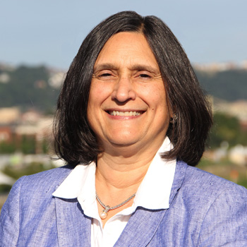 Mary L. Marazita合作论文数Department of Oral and Craniofacial Sciences, School of Dental Medicine, University of Pittsburgh6
 Darren M. Smith合作论文数6
 Kumar Anand合作论文数5
 Franklyn P. Cladis合作论文数5
 James Cray合作论文数5Lisa Vecchione合作论文数5Frederic W-B Deleyiannis合作论文数4
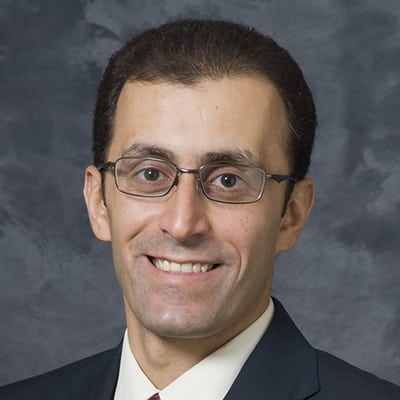 Ahmed M Afifi合作论文数Division of Plastic Surgery, Department of Surgery, University of Wisconsin-Madison3Rottgers S Alex合作论文数University of Pittsburgh3Dudas Jason R合作论文数3
 Leslie Elizabeth合作论文数3Otteson Todd合作论文数3Nayar Harry S合作论文数3
 Mark P. Mooney合作论文数McGowan Institute for Regenerative Medicine, University of Pittsburgh;Department of Anthropology, Faculty of Arts and Science, University of Pittsburgh3
 Seth Weinberg合作论文数Center for Craniofacial and Dental Genetics, School of Dental Medicine, University of Pittsburgh;Department of Oral and Craniofacial Sciences, School of Dental Medicine, University of Pittsburgh3
 Michael R Bykowski合作论文数Department of Neuroscience, University of Pittsburgh3Sukgi S. Choi合作论文数Children's National Medical Center, George Washington University Medical Center3
 Bernard J Costello合作论文数3Jeffrey P. Simons合作论文数3joseph e losee合作论文数2Regina Fenton合作论文数2Allison B Tobey合作论文数2Fasil Mathews合作论文数2Anne E Argenta合作论文数2
 Osama Basri合作论文数2Liliana Camison合作论文数2ellen cohn合作论文数2
 Alexander J Davit合作论文数2Matthew W Georg合作论文数Murtha Canc Ctr Res Program, Uniformed Serv Univ Hlth Sci2Madeleine K Bruce合作论文数2Decesare Gary E合作论文数2
 Jacqueline T. Hecht合作论文数Department of Pediatrics, McGovern Medical School, The University of Texas Health Science Center at Houston;Pediatric Research Center, The University of Texas Medical School at Houston2Pfaff Miles J合作论文数Department of Plastic and Reconstructive Surgery, Yale University2
 Shakir Sameer合作论文数University of Pittsburgh2
 Jake J. Lee合作论文数School of Medicine, University of Pittsburgh2
 Jennifer L McCoy合作论文数Pittsburgh Institute for Neurodegenerative Diseases and Department of Neurology, University of Pittsburgh2
 Lindsay Schuster合作论文数2Christopher R Kinsella合作论文数2Anne Argenta合作论文数2Richard E. Kirschner合作论文数2Katherine Neiswanger合作论文数1




