Undifferentiated malignant neoplasms pose diagnostic challenges, and reliable immunohistochemical markers with well-characterized staining profiles are desirable when characterizing them. Our initial observation of erythroblast transformation specific regulated gene-1 (ERG) reactivity in myeloid sarcomas led us to broadly explore the utility of ERG as a marker of immature hematolymphoid neoplasms presenting in extramedullary sites. We stained 207 immature and mature hematolymphoid lesions as well as 39 benign hematolymphoid tissues and found weak-to-moderate ERG immunopositivity in 15 of 16 (94%) acute myeloid leukemias/myeloid sarcomas, including 4 of 5 (80%) CD34-negative/CD117-negative acute myeloid leukemias/myeloid sarcomas. ERG positivity was also seen in all 9 cases of B-lymphoblastic and T-lymphoblastic leukemia/lymphoma, all 3 cases of hematogone hyperplasia, and all 4 cases of systemic mastocytosis. ERG was negative in 148 mature B-cell and T-cell lymphomas, including 2 high-grade B-cell lymphomas and 2 blastoid variant mantle cell lymphomas; 23 histiocytic/dendritic cell neoplasms; 2 indolent T-lymphoblastic proliferations; and 2 blastic plasmacytoid dendritic cell neoplasms. We conclude that ERG immunoreactivity may pose a significant diagnostic pitfall in the workup of undifferentiated malignant neoplasms, particularly those presenting in extramedullary sites.
The diagnosis of angioimmunoblastic T-cell lymphoma (AITL) is complex and requires the demonstration of a T-follicular helper (TFH) phenotype. Immunophenotypic markers that detect the TFH phenotype are highly variable, thereby necessitating the use of 3 to 5 TFH markers to substantiate a TFH phenotype. We tested the utility of germinal center markers human germinal center–associated lymphoma (HGAL) and LIM-domain only 2 (LMO2) in detecting a TFH phenotype. We compared their staining to that of 6 TFH markers in current use, PD-1, ICOS, CXCL13, SAP, CD10, and BCL6, in a cohort of 23 AITL. Our results show that although both markers can detect a TFH phenotype, HGAL was superior to LMO2 in the percent of cells stained and the intensity of staining, 2 variables used to generate H -scores. Using H -scores as the metric, HGAL was most comparable to BCL6 among the currently used TFH markers and was more sensitive than CXCL13, SAP, CD10, and LMO2. PD-1 and ICOS emerged as the most robust of the 8 markers tested in this study in detecting a TFH phenotype. We conclude that HGAL is a reliable marker of TFH cells and can aid in the diagnosis of lymphomas of TFH derivation, particularly in the recognition of early patterns of AITL.
Historically recognized by their characteristic histopathologic features, Spitz neoplasms are now known to be molecularly defined by mutually exclusive recurrent abnormalities that cause activation of the MAPK pathway. Spitz neoplasms with ALK rearrangements frequently demonstrate polypoid growth with a plexiform arrangement of nested, fusiform melanocytes in intersecting fascicles. Although neurotropism has been described in indolent Spitz neoplasms, this feature is not frequently mentioned in publications on histopathologic assessment of this group of melanocytic tumors. Here, we present an unusual case of a 3-year-old female with an ALK-positive compound Spitz nevus with extensive perineural and intraneural neurotropism occurring on the vermilion border of the lower lip.
 Yasodha Natkunam合作论文数Department of Pathology, Stanford University5
 Jingjing Zhang合作论文数Department of Immunology, University of Colorado Health Sciences Center and National Jewish Health;Department of Immunology, University of Colorado Health Sciences Center and National Jewish Health2
 Lhara Lezama合作论文数1Jennifer Y Wang合作论文数1
 Carlos Jose Suarez合作论文数Department of Pathology, Stanford University School of Medicine1Charu Vivek合作论文数Fogarty International Center, National Institutes of Health1
 Roberto Novoa合作论文数Department of Pathology, Stanford University;Department of Dermatology, Stanford University1
 Roger Allen Warnke合作论文数Department of Pathology, School of Medicine, Stanford University1
 Kerri E. Rieger合作论文数Department of Pathology, Stanford University1
 Brent Tan合作论文数Department of Pathology, School of Medicine, Stanford University;Division of Hematology, School of Medicine, Stanford University1
 Izidore S. Lossos合作论文数Division of Hematology, Department of Medicine, Sylvester Comprehensive Cancer Center, University of Miami1Shuchun Zhao合作论文数1
 Dita Gratzinger合作论文数Department of Pathology, Stanford University1
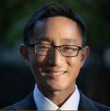 Derrick Wan合作论文数Department of Surgery, School of Medicine, Stanford University1
 Ryanne Ashley Brown合作论文数Department of Pathology, Stanford University1
 Florette K. Gray Hazard合作论文数Department of Pathology, Stanford University1
 Shyam S. Raghavan合作论文数Department of Chemistry, MIT1
 Donald E. Born合作论文数Department of Pathology, Stanford University1
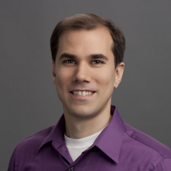 Jason Kurzer合作论文数Department of Pathology, Stanford Medicine, Stanford University1




