BACKGROUND:Schizophrenia is associated with widespread functional dysconnectivity, but the spatial scale and structural correlates of these alterations remain unclear. While relevant to local dysfunction, short-range connectivity is not well captured by standard approaches due to methodological constraints. METHODS:We applied a vertex-wise, distance-dependent analysis of functional connectivity strength (FCS) to resting-state fMRI data from 86 schizophrenia patients and 99 healthy controls across two datasets. FCS was partitioned by geodesic distance on the cortical surface and analyzed by cortical hierarchy. We also assessed two proxies of intracortical microstructure: T1/T2 ratio and a novel signal-detection-based measure of individualized data-driven functional connectivity density (idFCD). RESULTS:Schizophrenia patients exhibited reductions in short-range FCS within the dorsal primary somatosensory cortex. These functional alterations colocalized with abnormalities in both microstructural proxies and were not evident in global FCS analysis. In contrast, longer-range FCS was increased in transmodal regions, particularly the precuneus, without associated microstructural differences. Hierarchical analysis confirmed this dissociation, with structure-function disruption in primary networks and increased relative FCS in transmodal regions without microstructural association. CONCLUSIONS:Our findings support two distinct patterns of cortical dysconnectivity in schizophrenia: short-range reductions in primary sensory areas that colocalize with microstructural abnormalities, and longer-range increases in transmodal regions that appear structurally decoupled at the local level. By integrating distance-dependent functional measures with independent proxies of intracortical microstructure, this study highlights the role of short-range connectivity disruptions in primary areas and provides a complementary framework to conventional approaches based on regional or global analyses and diffusion-weighted imaging.
Over the past two decades, consciousness science has advanced from descriptive correlations to mechanistic predictions. Core requirements for conscious states have been clarified, and new diagnostic tools — most notably the Perturbational Complexity Index (PCI) derived from TMS–EEG — are moving toward clinical use. PCI indicates about a 95% probability of detecting consciousness in non-communicative patients when purposeful behavior is present. Recent studies in neurotypical individuals and communicative patients have adopted refined statistical methods accounting for regression-to-the-mean effects, along with rigorous awareness scales directly validated by subjective reports. Contrary to mainstream beliefs, these approaches consistently suggest that some degree of stimulus awareness is necessary for above-chance performance. Consciousness thus emerges not as an epiphenomenon but as a prerequisite for most purposeful behaviors in humans. This methodological progress opens new opportunities to bridge the long-standing divide between consciousness and cognitive sciences. At the same time, consciousness, intelligence and cognitive abilities are not identical: they can dissociate in revealing ways. Subjective experience exceeds its behavioral expression and remains accessible only to the experiencer. These observations motivate Integrated Information Theory, an explanatory framework that generate testable predictions about conscious states and qualitative contents independently of behavior, while also predicting that energy efficiency requirements may give rise to a tight link between consciousness and cognition in biological systems. Clinical and neuroscientific implications of such framework are discussed.
Although much is known about the molecular and cellular effects of general anesthetics, the neural mechanisms underlying loss and recovery of consciousness during anesthesia remain elusive. We provide the first report of human single neuron activity recorded throughout emergence from general anesthesia. Following cessation of propofol infusion, emergence was assessed as motor response to verbal command (RC). In most cells, firing rates increased during the course of emergence. Analysis of pooled firing rate changes indicated a shift towards criticality during emergence and attractor states during and immediately following anesthesia and immediately after RC. Changes in activity in some regions occurred prior to overt RC, with shortest latencies in the hippocampus, parahippocampal gyrus, and amygdala, followed by cingulate and then insular cortex. This work reveals that brain activity in medial temporal regions may presage restoration of responsiveness in humans following general anesthesia.
Study Objectives:Sleep spindles are electroencephalographic elements characteristic of non-rapid eye movement sleep (NREM) generated by thalamo-cortical interactions. Spindles have been linked to some of the cognitive benefits afforded by sleep and increased arousal threshold. This proof-of-concept study demonstrates that targeting the thalamus with Transcranial Electrical Stimulation with Temporal Interference (TES-TI) can enhance spindle activity. Materials and Methods:46 participants (24 ± 9.5 years; 58.7% F) underwent thalamic TES-TI stimulation during daytime naps. Three stimulation protocols with 15 kHz carrier frequency were tested during stage NREM 2: fixed difference frequency of 10 Hz (TES15kHz-TI10Hz), difference frequency matched to individual spindle peak, and no difference frequency. Spindle (sigma) band power and integrated spindle activity (ISA) were compared before and during the stimulation, and across stimulation protocols. Results:TES15kHz-TI10Hz stimulation was associated with increased sigma power (∆x~ STIM-PRE = 0.46 log10 μV2, p = .0042) and ISA (∆x~ STIM-PRE = 4.064 μV/s, p = .030). Cluster-based analysis localized the increase in sigma power across the entire scalp (p = .008). Linear mixed-effects models showed that both sigma power and ISA during stimulation increased significantly more in TES15kHz-TI10Hz compared to the other stimulation protocols. Conclusions:This study provides evidence supporting the successful use of TES-TI targeting the thalamus to enhance sleep spindle activity. Stimulation at a fixed difference frequency of 10 Hz increased sigma band power and ISA, whereas neither stimulation matched to individual sigma band peak nor TES alone produced comparable effects. These promising results warrant further investigations into the cognitive and clinical impact of TES-TI.
Abstract Identifying neural signatures of consciousness remains a central challenge in neuroscience. Sleep offers a tractable model for comparing brain activity in the presence or absence of subjective experience while minimizing behavioral responsiveness confounds. Using overnight 256-electrode high-density EEG in 140 participants and a serial-awakening paradigm, we analyzed 699 non-rapid eye movement (NREM) sleep stage 2 and 3 awakenings (351 dreaming experience, 348 no experience). Features from the 60s preceding awakening included regional spectral power, lagged-coherence connectivity, graph-theoretic metrics and gamma-to-alpha power ratios. Dreaming experiences were associated with shifts in posterior spectral balance, particularly reduced alpha and delta power and increased gamma-related measures, together with altered large-scale network organization. In participant-level cross-validated machine-learning analyses, all classifiers performed above chance, with the best ensemble model reaching an ROC-AUC of 0.80 and average precision of 0.80. These findings identify reproducible posterior electrophysiological and network-level signatures of conscious states during NREM sleep.
Given the importance of sleep for the regulation of cortical excitability, altered sleep homeostasis may contribute to the development and persistence of focal epilepsy. The goal of the present study was to quantify high-density EEG markers of sleep homeostasis in patients with focal epilepsy compared to healthy age- and gender-matched controls. In particular, we investigated the ability of sleep homeostasis markers to differentiate focal epilepsy versus healthy controls and quantified the deleterious effects of nocturnal epileptiform discharges on sleep homeostasis. Thirty-nine patients with focal epilepsy and 48 healthy controls underwent overnight 256-electrode high-density EEG recordings. Statistical analysis tested for differences in overnight slow-wave activity (i.e. delta power, 1-4 Hz, and theta power, 4-8 Hz) and average slope of slow waves between patients and healthy controls. We assessed the predictive value of sleep homeostasis parameters to identify focal epilepsy at the individual level. Finally, we investigated the relationship between the frequency of epileptiform discharges and abnormal overnight declines in sleep slow-wave activity and slow-wave slope. Patients with focal epilepsy displayed higher slow-wave activity and a steeper average slope of slow wave compared to healthy controls, exhibiting slow-wave activity values at the end of the night similar to those observed in healthy controls at the beginning of the night. Among sleep homeostasis markers, the presence of high local extremes in the topographical distribution of theta values proved to be discriminative at the single-subject level for distinguishing patients from healthy controls (area under the curve = 0.84). A significant negative association was observed between left-lateralized nocturnal epileptiform discharges and the overnight decline in slow-wave activity and slow wave slope, with the most pronounced effects over the left fronto-temporal leads. Local abnormalities in sleep homeostasis may constitute a reliable biomarker for focal epilepsy. Further studies are needed to determine whether it could also be used to identify focal epilepsy early in the disease course. Furthermore, frequent epileptiform discharges during sleep may contribute to abnormalities in sleep homeostasis and thus to the persistence of cortical hyperexcitability in focal epilepsy.
We investigate the subjective experience of space around the visual blind spot area, the cortical representation of which is missing feedforward connectivity from one eye. We performed these experiments as part of an adversarial collaboration to test contrasting theories of consciousness; Integrated Information Theory (IIT), Predictive Processing Active Inference (AI), and Predictive Processing Neurorepresentationalism (NREP) accounts. According to the Integrated Information Theory of consciousness, non-activatable retinotopic cortical regions, such as the blind spot region for the ipsilateral eye, create a different cause-effect structure and therefore should contribute differently to the perceived quality of space of activatable retinotopic regions. The two Predictive Processing accounts, in contrast, posit that internal models will accommodate structural deviations around the blindspot based on the available sensory evidence (particulars of this accommodation differ between the two accounts). We present a series of paradigms in which participants evaluate distances and areas that either include the blind spot or not (without stimulating it directly), as well as illusory motion that is either adjacent to the blind spot or not. We model psychometric functions relating perceived and objective space. These models vary in terms of bias and precision according to the experimental conditions (blind spot involved vs. not involved, ipsilateral vs. contralateral eye), making it possible to quantify the potential disruption of subjective spatial extendedness induced by the blind spot. We present simulated results for each experiment corresponding to the predictions of each account, and conclude by discussing challenges and plans for dissemination.
We aim to investigate the spatial experience of patients with chronic scotomas caused by lesion to early visual cortex, including primary visual cortex (V1) and adjacent extrastriate visual areas. These experiments are conducted as part of an adversarial collaboration testing contrasting theories of consciousness: Integrated Information Theory (IIT) and two Predictive Processing accounts, Active Inference (AI) and Neurorepresentationalism (NREP). The central question is whether lesions to early visual cortex alter the experienced extent of visual space itself, or instead primarily disrupt stimulus content within an otherwise preserved visual space. To address this, we use paradigms in which patients estimate distances or spatial extents that either span a scotomatous region or fall entirely within intact visual field locations. Psychometric functions relating perceived and physical extent are modeled to estimate shifts in the point of subjective equality (PSE). According to IIT, lesions to early visual cortex, including V1 and occipital exstrastriate cortex, should lead to systematic reductions in perceived spatial extent across the scotoma (negative PSE shifts), reflecting a contraction of experienced space. In contrast, Predictive Processing accounts posit that higher-level predictive mechanisms preserve spatial structure despite loss of early input, predicting little or no systematic contraction (with NREP allowing limited context-dependent effects). By quantifying distortions in perceived spatial extent, this protocol aims to distinguish between these competing theoretical predictions.
BACKGROUND:Slow waves are oscillations that reflect rhythmic alternation of neuronal activity and mediate key restorative functions of non-rapid eye movement (NREM) sleep. Left ventromedial prefrontal cortex regions are a "hot spot" for slow wave generation. The enhancement of slow wave activity (SWA, 0.5-4 Hz) has been shown to be beneficial, as in improving memory performance. To overcome limitations of current techniques, we assessed the ability of a non-invasive neuromodulatory tool, Transcranial Electrical Stimulation with Temporal Interference (TES-TI), to enhance SWA during NREM sleep overnight in healthy humans. METHODS:The current study is an interim analysis focused on the effects of TES-TI during NREM sleep in a laboratory setting. Two high frequency carriers with 1 Hz difference (TES15kHz-TI1Hz) produced amplitude-modulated temporal interference at 1Hz. Data were collected at the University of Wisconsin-Madison as part of the STRENGTHEN study (ClinicalTrials.gov, NCT06267521, 02/12/2024, single-blind, non-random allocation). Eligible participants were healthy adults (ages 18-50) assigned in parallel to one of four groups: (Group 1) TES15kHz (0 Hz difference frequency) 2 nights per week and meditation; (Group 2) TES15kHz-TI1Hz (1 Hz difference frequency) 2 nights per week and sham meditation; (Group 3) TES15kHz-TI1Hz 1 night per week and meditation; (Group 4) TES15kHz-TI1Hz 2 nights per week and meditation. Stimulation targeting left ventromedial prefrontal cortex occurred during NREM sleep over repeated nights ( ~ 10 stimulation periods, 3-min each, first half of night, 1-2 nights/week, 4-week protocol). Twenty-one participants (Groups 2, 3, 4) received TES15kHz-TI1Hz and seven participants received TES15kHz (Group 1). SWA was measured using simultaneous high-density electroencephalography with polysomnography. RESULTS:We show that SWA is increased with TES15kHz-TI1Hz (N = 21 total; N = 5 Group 2, N = 10 Group 3, N = 6 Group 4), and the effect outlasts the stimulation period; higher frequencies (sigma and beta) decrease. During stimulation, SWA is greater in TES15kHz-TI1Hz than pure TES15kHz (N = 7 Group 1). The incremental effects of TES15kHz-TI1Hz on SWA between first and last intervention night are positively associated with subjective ratings of restorative sleep. CONCLUSIONS:To our knowledge, this is the first study demonstrating that TES-TI enhances SWA and potentially its restorative function.
IntroductionFocal lesions such as a stroke can cause not only local effects but also distant effects in anatomically intact regions. The impact of stroke lesions on brain networks has been mapped using neuroimaging techniques such as functional magnetic resonance imaging (fMRI) and diffusion tensor imaging (DTI). In the present study, we established the feasibility of detecting network dysfunction at the electrocortical level using high-density electroencephalography (HD-EEG).Case reportWe studied brain function using HD-EEG in a patient with an acute left middle cerebral artery stroke. Slowing in the delta range was present beyond the ischemic focus, extending to the perilesional regions as well as distant regions. There was also delta connectivity in the stroke hemisphere with slowing affecting mainly distant regions that were interconnected with the site of the ischemic region (i.e., network-level diaschisis), although this was not statistically significant.ConclusionThis case study illustrates the feasibility of using HD-EEG to map local and distant electrical activity consequences of an acute stroke on cortical functions. Such a technique could be clinically useful to improve personalized stroke-network mapping in patients with acute cortical lesions.
Cannabis use is increasingly prevalent among individuals with epilepsy, yet its impact on seizure control remains poorly understood. While cannabidiol (CBD) has demonstrated antiseizure properties and gained FDA approval for specific epileptic syndromes, tetrahydrocannabinol (THC), the primary psychoactive compound in cannabis, may alter neuronal excitability and potentially exacerbate seizure activity. We present two illustrative case reports of male patients with focal epilepsy and chronic cannabis use who underwent treatment with antiseizure medications and responsive neurostimulation (RNS). In both cases, cannabis use was temporally associated with breakthrough seizures and poor seizure control. These cases highlight the complex and multifactorial relationship between cannabis use and seizure outcomes, including potential pharmacokinetic interactions with antiseizure medications (ASM) and the possibility that cannabis may blunt the neuromodulation effects of RNS. Given the retrospective data and limited detail on cannabis use, these findings should be interpreted with caution. As cannabis use rises among individuals with epilepsy, further research is needed to clarify its potential effects on seizures and treatment response, including neuromodulation.
Different theories explain how subjective experience arises from brain activity1,2. These theories have independently accrued evidence, but have not been directly compared3. Here we present an open science adversarial collaboration directly juxtaposing integrated information theory (IIT)4,5 and global neuronal workspace theory (GNWT)6-10 via a theory-neutral consortium11-13. The theory proponents and the consortium developed and preregistered the experimental design, divergent predictions, expected outcomes and interpretation thereof12. Human participants (n = 256) viewed suprathreshold stimuli for variable durations while neural activity was measured with functional magnetic resonance imaging, magnetoencephalography and intracranial electroencephalography. We found information about conscious content in visual, ventrotemporal and inferior frontal cortex, with sustained responses in occipital and lateral temporal cortex reflecting stimulus duration, and content-specific synchronization between frontal and early visual areas. These results align with some predictions of IIT and GNWT, while substantially challenging key tenets of both theories. For IIT, a lack of sustained synchronization within the posterior cortex contradicts the claim that network connectivity specifies consciousness. GNWT is challenged by the general lack of ignition at stimulus offset and limited representation of certain conscious dimensions in the prefrontal cortex. These challenges extend to other theories of consciousness that share some of the predictions tested here14-17. Beyond challenging the theories, we present an alternative approach to advance cognitive neuroscience through principled, theory-driven, collaborative research and highlight the need for a quantitative framework for systematic theory testing and building.
This overview of integrated information theory (IIT) emphasizes IIT's "consciousness-first" approach to what exists. Consciousness demonstrates to each of us that something exists–experience–and reveals its essential properties–the axioms of phenomenal existence. IIT formulates these properties operationally, yielding the postulates of physical existence. To exist intrinsically or absolutely, an entity must have cause-effect power upon itself, in a specific, unitary, definite and structured manner. IIT's explanatory identity claims that an entity's cause-effect structure accounts for all properties of an experience–essential and accidental–with no additional ingredients. These include the feeling of spatial extendedness, temporal flow, of objects binding general concepts with particular configurations of features, and of qualia such as colors and sounds. IIT's intrinsic ontology has implications for understanding meaning, perception, and free will, for assessing consciousness in patients, infants, other species, and artifacts, and for reassessing our place in nature.
As the field of consciousness science matures, the research agenda has expanded from an initial focus on the neural correlates of consciousness, to developing and testing theories of consciousness. Several theories have been put forward, each aiming to elucidate the relationship between consciousness and brain function. However, there is an ongoing, intense debate regarding whether these theories examine the same phenomenon. And, despite ongoing research efforts, it seems like the field has so far failed to converge around any single theory, and instead exhibits significant polarization. To advance this discussion, proponents of five prominent theories of consciousness—Global Neuronal Workspace Theory (GNWT), Higher-Order Theories (HOT), Integrated Information Theory (IIT), Recurrent Processing Theory (RPT), and Predictive Processing (PP)—engaged in a public debate in 2022, as part of the annual meeting of the Association for the Scientific Study of Consciousness (ASSC). They were invited to clarify the explananda of their theories, articulate the core mechanisms underpinning the corresponding explanations, and outline their foundational premises. This was followed by an open discussion that delved into the testability of these theories, potential evidence that could refute them, and areas of consensus and disagreement. Most importantly, the debate demonstrated that at this stage, there is more controversy than agreement between the theories, pertaining to the most basic questions of what consciousness is, how to identify conscious states, and what is required from any theory of consciousness. Addressing these core questions is crucial for advancing the field towards a deeper understanding and comparison of competing theories.
We discuss evidence from anesthesia, sleep, and mind-wandering studies, demonstrating the frequent detachment of consciousness from external sensory inputs, resulting in states of sensory disconnection. The challenge of behaviorally distinguishing between conscious states with sensory perception (connected consciousness [CC]) and those without it (disconnected consciousness [DC]) is examined in light of current methods for assessing consciousness. We evaluate the effectiveness of objective measures and behavioral responsiveness versus subjective measures in differentiating between CC and DC states during sleep and anesthesia. We conclude that the most effective approach to study sensory disconnection in these states is to serially awaken participants during the same sleep stage or at similar anesthetic concentrations after sensory stimulation, while neuroimaging data is recorded. Upon awakening, subjective reports on consciousness and sensory perception are collected to identify CC and DC states. This methodology allows for a within-state examination of the neural signatures of sensory disconnection, minimizing biases from between-state comparisons.
Visual memory relies on synchronized interactions and rhythms between the medial temporal lobes and neocortical brain regions. Non-invasive manipulation of memory-related brain regions, specifically deeper temporal lobe regions, has been limited by the lack of precision of non-invasive neuromodulation - when targeting deeper structures, the cortex is always stimulated, never deeper structures in isolation. Temporal Interference (TI) stimulation, a novel non-invasive technique, uses high-frequency carrier fields to deliver targeted, physiologically relevant neuromodulation via amplitude-modulated envelopes at specific brain regions. Here, we investigate TI's impact on figure memory encoding in 70 healthy participants using the Rey-Osterrieth and Taylor Complex Figure tasks, with TI applied in several brain regions independently and simultaneously - allowing investigation of combinations of medial temporal lobe and neocortical brain regions. Interestingly, higher frequency TI envelopes (130 Hz offset) targeting bilateral hippocampi and temporal cortices significantly impair recall (p = 6.54e-04), while lower frequency TI envelopes (5 Hz offset) targeting only the bilateral hippocampi significantly enhance recall (p = 0.0447). Stimulation using other combinations of medial temporal lobe and neocortical regions showed no effect, underscoring the critical role of frequency and focality of non-invasive brain stimulation and correct target selection. Finally, functional MRI reveals strong differences between the effects of 130 Hz and 5 Hz envelopes, specifically in hippocampal BOLD signals, brain connectivity, default mode, and attentional networks. These findings demonstrate TI's ability to bidirectionally modulate memory encoding through precise frequency and target tuning, offering a powerful tool for cognitive neuroscience and potential therapeutic applications for memory disorders.
Objectives:To track multi-unit activity (MUA) using hybrid Behnke-Fried electrodes continuously throughout human seizures, and to characterize how both neuronal firing and high-gamma (HG) changes over the ictal course, within versus outside the seizure-onset zone (SOZ), and by seizure severity. Methods:We analyzed 32 seizures - 9 focal preserved consciousness (FPC), 17 focal impaired consciousness (FIC), 6 focal-to-bilateral tonic-clonic (FBTC) - from 8 patients implanted with Behnke-Fried hybrid depth electrodes. Spikes were sorted jointly across a 10-min pre-ictal baseline and ictal period, yielding 895 units trackable through seizures. Macroelectrode contacts within 15 mm of each microelectrode were classified by the SOZ status of the corresponding micro site. Firing rates from MUA, HG power and phase-locked high-gamma (PLHG) were baseline-normalized and compared across seizure types and periods (first/second half; pre/post-generalization). Results:Unit waveforms were identified throughout seizures. Within the SOZ, firing increased at onset in all types, then attenuated (FPC, FIC) or plateaued (FBTC). PLHG peaked early and declined across all seizure types, including post-generalization in FBTC. In contrast, HG stayed elevated at onset for all seizures, was highest in FBTC, and increased further after generalization. Outside the SOZ, MUA, HG, and PLHG stayed near baseline in FPC, and rose progressively in FIC. In FBTC they showed a temporal dissociation: HG/PLHG rose pre-generalization and remained high, while MUA increased sharply only after generalization. Significance:We demonstrated feasibility to track multi-unit activity throughout seizures and revealed seizure-type- and location-specific micro-macro dynamics. Collectively, early rise and later decline in PLHG that mostly aligns with neuronal firing was concordant within SOZ. Moreover, increases in all measures outside the SOZ are a hallmark of seizures with impaired consciousness, and may suggest presence of a third driver. These findings offer testable biomarkers for future larger clinical studies. KEY POINTS:MUA can be tracked continuously during seizures with Behnke-Fried hybrid electrodes.In the SOZ, firing rises at onset, then attenuates in FPC/FIC or plateaus in FBTC; HG rises while PLHG peaks early then declines.Outside SOZ, firing, and HG/PLHG progressively increase, specifically after generalization.Combined MUA/HG/PLHG provide candidate biomarkers for SOZ mapping and impaired consciousness.
 Steven Laureys合作论文数School of Basic Medical Sciences, Hangzhou Normal University;Harvard Medical School, Harvard University;CERVO Brain Research Centre, Université Laval;GIGA Consciousness Research Unit, University of Liège219
 Audrey Vanhaudenhuyse合作论文数Cyclotron Research Centre, University of Liège96
 Olivia Gosseries合作论文数93
 Giulio Tononi合作论文数Department of Psychiatry, School of Medicine and Public Health, University of Wisconsin-Madison89
 Caroline Schnakers合作论文数Cyclotron Research Center, University of Liège81
 Pierre Maquet合作论文数Département des Sciences Cliniques, Faculté de Médecine, University of Liège78
 Gustave Moonen合作论文数National Institute of Child Health and Human Development, National Institutes of Health73Pierre Boveroux合作论文数60Marie-Aurelie Bruno合作论文数54
 Marcello Massimini合作论文数Department of Clinical Sciences;University of Milan;Department of Clinical Sciences, University of Milan51
 Athena Demertzi合作论文数Coma Science Group and Cyclotron Research Centre, University of Liège43
 Noirhomme Quentin合作论文数Coma Sci Grp, Univ Liege41
 Didier Ledoux合作论文数General Intensive Care and Department, Nephrology University Hospital35
 Andrea Soddu合作论文数34
 Rosanova Mario Carmine Emiliano合作论文数Department of Biomedical and Clinical Sciences, School of Medicine, University of Milan33
 Steve Majerus合作论文数Département De Psychologie, Faculté De Psychologie, Logopédie et Sciences De l'Éducation, Universite De Liege32
 Martin Desseilles合作论文数Department of Psychiatry, Centre Hospitalier Universitaire28
 Manuel Schabus合作论文数Department of Psychology, University of Salzburg26
 Vincent Bonhomme合作论文数25Philippe Peigneux合作论文数24
 Christophe Phillips合作论文数Cyclotron Research Center, University of Liège24Marieaurelie Bruno合作论文数24André Luxen合作论文数23
 Thien Thanh Dang-Vu合作论文数Cyclotron Research Centre, University of Liege23Christian Degueldre合作论文数22
 Albouy Geneviève合作论文数Cyclotron Research Centre.20
 Marie-Elisabeth Faymonville合作论文数20Silvia Casarotto合作论文数Department of Biomedical and Clinical Sciences "L. Sacco", Università degli Studi di Milano20
 Craig Phillips合作论文数Faculty of Medicine and Health, The University of Sydney19
 Virginie Sterpenich合作论文数Cyclotron Research Centre, University of Lièege-Sart Tilman19
 Gilles Vandewalle合作论文数Unité de Neuroimagerie fonctionnelle, Centre de recherche de l’Institut universitaire de gériatrie de Montréal18
 Struck Aaron F合作论文数Department of Neurological Surgery, University of Wisconsin17
 Adenauer G. Casali合作论文数Department of Clinical Sciences;Università degli Studi di Milano;Department of Clinical Sciences, Università degli Studi di Milano17
 Chatelle Camille合作论文数Cyclotron Res Ctr, Univ Liege15
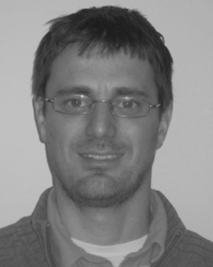 Riedner Brady A合作论文数15
 Luaba Tshibanda合作论文数15Annabelle Darsaud合作论文数15
 Jean-François Brichant合作论文数Centre Hospitalier Universitaire de Liège;Département des sciences cliniques, Faculté de Médecine, University of Liège15
 Thibaut Aurore合作论文数Cyclotron Research Centre and Neurology Department, University and University Hospital of Liège14
 Maurice Lamy合作论文数14
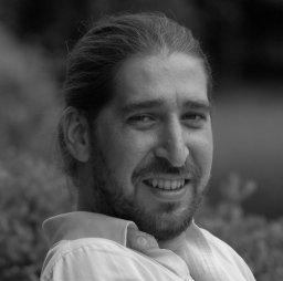 Simone Sarasso合作论文数Department of Psychiatry, University of Wisconsin–Madison14Evelyne Balteau合作论文数14
 Adrian M. Owen合作论文数Centre for Brain and Mind, Western University;Institute for Neuroscience, Western University13Bernard Lambermont合作论文数13
 Hustinx Roland合作论文数Service Médical de Médecine Nucléaire et Imagerie Onco, Université de Liège12
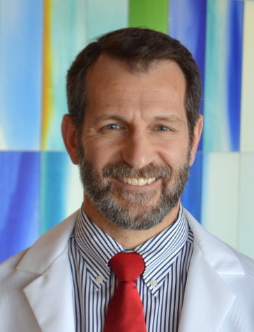 Joseph T. Giacino合作论文数Center for Bioethics, Harvard Medical School;Department of Physical Medicine and Rehabilitation, Harvard Medical School;Neurorehabilitation Laboratory, Spaulding Rehabilitation Hospital;Department of Psychiatry, Massachusetts General Hospital;Spaulding Rehabilitation Outcomes Center11
 Brinda Sevak合作论文数10Jitka Annen合作论文数Univ Liege, Coma Sci Grp, GIGA Consciousness, Liege, Belgium10




