Abstract Movement-related beta event-related synchronization (ERS) has been linked to motor control and learning, showing potential as a therapeutic target for those with movement deficits, such as stroke survivors. However, whether directly modulating beta ERS can causally influence motor performance remains unclear, largely due to the lack of methods designed to specifically target this neural activity. To address this gap, we developed a behaviourally-driven, closed-loop transcranial alternating current stimulation (tACS) approach to target movement-related beta ERS during a visuomotor adaptation task. We found that the behaviourally-driven, closed-loop beta-tACS specifically enhances beta ERS without affecting beta event-related desynchronization (ERD). Critically, this targeted enhancement significantly improves retention of motor adaptation. These findings support a functional role of beta ERS in motor behaviour and suggest that behaviourally-driven beta-tACS may provide a promising approach to further test the mechanistic links between beta ERS and behavioural outcomes in clinical populations.
Abstract Background Transcranial direct current stimulation (tDCS) is increasingly used as an adjunct to rehabilitation for young people with cerebral palsy (CP), yet considerable variability exists in clinical response. Individualised electric field modelling provides an opportunity to estimate the distribution of electrical fields generated by the stimulation delivered to the brain and explore potential relationships with functional outcomes. Methods Structural MRI scans from nineteen participants (10-16 years) from a previously published randomised controlled trial ( ISRCTN74235136 ) investigating the effects of tDCS combined with motor training, underwent participant-specific finite element modelling using SimNIBS. Electric field strength was quantified within anatomically defined motor regions of interest, including the primary motor cortex (M1), dorsal premotor cortex (PMd), supplementary motor area (SMA), and a combined motor network. Global grey matter electric field metrics and stimulation focality were also extracted. Results Estimated electric field strength differed significantly across motor regions (p<0.001), with PMd receiving significantly greater stimulation than both M1 and SMA. Electric field strength within a control region (primary visual cortex) was significantly lower than within M1 (p<0.001). Despite inter-individual variability in regional and global electric field metrics, no significant associations were observed between estimated electric field strength or focality and changes in function following intervention. Conclusion Individualised electric field modelling demonstrated that an M1-targeted tDCS montage preferentially stimulated PMd rather than M1 in young people with CP. These findings highlight the importance of subject-specific modelling when characterising current distribution and suggest that variability in electric field strength alone does not explain variability in behavioural response. Highlights Individualised modelling characterised tDCS electric fields in paediatric CP. M1-targeted tDCS produced greater electric field strength in PMd than M1. Motor regions received greater electric field strength than control visual cortex. Electric field strength did not predict upper- or lower-limb functional change.
Objectives: Non-invasive brain stimulation (NIBS) holds significant promise for treating neurological and neuropsychiatric conditions, yet translation into routine clinical practice remains limited. We aimed to explore stakeholder perceptions of NIBS and barriers to its clinical adoption. Methods: We conducted focus‑group interviews with 33 participants across three key stakeholder groups in the UK: (1) people with lived experience of brain injury, depression, or dementia; (2) healthcare professionals; and (3) researchers. Reflexive thematic analysis was used to identify themes in the data. Findings: Seven key themes emerged spanning preferences, hope and disappointment, communication, accessibility, infrastructure, ethical/regulatory uncertainty, and the evidence base. Across groups, NIBS was viewed positively and with cautious optimism, but substantial barriers were highlighted, including limited public and clinical awareness, challenges in demonstrating cost-effectiveness, infrastructure constraints, and difficulties navigating regulatory and translational pathways. Participants emphasised the importance of clear communication, improved education, and stronger interdisciplinary collaboration to support adoption. Notably, stakeholders prioritised evidence of clinical efficacy and usability over detailed mechanistic understanding. Conclusions: These findings provide actionable insights into the translational gap in NIBS and highlight priorities for facilitating its integration into clinical care.
Sleep plays a crucial role in consolidating recently acquired memories and preparing the brain for learning new ones, but the relationship between these two processes is currently unclear. According to the prominent Active Systems Consolidation model, memory representations that are initially reliant on the hippocampus are redistributed to neocortex during sleep for long-term storage. An indirect assumption of this model is that sleep-associated memory processing paves the way for next-day learning by freeing up hippocampal encoding resources. In this review, we evaluate two central tenets of this ‘resource reallocation hypothesis’: (i) sleep-associated memory consolidation reduces hippocampal engagement during retrieval, and (ii) this reduction in hippocampal burden enhances the brain's capacity for new learning. We then describe recent work that has directly tested the relationship between sleep-associated memory processing and next-day learning. In the absence of clear evidence supporting the resource reallocation hypothesis, we consider alternative accounts in which efficient learning is not contingent on prior overnight memory processing, but rather that sleep-associated consolidation and post-sleep learning rely on overlapping or independent mechanisms. We conclude by outlining how future research can rigorously test the resource reallocation hypothesis.
Movement-related beta event-related synchronization (ERS) has been linked to motor control and learning, showing potential as a therapeutic target for those with movement deficits, such as stroke survivors. However, whether directly modulating beta ERS can causally influence motor performance remains unclear, largely due to the lack of methods designed to specifically target this neural activity. To address this gap, we developed a novel behaviourally-driven, closed-loop transcranial alternating current stimulation (tACS) approach to target movement-related beta ERS during a visuomotor adaptation task. We found that the behaviourally-driven, closed-loop beta-tACS specifically enhances beta ERS without affecting beta event-related desynchronization (ERD). Critically, this targeted enhancement significantly improves retention of motor adaptation. These findings establish a causal relationship between beta ERS and motor behaviour and highlight the potential of behaviourally-driven beta-tACS as a therapeutic approach for improving motor function in clinical populations characterized by impaired beta activity. ### Competing Interest Statement The authors have declared no competing interest. Wellcome Trust, 224430/Z/21/Z NWO Rubicon, 04520232310005 Wellcome Trust, 222446/Z/21/Z NIHR Oxford Health Biomedical Research Centre, NIHR203316 Wellcome Trust, 203139/Z/16/Z Wellcome Trust, 203139/A/16/Z
Background: Spasticity is a motor phenomenon occurring in disorders of the central nervous system that impacts on active and passive function, and quality of life. Pharmacological, physical and surgical management options are available, each of which have limitations. Cryoneurolysis is a technique developed for the treatment of pain which involves the controlled freezing and thawing of peripheral nerves. Recent case reports and series have suggested it may offer a novel treatment approach for pain associated with spasticity. Objectives: To report on the evaluation of cryoneurolysis in the first cohort of patients treated in a UK spasticity clinic. Methods: Eight patients with a variety of neurological conditions (aged 25-75 years) underwent cryoneurolysis. Each had been receiving regular botulinum toxin injections and had ongoing treatment goals. All patients first underwent diagnostic nerve blocks with local anaesthetic to determine their appropriateness for the treatment. Cryoneurolysis was then performed with ultrasound and nerve stimulator guidance. Assessments included goal attainment, Modified Ashworth Scale (MAS), ArmA, LegA and the patient reported impact of spasticity scale (PRISM), alongside patient satisfaction and side effect questionnaires. Assessments were at baseline and at regular intervals over 9 to 12 months. Results: All patients attained at least one of their goals, with sustained effect for more than 6 months. MAS demonstrated mixed or modest improvements. Functional outcome measures (ArmA/LegA) showed several meaningful improvements, particularly in passive function. There was an indication of an improvement in PRISM across domains, which plateaued at 6 months. Post-procedure pain was the most common side effect but subsided in all affected patients by 3 months. Patient satisfaction was positive. Conclusions: Our findings contribute to a growing base of case reports and series suggesting that cryoneurolysis could be a potentially useful treatment modality for spasticity. Future controlled studies should aim to evaluate cost-effectiveness and compare with existing treatments.
Sleep disturbance is common but often overlooked after stroke. Regular sleep is increasingly recognised as important for overall health, yet little is known about how sleep regularity changes after stroke. This study examined differences in the Sleep Regularity Index (SRI) between stroke survivors and healthy controls using actigraphy data from an existing dataset (~ 1 week per participant). Data were analysed for 162 stroke survivors (mean age 61 ± 14 years, 5 ± 5 years post-stroke, 89 males) and 60 controls (mean age 57 ± 17 years, 32 males). Stroke survivors had significantly lower SRI scores than controls (p = 0.001), indicating less regular sleep. In the stroke group, higher SRI correlated with longer total sleep time (p = 0.003) and better self-reported sleep quality (p = 0.001) but not with other sleep metrics. Lower SRI was associated with worse depressive symptoms (p = 0.006) and lower quality of life (p = 0.001) but not with disability (p = 0.886) or time since stroke (p = 0.646). These findings suggest that sleep regularity is disrupted post-stroke and may influence well-being. Future research should explore interventions to improve sleep regularity and related health outcomes in stroke survivors.
Sleep is often disrupted after stroke. However, little is known about how stroke lesion extent or location influences sleep, particularly at the chronic stage of recovery. In this pragmatic study, we aimed to explore whether lesion characteristics could explain sleep variability in chronic stroke survivors. We analysed previously collected structural brain images (Magnetic Resonance Imaging) from 38 stroke participants (11 female, mean (SD) age 64 (12), mean (SD) time since stroke 95 (66) months) and sleep data (questionnaires (N=38, actigraphy (N=37), and electrophysiology (N=18)) which were collected in their home environment. Neither lesion volume nor lesion overlap with regions of interest (brainstem, basal ganglia, amygdala, hippocampus, thalamus) significantly predicted interindividual variability in subjective or objective sleep measures. However, a data-driven approach revealed clusters of voxels disconnected by the stroke lesions were linked to lower spindle density and amplitude (threshold free cluster enhancement p < 0.050). Overall, these results provide preliminary insights that lesion induced brain disconnection, rather than the extent and overlap of the lesion with grey matter regions, may be more informative when explaining sleep variability. However, larger prospective studies are needed to fully understand the effect of stroke lesions on sleep. ### Competing Interest Statement The authors have declared no competing interest. ### Funding Statement This work is supported by the Wellcome Trust and the NIHR Oxford Health Biomedical Research Centre (NIHR203316). The views expressed are those of the authors and not necessarily those of the NIHR or the Department of Health and Social Care. MKF is Funded by Guarantors of Brain and HJB is funded by the Wellcome Trust (222446/Z/21/Z). CJS holds a Senior Research Fellowship, funded by the Wellcome Trust (224430/Z/21/Z). The Wellcome Centre for Integrative Neuroimaging is supported by core funding from the Wellcome Trust (203139/Z/16/Z and 203139/A/16/Z). ### Author Declarations I confirm all relevant ethical guidelines have been followed, and any necessary IRB and/or ethics committee approvals have been obtained. Yes The details of the IRB/oversight body that provided approval or exemption for the research described are given below: The study was approved by the Central University Research Ethics Committee, University of Oxford (reference R85306) I confirm that all necessary patient/participant consent has been obtained and the appropriate institutional forms have been archived, and that any patient/participant/sample identifiers included were not known to anyone (e.g., hospital staff, patients or participants themselves) outside the research group so cannot be used to identify individuals. Yes I understand that all clinical trials and any other prospective interventional studies must be registered with an ICMJE-approved registry, such as ClinicalTrials.gov. I confirm that any such study reported in the manuscript has been registered and the trial registration ID is provided (note: if posting a prospective study registered retrospectively, please provide a statement in the trial ID field explaining why the study was not registered in advance). Yes I have followed all appropriate research reporting guidelines, such as any relevant EQUATOR Network research reporting checklist(s) and other pertinent material, if applicable. Yes Data derivatives and analysis scripts will be made available here: https://osf.io/vbu7c/. Raw data are available upon reasonable request from the corresponding author.
PURPOSE:Cerebral Palsy (CP) is the commonest cause of childhood motor disability. Transcranial direct current stimulation (tDCS) is a promising adjuvant therapy, but research targeting upper and lower limbs simultaneously is needed. We aimed to pilot tDCS with upper/lower limb motor training, estimate the potential effect on motor function, and investigate brain imaging correlates of function. MATERIALS AND METHODS:Participants (10-16 years) with CP affecting upper and/or lower limbs were randomised (online software) to 10 sessions of active (n = 14) or sham (n = 13) tDCS combined with motor training. The primary outcomes were upper and lower limb function assessed at 1-week post-intervention using the Jebson Taylor hand function (JTT) and Timed Up and Go (TUG) tests. Secondary, imaging outcomes included baseline tractography, grey matter volume, and resting state connectivity. RESULTS:Adherence was good: 74% completed all intervention sessions, 100% completed the primary outcome assessment. There were no between-group differences (1-week post-intervention, intention-to-treat; group-by-time JTT: F(1,25)=1.189,p = 0.286, partial-eta-squared = 0.05; TUG: F(1,25)=1.605,p = 0.217, partial-eta-squared = 0.06). Imaging showed subtle associations between better JTT at baseline and higher grey matter volume (caudate nucleus) and stronger sensorimotor resting state connectivity. CONCLUSIONS:The trial was well tolerated, but effect sizes were small. Larger studies are needed to further explore tDCS for CP.
Introduction Consolidation of motor skill learning, a key component of rehabilitation post-stroke, is known to be sleep dependent. However, disrupted sleep is highly prevalent after stroke and is often associated with poor motor recovery and quality of life. Previous research has shown that digital cognitive behavioural therapy (dCBT) for insomnia can be effective at improving sleep quality after stroke. Therefore, the aim of this trial is to evaluate the potential for sleep improvement using a dCBT programme, to improve rehabilitation outcomes after stroke. Methods and analysis We will conduct a parallel-arm randomised controlled trial of dCBT (Sleepio) versus treatment as usual among individuals following stroke affecting the upper limb. Up to 100 participants will be randomly allocated (2:1) into either the intervention (6–8 week dCBT) or control (continued treatment as usual) group. The primary outcome of the study will be change in insomnia symptoms pre to post intervention compared with treatment as usual. Secondary outcomes include improvement in overnight motor memory consolidation and sleep measures between intervention groups, correlations between changes in sleep behaviour and overnight motor memory consolidation in the dCBT group and changes in symptoms of depression and fatigue between the dCBT and control groups. Analysis of covariance models and correlations will be used to analyse data from the primary and secondary outcomes. Ethics and dissemination The study has received approval from the National Research Ethics Service (22/EM/0080), Health Research Authority (HRA) and Health and Care Research Wales (HCRW), IRAS ID: 306 291. The results of this trial will be disseminated via presentations at scientific conferences, peer-reviewed publication, public engagement events, stakeholder organisations and other forms of media where appropriate. Trial registration number NCT05511285 .
Introduction There is growing evidence that sleep is disrupted after stroke, with worse sleep relating to poorer motor outcomes. It is also widely acknowledged that consolidation of motor learning, a critical component of poststroke recovery, is sleep-dependent. However, whether the relationship between disrupted sleep and poor outcomes after stroke is related to direct interference of sleep-dependent motor consolidation processes, is currently unknown. Therefore, the aim of the present study is to understand whether measures of motor consolidation mediate the relationship between sleep and clinical motor outcomes post stroke.Methods and analysis We will conduct a longitudinal observational study of up to 150 participants diagnosed with stroke affecting the upper limb. Participants will be recruited and assessed within 7 days of their stroke and followed up at approximately 1 and 6 months. The primary objective of the study is to determine whether sleep in the subacute phase of recovery explains the variability in upper limb motor outcomes after stroke (over and above predicted recovery potential from the Predict Recovery Potential algorithm) and whether this relationship is dependent on consolidation of motor learning. We will also test whether motor consolidation mediates the relationship between sleep and whole-body clinical motor outcomes, whether motor consolidation is associated with specific electrophysiological sleep signals and sleep alterations during subacute recovery.Ethics and dissemination This trial has received both Health Research Authority, Health and Care Research Wales and National Research Ethics Service approval (IRAS: 304135; REC: 22/LO/0353). The results of this trial will help to enhance our understanding of the role of sleep in recovery of motor function after stroke and will be disseminated via presentations at scientific conferences, peer-reviewed publication, public engagement events, stakeholder organisations and other forms of media where appropriate.Trial registration number ClinicalTrials.gov: NCT05746260, registered on 27 February 2023.
Background and Aims: The purpose of this Third Stroke Recovery and Rehabilitation Roundtable (SRRR3) was to develop consensus recommendations to address outstanding barriers for the translation of preclinical and clinical research using the non-invasive brain stimulation (NIBS) techniques Transcranial Magnetic Stimulation (TMS) and Transcranial Direct Current Stimulation (tDCS) and provide a roadmap for the integration of these techniques into clinical practice.Methods: International NIBS and stroke recovery experts (N = 18) contributed to the consensus process. Using a nominal group technique, recommendations were reached via a five-stage process, involving a thematic survey, two priority ranking surveys, a literature review and an in-person meeting.Results and Conclusions: Results of our consensus process yielded five key evidence-based and feasibility barriers for the translation of preclinical and clinical NIBS research, which were formulated into five core consensus recommendations. Recommendations highlight an urgent need for (1) increased understanding of NIBS mechanisms, (2) improved methodological rigor in both preclinical and clinical NIBS studies, (3) standardization of outcome measures, (4) increased clinical relevance in preclinical animal models, and (5) greater optimization and individualization of NIBS protocols. To facilitate the implementation of these recommendations, the expert panel developed a new SRRR3 Unified NIBS Research Checklist. These recommendations represent a translational pathway for the use of NIBS in stroke rehabilitation research and practice.
Background: Intermittent theta-burst stimulation (i) (TBS) is a transcranial magnetic stimulation (TMS) plasticity protocol. Conventionally, TBS is applied using biphasic pulses due to hardware limitations. However, mono phasic pulses are hypothesised to recruit cortical neurons more selectively than biphasic pulses, predicting stronger plasticity effects. Monophasic and biphasic TBS can be generated using a custom-made pulse-width modulation-based TMS device (pTMS).Objective: Using pTMS, we tested the hypothesis that monophasic iTBS would induce a stronger plasticity effect than biphasic, measured as induced increases in motor corticospinal excitability.Methods: In a repeated-measures design, thirty healthy volunteers participated in three separate sessions, where monophasic and biphasic iTBS was applied to the primary motor cortex (M1 condition) or the vertex (control condition). Plasticity was quantified as increases in motor corticospinal excitability after versus before iTBS, by comparing peak-to-peak amplitudes of motor evoked potentials (MEP) measured at baseline and over 60 min after iTBS.Results: Both monophasic and biphasic M1 iTBS led to significant increases in MEP amplitude. As predicted, linear mixed effects (LME) models showed that the iTBS condition had a significant effect on the MEP amplitude (chi(2) (1) = 27.615, p < 0.001) with monophasic iTBS leading to significantly stronger plasticity than biphasic iTBS (t (693) = 2.311, p = 0.021). Control vertex iTBS had no effect. Conclusions: In this study, monophasic iTBS induced a stronger motor corticospinal excitability increase than biphasic within participants. This greater physiological effect suggests that monophasic iTBS may also have potential for greater functional impact, of interest for future fundamental and clinical applications of TBS.
The activation of nitric oxide synthase (NOS) has been linked to excitatory input via NMDA receptors, We hypothesized that NOS-positive neurons that have NMDA receptors on their surface would have high levels of cytochrome oxidase (C.O.) as energy generator for membrane repolarization. In order to compare the distribution of these markers on the same section, we reacted rat brain sections for C.O. histochemistry followed by NOS immunogold silver staining (IGSS). Adjacent sections were reacted for NOS IGSS followed by indirect immunoperoxidase for NMDA receptor subunit RI (NMDARI). We found that the staining pattern varied among regions but were consistent within each region examined, There are three types of NOS immunoreactive (NOS-ir) cells: (1) NOS-ir neurons that had moderate to high levels of both NMDARI and C.O. staining, such as the pontine reticular nuclei, motor and mesencephalic nuclei of the trigeminal nerve, and some motor neurons in the spinal cord, (2) NOS-ir neurons that were immunoreactive for NMDARI (NMDARI-ir) but had low levels of C.O. activity in their somata. Their dendrites, however, were both NMDARI-ir and rich in C.O. Examples of this type include neurons in the caudate and putamen, and periglomerular cells in the olfactory bulb, (3) We also found that some NOS-ir neurons were not NMDARI-ir and had low C.O. activity, In addition to postsynaptic neurons, C.O. and NOS levels were both high in the inner segments of retinal photoreceptor cells, where energy-demanding active ion transport maintains the dark current and where NO presumably activates guanylate cyclase for the production of cGMP, which keeps the Na+ channels open in the dark. Our findings suggest that NMDA receptors are available for the majority of NOS-ir neurons, which comprise a heterogenous population with varying energy demands,
Sleep plays a key role in supporting brain function and resilience to brain decline. It is well known that sleep changes substantially with aging and that aging is associated with deterioration of brain structure. In this study, we sought to characterize the relationship between slow wave slope (SWslope)-a key marker of sleep architecture and an indirect proxy of sleep quality-and microstructure of white matter pathways in healthy adults with no sleep complaints. Participants were 12 young (24-27 years) and 12 older (50-79 years) adults. Sleep was assessed with nocturnal electroencephalography (EEG) and the Pittsburgh Sleep Quality Index (PSQI). White matter integrity was assessed using tract-based spatial statistics (TBSS) on tensor-based metrics such as Fractional Anisotropy (FA) and Mean Diffusivity (MD). Global PSQI score did not differ between younger (n = 11) and older (n = 11) adults (U = 50, p = 0.505), but EEG revealed that younger adults had a steeper SWslope at both frontal electrode sites (F3: U = 2, p < 0.001, F4: U = 4, p < 0.001, n = 12 younger, 10 older). There were widespread correlations between various diffusion tensor-based metrics of white matter integrity and sleep SWslope, over and above effects of age (n = 11 younger, 9 older). This was particularly evident for the corpus callosum, corona radiata, superior longitudinal fasciculus, internal and external capsule. This indicates that reduced sleep slow waves may be associated with widespread white matter deterioration. Future studies should investigate whether interventions targeted at improving sleep architecture also impact on decline in white matter microstructure in older adults.
The recruitment of disabled participants for conducting usability evaluation of accessible information and communication technologies (ICT) is a challenge that current research faces. To overcome these challenges, researchers have been calling upon able-bodied participants to undergo disability simulations. However, this practice has been criticized due to the different experiences and expectations that disabled and able-bodied participants may have with ICT. This paper presents the methodology and lessons learned from ongoing mixed method-based usability evaluation of a suboptimal conventional computer mouse and an assistive gesture-based interface (i.e., the Leap Motion Controller) by stroke patients with upper-limb impairment and able-bodied participants experiencing a motor dysfunction simulation. The paper concludes with recommendations for future multidisciplinary research on ICT accessibility by people with disabilities.
Abstract Real-time functional MRI neurofeedback allows individuals to self-modulate their ongoing brain activity. This may be a useful tool in clinical disorders that are associated with altered brain activity patterns. Motor impairment after stroke has previously been associated with decreased laterality of motor cortex activity. Here we examined whether chronic stroke survivors were able to use real-time fMRI neurofeedback to increase laterality of motor cortex activity and assessed effects on motor performance and on brain structure and function. We carried out a randomized, double-blind, sham-controlled trial (ClinicalTrials.gov: NCT03775915) in which 24 chronic stroke survivors with mild to moderate upper limb impairment experienced three training days of either Real (n = 12) or Sham (n = 12) neurofeedback. Assessments of brain structure, brain function and measures of upper-limb function were carried out before and 1 week after neurofeedback training. Additionally, measures of upper-limb function were repeated 1 month after neurofeedback training. Primary outcome measures were (i) changes in lateralization of motor cortex activity during movements of the stroke-affected hand throughout neurofeedback training days; and (ii) changes in motor performance of the affected limb on the Jebsen Taylor Test (JTT). Stroke survivors were able to use Real neurofeedback to increase laterality of motor cortex activity within (P = 0.019), but not across, training days. There was no group effect on the primary behavioural outcome measure, which was average JTT performance across all subtasks (P = 0.116). Secondary analysis found improvements in the performance of the gross motor subtasks of the JTT in the Real neurofeedback group compared to Sham (P = 0.010). However, there were no improvements on the Action Research Arm Test or the Upper Extremity Fugl–Meyer score (both P > 0.5). Additionally, decreased white-matter asymmetry of the corticospinal tracts was detected 1 week after neurofeedback training (P = 0.008), indicating that the tracts become more similar with Real neurofeedback. Changes in the affected corticospinal tract were positively correlated with participants neurofeedback performance (P = 0.002). Therefore, here we demonstrate that chronic stroke survivors are able to use functional MRI neurofeedback to self-modulate motor cortex activity in comparison to a Sham control, and that training is associated with improvements in gross hand motor performance and with white matter structural changes.
 Heidi Johansen-Berg合作论文数Wellcome Centre for Integrative Neuroimaging, University of Oxford;Plasticity Group, Nuffield Department of Clinical Neurosciences, Medical Sciences Division, University of Oxford;Department of Clinical Neurology, University of Oxford14
 Cathy Stinear合作论文数Clinical Neuroscience Laboratory, Department of Medicine, School of Medicine, Faculty of Medical and Health Sciences, University of Auckland9
 Winston Byblow合作论文数Department of Exercise Sciences, Faculty of Science, The University of Auckland9
 Matthew Weightman合作论文数Sch Psychol, Univ Birmingham6
 Di Newham合作论文数6
 Charlotte Stagg合作论文数Nuffield Department of Clinical Neurosciences, Medical Sciences Division, University of Oxford6
 Tom Smejka合作论文数Maastricht University6
 Barbara Robinson合作论文数5
 James Coxon合作论文数Turner Institute for Brain and Mental Health, School of Psychological Sciences, Monash University5
 Isaac Sorinola合作论文数School of Population Health & Environmental Sciences, Faculty of Life Sciences & Medicine, King’s College London4
 Alan Barber合作论文数Faculty of Medical and Health Sciences, University of Auckland;Centre for Brain Research, University of Auckland4
 Sarah F Roberts-Lewis合作论文数3
 Zich Catharina合作论文数European Medical School, University of Oldenburg3Emma Garratt合作论文数2
 Veerle van Gils合作论文数Alzheimer Ctr Limburg, Maastricht Univ2
 Marilien Marzolla合作论文数2
 Zeena Sanders合作论文数Wellcome Ctr Integrat Neuroimaging, Univ Oxford2Michael Lührs合作论文数2
 James T H Teo合作论文数Kings Coll Hosp NHS Fdn Trust, London, England2
 James Newham合作论文数King's College London2
 Cassandra Sampaio-Baptista合作论文数2
 David Henderson Slater合作论文数2
 Alasdair L Henry合作论文数Institute of Inflammation &University of Manchester;Institute of Inflammation & Repair, University of Manchester2
 Salvan Piergiorgio合作论文数Institute of Psychiatry, King's College London2
 Colin A. Espie合作论文数Nuffield Department of Clinical Neurosciences, Medical Sciences Division, University of Oxford2Tim Theologis合作论文数Oxford Gait Laboratory, Nuffield Orthopaedic Centre2
 Sebastian W. Rieger合作论文数Department of Psychiatry, University of Oxford2
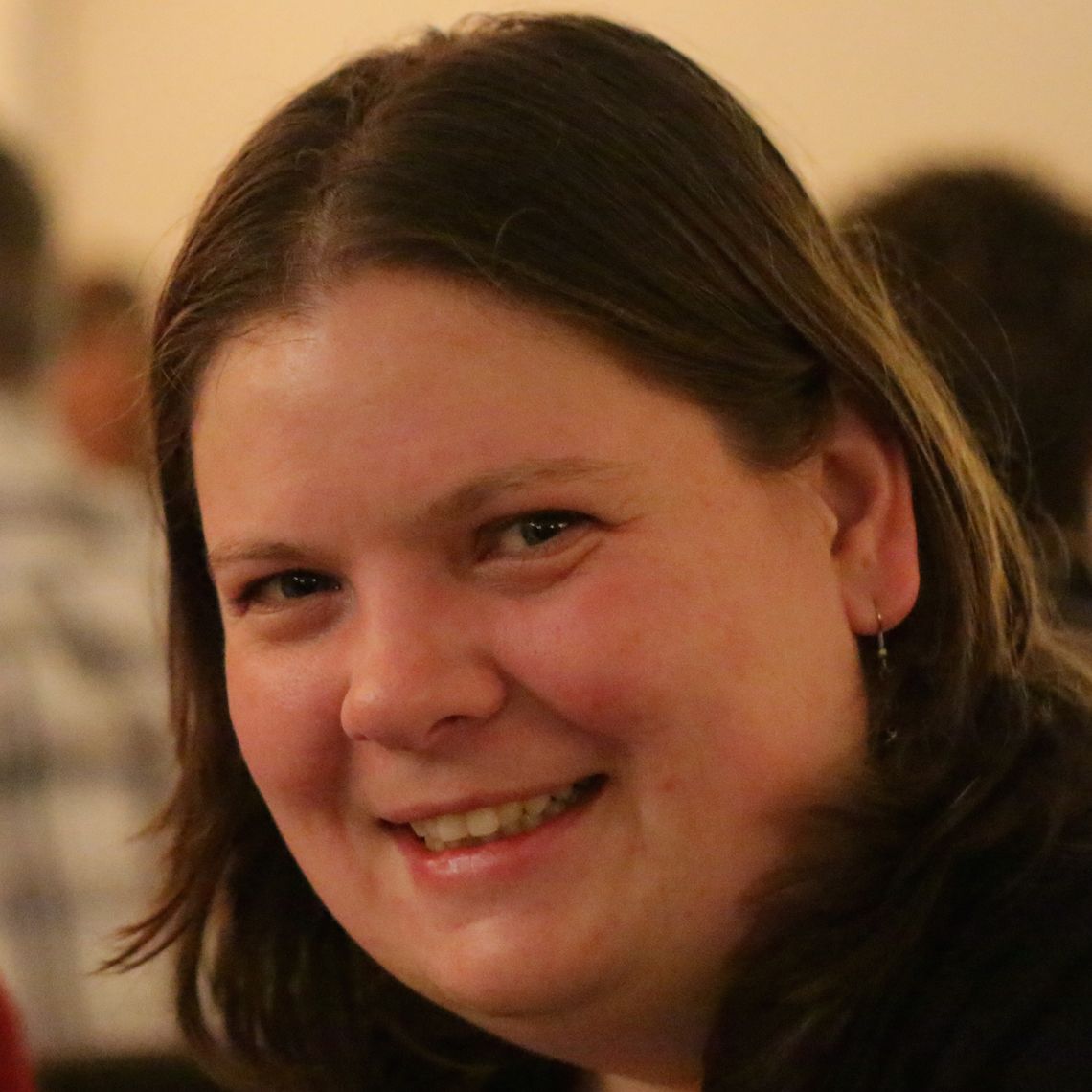 Nele Demeyere合作论文数Department of Experimental Psychology, University of Oxford2
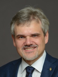 Rainer Goebel (R.W.)合作论文数Department of Cognitive Neuroscience, Faculty of Psychology and Neuroscience, Maastricht University;Maastricht Brain Imaging Centre2
 Hanna Willis合作论文数1Ece Yilmaz Kara合作论文数1Nathan Steadman合作论文数1Daan Meester合作论文数1Janet Cockburn合作论文数1Andrew Meaney合作论文数1Rachel Buckingham合作论文数1
 Jonathan Sanching Tsay合作论文数1Cosima Graef合作论文数1Lasbareille Camille合作论文数1
 Loic Couture合作论文数1Valentina Azzollini合作论文数1Ioana-Florentina Grigoras合作论文数1Emad Al-Yahya合作论文数1
 Alice Lagas合作论文数School of Optometry and Vision Science, University of Auckland1Kausar Raheel合作论文数1
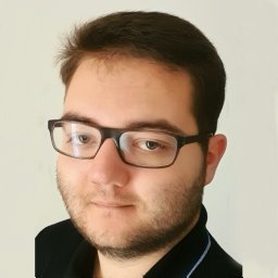 Raul C. Sîmpetru合作论文数Department Artificial Intelligence in Biomedical Engineering, Friedrich-Alexander-Universität Erlangen-Nürnberg1
 Ronan Mooney合作论文数Department of Physical Medicine and Rehabilitation, Johns Hopkins University School of Medicine1
 Jinpei Han合作论文数Brain & Behaviour Lab, Royal School of Mines, Imperial College London;AstraZeneca;Faisal Lab1
 Fanny Quandt合作论文数1




