Tumor-infiltrating lymphocytes (TIL) often fail to restrain tumor growth due to progressive differentiation into an "exhausted" state. Tissue-resident memory T cells (TRM) maintain protection from infection for years in healthy tissues, and patient tumors that contain TIL with TRM features are associated with better prognosis. Proteomic and transcriptomic profiling of T cell populations identified proteostasis as a significant factor distinguishing TRM and progenitor-exhausted TIL from terminally exhausted TIL, including loss of E3 ubiquitin ligases NEURL3, RNF149, and WSB1, with accumulation of unfolded proteins despite functional proteasome activity. Enforced expression of these ligases in T cells preserved stem-like TCF1+ populations and improved function in tumors and chronic infection, whereas deficiency impaired TIL and altered T cell differentiation during acute infection. Sustained ligase expression rescued the accumulation of unfolded proteins in TIL and improved immunotherapy outcomes in preclinical models, underscoring the critical role of proteostasis in TIL function and highlighting a promising avenue for advancing cancer immunotherapy.
Differential expression between normal chr1 and gain(1q) MM patients with t(4;14) translocation
Impact of additional chromosome 1 structural events on outcome A.Effect of amp(1q) with or without a total (TT) gain on PFS B. Effect of amp(1q) on OS
Differential expression between patients with no 1q gain and those with a gain(1q:CKS1B). A. All genes that are differentially expressed
Differential expression between normal chr1 and gain(1q) MM patients with t(11;14) translocation
Methylation probe intensity across chromosome 1. Heatmap representation of the beta value for all probes across chromosome 1, the probes a presented in a genomic coordinate sorted fashion. The recurrent regions of deletion, gain, templated insertion, and chromothripsis have be highlighted along with the cytoband information for reference.
Supplemental Spreadsheet 8: Differential expression between normal chr1 and whole-arm gains group MM patients
List of dysregulated oncogenes, tumor suppressor genes, and fusion proteins in the presence or absence of gain(1q) based on the Cancer Gene Census
Genes with increased dependency in cell lines with del(1p) [part A] Genes with increased dependency in cell lines with del(1p) [part B]
Bayes Factor output between genetic variables Correlation analysis between deletion region, gain regions, and other genetic events
Recurrent regions of chromothripsis and chromoplexy together with the candidate genes located within the regions. A. CHromothripsis B. Templated Instertion
Concurrent Hypomethylation and Overexpression Probes. The dot plot (top) displays the distribution of statistically significant CpG probes with difference in group mean methylation beta value, where the greater the value is a proxy for loss of methylation at the CpG probe. The probes are oriented by genomic locus across chromosome 1 with annotations for the midpoint of the recurrent gained regions on the 1q arm. The area graph (bottom) displays the ratio of probes that are significantly differentially methylated in any given 2MB window across chromosome 1. The highlighted region corresponds to the 1q arm.
Chromatin state of different SV regions. The CT1 region at 1p36.11-1p34.3 is comprised of greater than 74% A-compartment (range 74-100%); CT2 at 1p33-1p32.3 comprises both inactive (range 35-59%) and active chromatin (range 41-65%) and, CT3 at 1p13.3-1p12, overlaps with TI1, had greater variability with 86% A-compartment in U266 while only 1% in KMS11. TI1 region predominantly laid in B-compartment (range 63-73%) and TI2 lay in an almost entirely active compartment (range 91-100%). NBC=Naïve B-cell, MBC=Memory B-cell, GCBC=Germinal Center B-cell, PC=plasma cell.
Recurrent regions of copy number gain and loss together with the candidate genes located within the regions.
Correlation between age, telomere length and 1q gain subtype. A. Correlation between telomere length and chromosome 1 rearrangement subtype B. Correlation between age and chromosome 1 rearrangement subtype
 Gareth J Morgan合作论文数Univ Arkansas Med Sci, Myeloma Inst, Little Rock, AR 72205 USA79
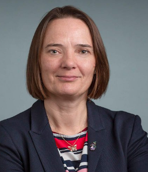 Faith E. Davies合作论文数Department of Medicine, NYU Grossman School of Medicine;Center for Blood Cancers, Perlmutter Cancer Center, NYU Langone Health70
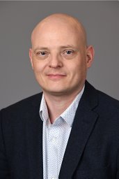 Brian Walker合作论文数Department of Medicine, Miller School of Medicine, University of Miami65
 Eileen Mary Boyle合作论文数54
 Cody Ashby合作论文数Arkansas State University50
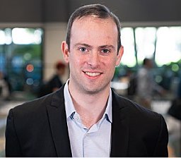 Louis Williams合作论文数Perlmutter Cancer Center, NYU Langone Health43
 Yubao Wang合作论文数Perlmutter Cancer Center, NYU Langone Health38
 Maura Francesco合作论文数Department of Clinical Sciences and Community Health, University of Milano and Hematology 1 CTMO38
 Hussein Ghamlouch合作论文数Department of Immunology, Université de Picardie Jules Verne38
 Marc Braunstein合作论文数37
 Barbara Bruno合作论文数University of Torino37Rhee Frits van合作论文数35Ariel Siegel合作论文数34Lydia Montes合作论文数32
 Aristotelis Tsirigos合作论文数Department of Pathology, Grossman School of Medicine, New York University32Corre Jill合作论文数32
 Carolina Schinke合作论文数32Alexander Li合作论文数31David Kaminetzky合作论文数31
 C. Ola Landgren合作论文数Division of Myeloma, Department of Medicine, Miller School of Medicine, University of Miami;Sylvester Comprehensive Cancer Center, University of Miami31
 Christopher Wardell合作论文数Center for Musculoskeletal Disease Research, College of Medicine, University of Arkansas for Medical Sciences;Department of Biomedical Informatics, College of Medicine, University of Arkansas for Medical Sciences;Myeloma Institute for Research and Therapy31
 Varma Gaurav合作论文数Department of International Health, Johns Hopkins Bloomberg School of Public Health31
 Sharmilan Thanendrarajan合作论文数Medizinische Klinik und Poliklinik III;Center for Integrated Oncology (CIO),, University of Bonn;Center for Integrated Oncology (CIO),, University of Bonn29
 Maurizio Zangari合作论文数29
 Bart Barlogie合作论文数Icahn School of Medicine at Mount Sinai23Weinhold Niels合作论文数20Shayu Deshpande合作论文数Univ Arkansas Med Sci, Myeloma Inst, Little Rock, AR 72205 USA19
 Yan Wang合作论文数Harbin Institute of Technology(Harbin Institute of Technology,Harbin Inst. of Technol.),Harbin,China15Patrick Blaney合作论文数Perlmutter Cancer Center, NYU Langone Health, New York, NY, USA.14Thakurta Anjan合作论文数14Sarah K. Johnson合作论文数14James Stoeckle合作论文数12Erin Flynt合作论文数12
 Leo Rasche合作论文数Department of Internal Medicine II, University Hospital Würzburg12
 Shweta S. Chavan合作论文数Pathomiq, Inc.12Tytarenko Ruslana G合作论文数11
 Erich Allen Peterson合作论文数University of Arkansas at Little Rock11Ruslana G. Tytarenko合作论文数11
 Even Holth Rustad合作论文数Mem Sloan Kettering Canc Ctr, Dept Med, Myeloma Serv, 1275 York Ave, New York, NY 10021 USA10
 Christoph Heuck合作论文数10Adam Rosenthal合作论文数10
 Robert E Belford合作论文数10Purvi Patel合作论文数9Arnaldo A. Arbini合作论文数9Rogers Lora J合作论文数9Lehal Rajwinder合作论文数9
 Daniel Berleant合作论文数Department of Information Science, University of Arkansas Little Rock9Antje Hoering合作论文数9L. Joseph Su合作论文数9




