Numerous endotherm species lose cardiac regenerative capacity shortly after birth, which is in contrast to many ectotherm species who regenerate throughout life. Whether the enhanced contractile function required for endothermy contributes to the cell-cycle exit remains to be explored. Herein, we use human cardiac organoids with advanced maturation combined with direct targeting of contraction using mavacamten and aficamten to enable exquisite control of active contraction over brief time windows. We show that transient inhibition of contraction re-activates the cell cycle. Multi-omics analyses demonstrated the cell cycle response to be mediated through a dedifferentiation-like process, which was swiftly reversed upon removal of the myosin inhibitors. Together these findings reveal that active contraction maintains differentiation including cell cycle arrest in cardiomyocytes. ### Competing Interest Statement E.R.P., R.J.M. and J.E.H. are co-inventors on a patent relating to the Heart-Dyno device and human cardiac organoid maturation used in this study (WO2018035574A1 filed by the University of Queensland) which is licensed to Dynomics. E.R.P., R.J.M. and J.E.H. are co-inventors on a patent for cardiac regeneration therapeutics (WO2020186283A1 filed by QIMR Berghofer). E.R.P. and J.E.H. are co-inventors on a patent for the serum-free conditions supporting the vascular population used in this study (WO2024016058 filed by MCRI and QIMR Berghofer). J.E.H. is co-inventor on licensed patents for cardiac differentiation and engineered heart muscle, some aspects of which are used in this study (WO2015040142A1 and WO2015025030A1), which are licensed to MyriaMed and Repairon. E.R.P., R.J.M. and J.E.H. are cofounders, scientific advisors, and stockholders in Dynomics. J.E.H. and R.J.M. are co-inventors on a provisional patent filed by QIMR Berghofer on the DM-hCO conditions (2024902826). Q-D.W. is an employee of AstraZeneca. The remaining authors declare no competing interests. Snow Medical Research Foundation, SMRF2019-060 National Health and Medical Research Council of Australia, GNT2008376 Diabets Australia, Y23G-KRYJ Novo Nordisk Foundation, NNF21CC0073729
Myeloid-derived suppressor cells (MDSCs) are myeloid cells with potent suppressive function. MDSC are generally associated with pathologies, and are otherwise rare, thereby restricting efforts to explore their biology. To address this limitation, we sought to generate and characterize MDSC-derived from human induced-pluripotent stem cells (iPSC). Embryoid bodies were used to facilitate generation of CD34+ hematopoietic progenitor cells from iPSC. CD34+ cells were further expanded and differentiated into CD45+CD33+HLA-DR- cells, MDSCs, in medium supplemented with GM-CSF and G-CSF or M-CSF. The predominant subsets of MDSCs, neutrophilic MDSCs (PMN-MDSCs, CD14-CD15+) and monocytic MDSCs (M-MDSCs, CD14+CD15-), were successfully generated in response to GM-CSF and G-CSF or M-CSF stimulation, respectively. Both MDSC populations suppressed proliferation of CD4+ and CD8+ T cells in vitro, dampening T cell secretion of GM-CSF, IL-6, IFN-γ, and TNF-α. Further planned characterization includes single cell RNA-seq, as well as more thorough quantification of MDSC subpopulation secretions and suppressive function. We anticipate that iPSC-derived MDSCs could be used as a tool to facilitate modeling of pathologies, such as tumor biology, or to assess medicines intended to target these cells. Technological Innovations in Immunology (TECH)
Chondrogenic induction of bone-marrow-derived stromal cells (BMSCs) is typically accomplished with medium supplemented with growth factors (GF) from the transforming growth factor-beta (TGF-β)/bone morphogenetic factor (BMP) superfamily. In a previous study, we demonstrated that brief (1–3 days) stimulation with TGF-β1 was sufficient to drive chondrogenesis and hypertrophy using small-diameter microtissues generated from 5000 BMSC each. This biology is obfuscated in typical large-diameter pellet cultures, which suffer radial heterogeneity. Here, we investigated if brief stimulation (2 days) of BMSC microtissues with BMP-2 (100 ng/mL) or growth/differentiation factor (GDF-5, 100 ng/mL) was also sufficient to induce chondrogenic differentiation, in a manner comparable to TGF-β1 (10 ng/mL). Like TGF-β1, BMP-2 and GDF-5 are reported to stimulate chondrogenic differentiation of BMSCs, but the effects of transient or brief use in culture have not been explored. Hypertrophy is an unwanted outcome in BMSC chondrogenic differentiation that renders engineered tissues unsuitable for use in clinical cartilage repair. Using three BMSC donors, we observed that all GFs facilitated chondrogenesis, although the efficiency and the necessary duration of stimulation differed. Microtissues treated with 2 days or 14 days of TGF-β1 were both superior at producing extracellular matrix and expression of chondrogenic gene markers compared to BMP-2 and GDF-5 with the same exposure times. Hypertrophic markers increased proportionally with chondrogenic differentiation, suggesting that these processes are intertwined for all three GFs. The rapid action, or “temporal potency”, of these GFs to induce BMSC chondrogenesis was found to be as follows: TGF-β1 > BMP-2 > GDF-5. Whether briefly or continuously supplied in culture, TGF-β1 was the most potent GF for inducing chondrogenesis in BMSCs.
The heart is a metabolic "omnivore" and adjusts its energy source depending on the circulating metabolites. Human cardiac organoids, a three-dimensional in vitro model of the heart wall, are a useful tool to study cardiac physiology and pathology. However, cardiac tissue naturally experiences shear stress and nutrient fluctuations via blood flow in vivo, whilst in vitro models are conventionally cultivated in a static medium. This necessitates the regular refreshing of culture media, which creates acute cellular disturbances and large metabolic fluxes. To culture human cardiac organoids in a more physiological manner, we have developed a perfused bioreactor for cultures in a 96-well plate format. The designed bioreactor is easy to fabricate using a common culture plate and a 3D printer. Its open system allows for the use of traditional molecular biology techniques, prevents flow blockage issues, and provides easy access for sampling and cell assays. We hypothesized that a perfused culture would create more stable environment improving cardiac function and maturation. We found that lactate is rapidly produced by human cardiac organoids, resulting in large fluctuations in this metabolite under static culture. Despite this, neither medium perfusion in bioreactor culture nor lactate supplementation improved cardiac function or maturation. In fact, RNA sequencing revealed little change across the transcriptome. This demonstrates that cardiac organoids are robust in response to fluctuating environmental conditions under normal physiological conditions. Together, we provide a framework for establishing an easily accessible perfusion system that can be adapted to a range of miniaturized cell culture systems.
Adeno-associated virus (AAV) vectors can mediate long-term expression of immunogenic transgenes in vivo through transduction of tolerogenic cells in the liver. Tissue-targeted AAV vectors allow transduction of non-hepatic cells, but this necessitates development of strategies to minimize transgene immunogenicity. Here, we first validated that AAV capsids with tissue-specific tropism and transgene promoters enabled expression of the immunogenic protein, firefly luciferase, in liver, muscle, or adipose tissue. Cellular immunity was detectable in animals where luciferase was expressed in muscle or adipose, but not liver tissue. With the objective of enhancing tolerance of transduced non-hepatic cells, AAV vectors were engineered to co-express luciferase plus the immune checkpoint protein, PD-L1. In animals where transduced cells expressed luciferase but not PD-L1, there was incremental depletion of transduced cells over time. By contrast, the bioluminescent signal increased incrementally over the study, and was significantly greater, in the muscle and adipose tissue of animals where PD-L1 was co-expressed with luciferase. Our data demonstrate that PD-L1 co-expression facilitates persistent, tissue-targeted expression of immunogenic transgenes without transducing tolerogenic hepatic cells. Our strategy of PD-L1 co-expression may provide a versatile platform for sustained expression of immunogenic transgenes in gene and cell therapies.
Engineered heart tissues (EHTs) have become a well-established model of human cardiac muscle. Metabolic maturity, improved sarcoplasmic reticulum (SR) Ca2+ handling, and the incorporation of critical non-myocyte cell populations are advantages that EHTs provide over immature 2D cardiomyocyte models. Our human cardiac organoid-EHT hybrid (hCO) platform was developed to be compatible with standardized 96-well tissue culture formats to enable high-throughput functional screening, while retaining the physiological advantages of larger EHTs. Here, we explored the applicability of hCOs as a model of various arrhythmic mechanisms. QT-prolongation could be mimicked with LTCC agonist BayK8644 or hERG blocker dofetilide, which increased the relaxation time of the hCOs and induced arrhythmia at high concentrations. Ectopy was induced by perturbing SR Ca2+ cycling with ryanodine or caffeine. HCN4 blocker ivabradine slowed rate, and at high concentrations produced a complex pause-burst phenotype. Increasing the cardiomyocyte-to-fibroblast ratio with growth factors, or the addition of extra fibroblasts prolonged contraction duration and induced ectopy. Inflammatory cytokines also increased relaxation time and provoked ectopy. From this data we propose that hCOs are a sensitive and reliable platform for studying arrhythmias and we are currently exploring the impact of CPVT-linked RYR2 and CASQ2 mutations on hCO arrhythmogenesis.
The financial viability of a cell and tissue-engineered therapy may depend on the compatibility of the therapy with mass production and cryopreservation. Herein, we developed a method for the mass production and cryopreservation of 3D cartilage microtissues. Cartilage microtissues were assembled from either 5000 human bone marrow-derived stromal cells (BMSC) or 5000 human articular chondrocytes (ACh) each using a customized microwell platform (the Microwell-mesh). Microtissues rapidly accumulate homogenous cartilage-like extracellular matrix (ECM), making them potentially useful building blocks for cartilage defect repair. Cartilage microtissues were cultured for 5 or 10 days and then cryopreserved in 90% serum plus 10% dimethylsulfoxide (DMSO) or commercial serum-free cryopreservation media. Cell viability was maximized during thawing by incremental dilution of serum to reduce oncotic shock, followed by washing and further culture in serum-free medium. When assessed with live/dead viability dyes, thawed microtissues demonstrated high viability but reduced immediate metabolic activity relative to unfrozen control microtissues. To further assess the functionality of the freeze-thawed microtissues, their capacity to amalgamate into a continuous tissue was assess over a 14 day culture. The amalgamation of microtissues cultured for 5 days was superior to those that had been cultured for 10 days. Critically, the capacity of cryopreserved microtissues to amalgamate into a continuous tissue in a subsequent 14-day culture was not compromised, suggesting that cryopreserved microtissues could amalgamate within a cartilage defect site. The quality ECM was superior when amalgamation was performed in a 2% O-2 atmosphere than a 20% O-2 atmosphere, suggesting that this process may benefit from the limited oxygen microenvironment within a joint. In summary, cryopreservation of cartilage microtissues is a viable option, and this manipulation can be performed without compromising tissue function.
Arrhythmias are dangerous uncoordinated contractions of the heart. They are caused by disturbances in the initiation or propagation of the electrical signals that coordinate contraction of the hearts four chambers. Existing in vitro and in vivo research model fail to accurately recapitulate the molecular and cellular pathophysiology of arrhythmic, and so the development of effect antiarrhythmic therapeutics has been slow. In this thesis, we work towards characterizing the functional response of atrial and ventricular human cardiac organoids model to proarrhythmic insults.
(1) Background: There are no high-throughput microtissue platforms for generating bone marrow micro-ossicles. Herein, we describe a method for the assembly of arrays of microtissues from bone marrow stromal cells (BMSC) in vitro and their maturation into bone marrow micro-ossicles in vivo. (2) Methods: Discs with arrays of 50 microwells were used to assemble microtissues from 3 × 10⁵ BMSCs each on a nylon mesh carrier. Microtissues were cultured in chondrogenic induction medium followed by hypertrophic medium in an attempt to drive endochondral ossification, and then they were implanted in NOD.Cg-Prkdcscid Il2rgtm1Wjl/SzJ (NSG) mice, where they were remodeled into bone marrow micro-ossicles. Mice were transplanted with 105 human umbilical cord blood CD34+ cells. (3) Results: Micro-ossicles contained more human CD45+ cells, but fewer human CD34+ progenitor cells than mouse marrow. Human hematopoietic progenitor cells cycle rapidly at non-physiological rates in mouse marrow, and reduced CD34+ cell content in micro-ossicles is consistent with the notion that the humanized niche better controls progenitor cell cycling. (4) Conclusions: Assembling microtissues in microwells, linked by a nylon membrane carrier, provides an elegant method to manufacture and handle arrays of microtissues with bone organ-like properties. More generally, this approach and platform could aid bridging the gap between in vitro microtissue manipulation and in vivo microtissue implantation.
Early-career researchers in Australia report dissatisfaction, bullying and questionable research practices. We discuss how this may contribute to the replication crisis and suggest local and international strategies to improve the industry.
For bone marrow stromal cells (BMSC) to be useful in cartilage repair their propensity for hypertrophic differentiation must be overcome. A single day of TGF-β1 stimulation activates intrinsic signaling cascades in BMSCs which subsequently drives both chondrogenic and hypertrophic differentiation. TGF-β1 stimulation upregulates SP7, a transcription factor known to contribute to hypertrophic differentiation, and SP7 remains upregulated even if TGF-β1 is subsequently withdrawn from the chondrogenic induction medium. Herein, we stably transduced BMSCs to express an shRNA designed to silence SP7, and assess the capacity of SP7 silencing to mitigate hypertrophy. SP7 silencing dampened both hypertrophic and chondrogenic differentiation processes, resulting in diminished microtissue size, impaired glycosaminoglycan production and reduced chondrogenic and hypertrophic gene expression. Thus, while hypertrophic features were dampened by SP7 silencing, chondrogenic differentation was also compromised. We further investigated the role of SP7 in monolayer osteogenic and adipogenic cultures, finding that SP7 silencing dampened characteristic mineralization and lipid vacuole formation, respectively. Overall, SP7 silencing affects the trilineage differentiation of BMSCs, but is insufficient to decouple BMSC hypertrophy from chondrogenesis. These data highlight the challenge of promoting BMSC chondrogenesis whilst simultaneously reducing hypertrophy in cartilage tissue engineering strategies.
Spray nebulization is an elegant, but relatively unstudied, technique for scaffold production. Herein we fabricated mesh scaffolds of polycaprolactone (PCL) nanofibers via spray nebulization of 8% PCL in dichloromethane (DCM) using a 55.2 kPa compressed air stream and 17 ml h −1 polymer solution flow rate. Using a refined protocol, we tested the hypothesis that spray nebulization would simultaneously generate nanofibers and eliminate solvent, yielding a benign environment at the point of fiber deposition that enabled the direct deposition of nanofibers onto cell monolayers. Nanofibers were collected onto a rotating plate 20 cm from the spray nozzle, but could be collected onto any static or moving surface. Scaffolds exhibited a mean nanofiber diameter of 910 ± 190 nm, ultimate tensile strength of 2.1 ± 0.3 MPa, elastic modulus of 3.3 ± 0.4 MPa, and failure strain of 62 ± 6%. In vitro , scaffolds supported growth of human keratinocyte cell epithelial-like layers, consistent with potential utility as a dermal scaffold. Fourier-transform infrared spectroscopy demonstrated that DCM had vaporized and was undetectable in scaffolds immediately following production. Exploiting the rapid elimination of DCM during fiber production, we demonstrated that nanofibers could be directly deposited on to cell monolayers, without compromising cell viability. This is the first description of spray nebulization generating nanofibers using PCL in DCM. Using this method, it is possible to rapidly produce nanofiber scaffolds, without need for high temperatures or voltages, yielding a method that could potentially be used to deposit nanofibers onto cell cultures or wound sites.
If it were possible to purchase tumour-spheroids as a standardised product, ready for direct use in assays, this may contribute to greater research reproducibility, potentially reducing costs and accelerating outcomes. Herein, we describe a workflow where uniformly sized cancer tumour-spheroids are mass-produced using microwell culture, cryopreserved with high viability, and then cultured in neutral buoyancy media for drug testing. C4-2B prostate cancer or MCF-7 breast cancer cells amalgamated into uniform tumour-spheroids after 48 h of culture. Tumour-spheroids formed from 100 cells each tolerated the cryopreservation process marginally better than tumour-spheroids formed from 200 or 400 cells. Post-thaw, tumour-spheroid metabolic activity was significantly reduced, suggesting mitochondrial damage. Metabolic function was rescued by thawing the tumour-spheroids into medium supplemented with 10 µM N-Acetyl-l-cysteine (NAC). Following thaw, the neutral buoyancy media, Happy Cell ASM, was used to maintain tumour-spheroids as discrete tissues during drug testing. Fresh and cryopreserved C4-2B or MCF-7 tumour-spheroids responded similarly to titrations of Docetaxel. This protocol will contribute to a future where tumour-spheroids may be available for purchase as reliable and reproducible products, allowing laboratories to efficiently replicate and build on published research, in many cases, making tumour-spheroids simply another cell culture reagent.
In 2019 we surveyed Australian early career researchers (ECRs) working in STEMM (science, technology, engineering, mathematics and medicine). ECRs almost unanimously declared a “love of research”, however, many reported frequent bullying and questionable research practices (QRPs), and that they intended to leave because of poor career stability. We replicated the survey in 2022 to determine the impact of the COVID-19 pandemic and sought more information on bullying and QRPs. Here, we compare data from 2019 (658 respondents) and 2022 (530 respondents), and detail poor professional and research conditions experienced by ECRs. Job satisfaction declined (62% versus 57%), workload concerns increased (48.6% versus 60.6%), more indicated “now is a poor time to commence a research career” (65% versus 76%) from 2019 to 2022, and roughly half reported experiencing bullying. Perhaps conditions could be tolerable if the ecosystem were yielding well-trained scientists and high-quality science. Unfortunately, there are signs of poor supervision and high rates of QRPs. ECRs detailed problems likely worthy of investigation, but few (22.4%) felt that their institute would act on a complaint. We conclude by suggesting strategies for ECR mentorship, training, and workforce considerations intended to maintain research excellence in Australia and improve ECR career stability.
Abstract Background While prostate cancer (PCa) cells most often metastasize to bone in men, species-specific differences between human and mouse bone marrow mean that this pattern is not faithfully replicated in mice. Herein we evaluated the impact of partially humanizing mouse bone marrow with human bone marrow-derived stromal cells (BMSC, also known as "mesenchymal stem cells") on human PCa cell behaviour. Methods BMSC are key cellular constituents of marrow. We used intrafemoral injection to transplant 5 × 10⁵ luciferase (Luc) and green fluorescence protein (GFP) expressing human BMSC (hBMSC-Luc/GFP) into the right femur of non-obese diabetic (NOD)-severe combined immunodeficiency (scid) interleukin (IL)-2γ−/− (NSG) mice. Two weeks later, 2.5 × 10⁶ PC-3 prostate cancer cells expressing DsRed (PC-3-DsRed) were delivered into the mice via intracardiac injection. PC-3-DsRed cells were tracked over time using an In Vivo Imaging System (IVIS) live animal imaging system, X-ray and IVIS imaging performed on harvested organs, and PC-3 cell numbers in femurs quantified using flow cytometry and histology. Results Flow cytometry analysis revealed greater PC-3-DsRed cell numbers within femurs of the mice that received hBMSC-Luc/GFP. However, while there were overall greater PC-3-DsRed cell numbers in these animals, there were not more PC-3-DsRed in the femurs injected with hBMSC-Luc/GFP than in contralateral femurs. A similar proportion of mice in with or without hBMSC-Luc/GFP had bone lessions, but the absolute number of bone lesions was greater in mice that had received hBMSC-Luc/GFP. Conclusion PC-3-DsRed cells preferentially populated bones in mice that had received hBMSC-Luc/GFP, although PC-3-DsRed cells not specifically localize in the bone marrow cavity where hBMSC-Luc/GFP had been transplanted. hBMSC-Luc/GFP appear to modify mouse biology in a manner that supports PC-3-DsRed tumor development, rather than specifically influencing PC-3-DsRed cell homing. This study provides useful insights into the role of humanizing murine bone marrow with hBMSC to study human PCa cell biology.
Bone morphogenetic protein (BMP) cascades are upregulated during bone marrow-derived stromal cell (BMSC) chondrogenesis, contributing to hypertrophy and preventing effective BMSC-mediated cartilage repair. Previous work demonstrated that a proprietary BMP inhibitor prevented BMSC hypertrophy, yielding stable cartilage tissue. Because of the significant therapeutic potential of a molecule capable of hypertrophy blockade, we evaluated the capacity of a commercially available BMP type I receptor inhibitor with similar properties, LDN 193189, to prevent BMSC hypertrophy. Using 14-day microtissue chondrogenic induction cultures we found that LDN 193189 permitted BMSC chondrogenesis but did not prevent hypertrophy. LDN 193189 was sufficiently potent to counter mineralization and adipogenesis in response to exogenous BMP-2 in osteogenic induction cultures. LDN 193189 did not modify BMSC behavior in adipogenic induction cultures. Although LDN 193189 is effective in countering BMP signaling in a manner that influences BMSC fate, this blockade is insufficient to prevent hypertrophy.
When repairing cartilage defects a major challenge is achieving high-quality integration between the repair tissue and adjacent native cartilage. Matrix-rich cartilage is not easily remodeled, motivating several studies to trial enzyme treatment of the tissue interface to facilitate remodeling and integration. Studying and optimizing such processes is tedious, as well as potentially expensive, and thus simpler models are needed to evaluate the merits of enzyme treatment on cartilage tissue integration. Herein, we used engineered cartilage microtissues formed from bone marrow-derived stromal cells (BMSC) or expanded articular chondrocytes (ACh) to study the impact of enzyme treatment on cartilage tissue integration and matrix remodeling. A 5-min treatment with collagenase appeared to improve cartilage microtissue integration, while up to 48 h treatment with hyaluronidase did not. Alcian blue and anti-collagen II staining suggested that collagenase treatment did facilitate near seamless integration of cartilage microtissues. Microtissue sections were stained with Picrosirius red and characterized using polarized light microscopy, revealing that individual microtissues contained a collagen network organized in concentric shells. While collagenase treatment appeared to improve tissue integration, assessment of the collagen fibers with polarized light indicated that enzymatically damaged networks were not remodeled nor restored during subsequent culture. This model and these data paradoxically suggest that collagen network disruption is required to improve cartilage tissue integration, but that the disrupted collagen networks are unlikely to subsequently be restored. Future studies should attempt to limit collagen network disruption to the surface of the cartilage, and we recommend using Picrosirius red staining and polarized light to assess the quality of matrix remodeling and integration.
 Kathryn Futrega合作论文数National Institute of Dental and Craniofacial Research (NIDCR), National Institutes of Health (NIH)/Centre for Biomedical Technologies (CBT), Queensland University of Technology (QUT)35
 William B. Lott合作论文数Molecular Biology Program, New Mexico State University;Department of Chemistry and Biochemistry, New Mexico State University;School of Life Sciences, Queensland University of Technology;New Mexico State University, Queensland University of Technology26
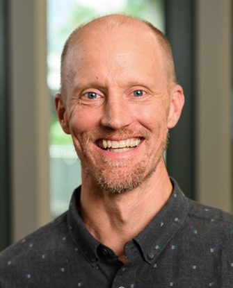 Travis Klein合作论文数School of Mechanical, Medical & Process Engineering, Faculty of Engineering, Queensland University of Technology;Centre for Biomedical Technologies, Queensland University of Technology12
 Ross Crawford合作论文数School of Mechanical, Medical and Process Engineering, Faculty of Engineering, Queensland University of Technology;Institute of Health Biomedical Innovation, Queensland University of Technology;Department of Orthopaedic Surgery, The Prince Charles Hospital;Department of Orthopaedic Surgery, Holy Spirit Northside Private Hospital10Kerry Atkinson合作论文数School of Optometry and Vision Science, Faculty of Medical and Health Sciences, The University of Auckland8
 Eamonn McKenna合作论文数7
 Justin John Cooper White合作论文数School of Chemical Engineering, Faculty of Engineering, Architecture and Information Technology, The University of Queensland7
 Mahboubeh Kabiri合作论文数Department of Biotechnology, College of science, University of Tehran7
 Judith Clements合作论文数School of Biomedical Sciences, Faculty of Health, Queensland University of Technology;Kallikrein Protease & Tumour Microenvironment Research Group, Institute of Health and BioMedical Innovation, Faculty of Health, Queensland University of Technology7
 Karen Chambers合作论文数Newport City School System7
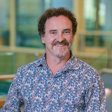 Justin J. Cooper White合作论文数Australian Institute for Bioengineering and Nanotechnology, University of Queensland7
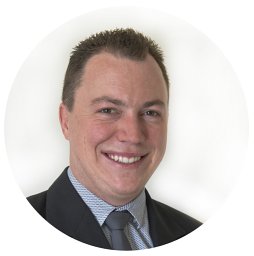 Thomas Danny Michl合作论文数Ian Wark Research Institute, University of South Australia6Hans J. Griesser合作论文数University of South Australia6Gary Brooke合作论文数6
 Pamela Robey合作论文数Skeletal Biology Section, National Institute of Dental and Craniofacial Research, National Institutes of Health6
 Pamela J. Russell合作论文数5
 Md. Shafiullah Shajib合作论文数School of Biomedical Science, Queensland University of Technology5Christian K合作论文数Cancer Research Division, Cancer Council New South Wales5
 Larkins Jo-Ann合作论文数School of Applied and Biomedical Sciences, Federation University5Babur Betul Kul合作论文数5Parisa Ghanavi合作论文数5
 Bryan R. Coad合作论文数5
 Rose Ann Franco合作论文数College of Medicine, Soonchunhyang University5Osiecki Michael合作论文数4Lor Mary合作论文数4
 Mosaad Eman M O合作论文数Stem Cell Therapies Laboratory, Queensland University of Technology at the Translational Research Institute;Institute of Health and Biomedical Innovation, Queensland University of Technology;Australian Prostate Cancer Research Centre, Translational Research Institute;Queensland University of Technology, Queensland University of Technology at the Translational Research Institute4
 James I. Hudson合作论文数Biological Psychiatry Laboratory, McLean Hospital;Harvard Medical School4
 Krasimir Vasilev合作论文数Biomedical Nanoengineering Laboratory, College of Medicine and Public Health, Flinders University4
 Brandon Markway合作论文数4
 Lars Nielsen合作论文数Nielsen Group, Australian Institute for Bioengineering and Nanotechnology, University of Queensland4
 Charles Sawyers合作论文数Memorial Sloan Kettering Cancer Center;Howard Hughes Medical Institute4Carolyn Johnstone合作论文数3Bianca Guillesser合作论文数3Anastasia Brooks合作论文数3
 Xin Liu合作论文数Therapeutics Research Centre, School of Medicine, The University of Queensland3
 Xiaowen Liang合作论文数Diamantina Institute, Faculty of Medicine, The University of Queensland3
 Jean-Pierre Levesque合作论文数Mater Research Institute, Faculty of Medicine, The University of Queensland3Wang Hao-lu合作论文数3Mills Richard J合作论文数3Roberts Michael S合作论文数3
 Yasuhiro Ikeda合作论文数3
 Elizabeth Williams合作论文数School of Biomedical Sciences, Faculty of Health, Queensland University of Technology3
 Tristan I. Croll合作论文数Australian Institute for Bioengineering and Nanotechnology, University of Queensland3John Wongvipat合作论文数3
 Hans David Ulmert合作论文数Department of Molecular and Medical Pharmacology, University of California Los Angeles3
 Wendy Wright合作论文数Institute of Innovation, Science and Sustainability, Federation University3
 Rebecca Lee Fitzsimmons合作论文数Institute for Molecular Bioscience, The University of Queensland3
 Trent P. Munro合作论文数Department of Cell Biology, Harvard Medical School3Peter A. George合作论文数3




