This seminar course covers pioneering physics research in optics, astron- omy, and nuclear physics, with an emphasis on discoveries by women physicists, African- American physicists, and other groups who are underrepresented in physics.
This first-year seminar course covers the history of modern physics using biomedical imaging as an overarching theme, focusing on both the scientists who made key discoveries and those unrecognized for their work.
In a temporal focusing microscope, dispersion can remotely shift the temporal focal plane axially, but only a single depth can be in focus at a time on a fixed camera. In this paper, we demonstrate remote focusing in a temporal focusing microscope. Dispersion tuning with an electrically tunable lens (ETL) in a 4 f pulse shaper scans the excitation plane axially, and another ETL in the detection path keeps the shifted excitation plane in focus on the camera. Image stacks formed using two ETLs versus a traditional stage scan are equivalent.
This paper presents an intermediate-level undergraduate course on the physical principles of biomedical optics and imaging. Through in-class labs, Mathematica simulations, field trips, and group presentations, students learn about fundamental imaging concepts in optical microscopes. After developing an understanding of the role of the Fourier transform in image formation, the course shifts to non-optical imaging, including x-ray computed tomography, ultrasound, and magnetic resonance imaging. The significance of this course is its hands-on nature, and this paper offers examples of laboratory exercises and simulations to promote active learning in the classroom.
We present dispersion compensation by a liquid lens (DisCoBALL), which provides tunable group-delay dispersion (GDD) that is high speed, has a large tuning range, and uses off-the-shelf components. GDD compensation is crucial for experiments with ultrashort pulses. With an electrically tunable lens (ETL) at the Fourier plane of a 4f grating pair pulse shaper, the ETL applies a parabolic phase shift in space and therefore a parabolic phase shift to the laser spectrum, i.e., GDD. The GDD can be tuned with a range greater than 2×10⁵ fs2 at a rate of 100 Hz while maintaining stable coupling into a single-mode fiber.
We implement high-speed axial scanning in a two-photon temporal focusing microscope by dispersion tuning with an electrically tunable lens. We remotely shift the temporal focus 100 μm axially at 100 Hz.
We show 3.0 ps pulses from 1877 nm to 2008 nm at variable repetition rates up to 18 GHz using time-lens compression of a tunable CW laser. The center wavelength is changed by tuning the CW seed laser, and the repetition rate is changed by electronically tuning the drive of the master RF clock. The repetition rate of 18 GHz represents a record speed for pulse generation in this spectral region. This simple all-fiber platform uses standard 1550 nm telecom components, offering a turn-key, flexible, robust alternative to pulse generation in the 2.0 mu m region with both wavelength and repetition rate tunability.
We characterize the focusing properties of the LP02 mode in a typical multiphoton microscope. Under varying back-aperture filling conditions, we measure vastly different point spread functions, including a null at the focus.
Simultaneous spatial and temporal focusing is used to acquire high speed (200Hz), chemically specific axial scans of mouse skin through a single-mode fiber. The temporal focus is remotely scanned by modulating the group delay dispersion (GDD) at the proximal end of the fiber. No moving parts or electronics are required at the distal end. A novel GDD modulation technique is implemented using a piezo bimorph mirror in a folded grating pair to achieve a large GDD tuning range at high speed.
We present multiphoton photothermal imaging of non-fluorescent, absorbing structures in scattering samples. Wide-field LED probe illumination is collected through the objective and de-scanned through a confocal pinhole. Demonstrations of nanoparticle and brain-slice imaging are presented.
Remote axial scans of mouse tissue are taken at high speed in a temporal focusing setup. A piezo bimorph mirror is used for tunable dispersion, capable of ±2.5×10⁵ fs2 of GDD and 100 Hz modulation.
As a result of the large difference between scattering mean free paths and absorption lengths in brain tissue, scattering dominates over absorption by water and intrinsic molecules in determining the attenuation factor for wavelengths between 350 nm and 1300 nm. We propose using longer wavelengths for two-photon excitation, specifically the 1300-nm region, in order to reduce the effect of scattering and thereby increase imaging depth. We present two photon fluorescence microscopy images of cortical vasculature in in vivo mouse brain beyond 1 mm. We also explore the capabilities of the 1300-nm excitation for third harmonic generation microscopy of red blood cells in in vivo mouse brain.
We present a technique for tunable dispersion compensation that is low cost, high speed, and has a large tuning range. By rotating a cylindrical lens at the Fourier plane of a folded 4f grating pair system, the group-velocity dispersion can be tuned over a range greater than 10(5) fs(2), sufficient for compensating the dispersion of several meters of optical fiber.
We demonstrate enhanced axial confinement in a temporal focusing setup with a shaped spectrum and a narrow emission filter, achieving a reduction of 1 order of magnitude of the out-of-focus background when compared with conventional point-scanning two-photon microscopy. By rejecting the background in the optical domain, our technique circumvents the noise problems common in other background subtraction techniques.
We present a new technique for dispersion compensation with >105 fs2 range and kilohertz tuning speed, enabling high-speed focal plane scanning of two-photon excited fluorescence in a temporal focusing setup.
We compare the maximal two-photon fluorescence microscopy (TPM) imaging depth achieved with 775-nm excitation to that achieved with 1280-nm excitation through in vivo and ex vivo TPM of fluorescently-labeled blood vessels in mouse brain. We achieved high contrast imaging of blood vessels at approximately twice the depth with 1280-nm excitation as with 775-nm excitation. An imaging depth of 1 mm can be achieved in in vivo imaging of adult mouse brains at 1280 nm with approximately 1-nJ pulse energy at the sample surface. Blood flow speed measurements at a depth of 900 mum are performed.
Simultaneous spatial and temporal focusing (SSTF), when combined with nonlinear microscopy, can improve the axial excitation confinement of wide-field and line-scanning imaging. Because two-photon excited fluorescence depends inversely on the pulse width of the excitation beam, SSTF decreases the background excitation of the sample outside of the focal volume by broadening the pulse width everywhere but at the geometric focus of the objective lens. This review theoretically describes the beam propagation within the sample using Fresnel diffraction in the frequency domain, deriving an analytical expression for the pulse evolution. SSTF can scan the temporal focal plane axially by adjusting the GVD in the excitation beam path. We theoretically define the axial confinement for line-scanning SSTF imaging using a time-domain understanding and conclude that line-scanning SSTF is similar to the temporally-decorrelated multifocal multiphoton imaging technique. Recent experiments on the temporal focusing effect and its axial confinement, as well as the axial scanning of the temporal focus by tuning the GVD, are presented. We further discuss this technique for axial-scanning multiphoton fluorescence fiber probes without any moving parts at the distal end. The temporal focusing effect in SSTF essentially replaces the focusing of one spatial dimension in conventional wide-field and line-scanning imaging. Although the best axial confinement achieved by SSTF cannot surpass that of a regular point-scanning system, this trade-off between spatial and temporal focusing can provide significant advantages in applications such as high-speed imaging and remote axial scanning in an endoscopic fiber probe.
Get PDF Email Share Share with Facebook Tweet This Post on reddit Share with LinkedIn Add to CiteULike Add to Mendeley Add to BibSonomy Get Citation Copy Citation Text M. Durst, G. Zhu, and C. Xu, "Effects of Refractive-Index Mismatch and Scattering on Simultaneous Spatial and Temporal Focusing," in Frontiers in Optics 2007/Laser Science XXIII/Organic Materials and Devices for Displays and Energy Conversion, OSA Technical Digest (CD) (Optica Publishing Group, 2007), paper FTuU2. Export Citation BibTex Endnote (RIS) HTML Plain Text Citation alert Save article
We show theoretically and experimentally that simultaneous spatial and temporal focusing can scan the temporal focal plane axially by adjusting the group velocity dispersion in the excitation beam path. When the group velocity dispersion is small, the pulse width at the temporal focal plane is transform-limited, and the amount of shift depends linearly upon the dispersion. By adding a meter of large mode area fiber into the system, we demonstrate this axial scanning capability in a fiber delivery configuration. Because a transform-limited pulse width is automatically recovered at the temporal focal plane, simultaneous spatial and temporal focusing negates the need for any dispersion pre-compensation, further facilitating its integration into a fiber delivery system. A highly promising application for simultaneous spatial and temporal focusing is an axial scanning multiphoton fluorescence fiber probe without any moving parts at the distal end and without dispersion pre-compensation. (c) 2006 Optical Society of America
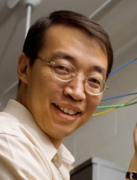 Chris Xu合作论文数School of Applied and Engineering Physics, Cornell University13
 Guanghao Zhu合作论文数4Demirhan Kobat合作论文数4
 Adam Straub合作论文数3Anthony Turcios合作论文数2
 Chris Schaffer合作论文数Meinig School of Biomedical Engineering, College of Engineering, Cornell University2
 Nozomi Nishimura合作论文数Schaffer-Nishimura Lab, Meinig School of Biomedical Engineering, College of Engineering, Cornell University2Angela W. Wong合作论文数2
 James Van Howe合作论文数School of Applied and Engineering Physics;Cornell University;School of Applied and Engineering Physics, Cornell University2Tw Schneider合作论文数1Robin Houston合作论文数1
 Jonathan Gorstein合作论文数1Joseph Moscatelli合作论文数1Samuel Yurak合作论文数1Ea Schmieman合作论文数1Jacky Knowles合作论文数1
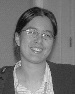 Jennifer H Lee合作论文数1
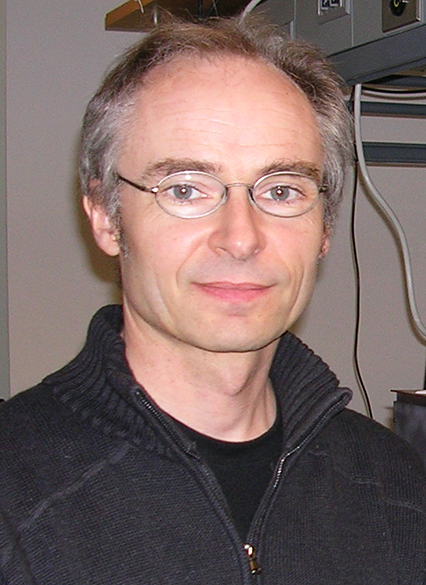 Jerome C. Mertz合作论文数Department of Biomedical Engineering, College of Engineering, Boston University;Center for Systems Neuroscience, Boston University1Codling Karen合作论文数1
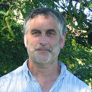 Warren R. Zipfel合作论文数Meinig School of Biomedical Engineering, College of Engineering, Cornell University1
 John F Currie合作论文数Engineering Physics Department, École Polytechnique1Pascal Belicard合作论文数1Valery Kulishenko合作论文数1A.R. Leeds合作论文数1
 Lars Grüner Nielsen合作论文数Department of Photonics Engineering, DTU Fotonik1Shruti M. Paranjape合作论文数1
 Elena Georgieva合作论文数1Edward Van Keuren合作论文数1




