BACKGROUND:Oral squamous cell carcinoma (OSCC) is the most common malignancy of the head and neck, associated with substantial morbidity and mortality worldwide. Oral epithelial dysplasia (OED) can precede OSCC, offering a critical window for preventive and therapeutic intervention. This review maps the historical and current landscape of in vitro and in vivo models of OED, providing insight into their strengths, limitations, and translational relevance. METHODS:A systematic review and temporal analysis were conducted following PRISMA guidelines, with literature searches performed across Medline, EMBASE, EBM Reviews, and Web of Science. RESULTS:From 4009 records, 292 studies from 26 countries were included, yielding 307 models of OED. Only a minority of studies (17.4%) focused primarily on dysplasia as a precancerous condition. In vivo models predominated (88.9%), while in vitro systems were comparatively scarce and largely limited to 2D cultures. Organoid-based approaches were rarely reported, highlighting a gap in advanced model development. Reproducibility data were available for 125 studies (45.8%). The hamster emerged as the most frequently used animal [n = 96, 35%], and 4-nitroquinoline-1-oxide (4-NQO) remained the most common carcinogen, particularly in murine models. A strong sex bias was observed, with male animals heavily over-represented. CONCLUSION:Overall, models of OED remain under-represented and under-developed, particularly in comparison to innovations in other fields of cancer research. Despite the central role of OED in oral carcinogenesis, current models do not adequately reflect clinical diversity or exploit modern 3D and patient-derived technologies. This review provides a critical reference point to guide future studies toward more accurate, reproducible, and clinically relevant models, with the potential to advance prevention, early detection, and targeted therapies for OSCC.
BACKGROUND:Oral Medicine is that specialist branch of dentistry concerned with the diagnosis, prevention, and predominantly non-surgical management of medically-related disorders and conditions affecting the oral and maxillofacial region, in particular oral mucosal disease and orofacial pain, as well as the oral health care of medically complex patients. Published national curricula for postgraduate Oral Medicine training exist in the United Kingdom (UK General Dental Council, 2010; revised 2023) and the United States (American Academy of Oral Medicine competency framework, 2015), and an international competency consensus was established by the Sixth World Workshop on Oral Medicine (2015). Despite this international momentum, no equivalent nationally published, competency-aligned postgraduate curriculum has been established for the Australasian region. This manuscript describes the development and structure of a contemporary 3-year Doctor of Clinical Dentistry (DClinDent) curriculum for Oral Medicine specialist training, developed by the Oral Medicine Academy of Australasia (OMAA) and aligned with the OMAA Essential Competencies (2026). METHODS:The curriculum was developed through a joint collaboration of all program convenors and heads of training of DClinDent (Oral Medicine) programs in Australia and New Zealand, together with the OMAA Curriculum Working Group, informed by the OMAA Essential Competencies (2026), benchmarked against the UK GDC Oral Medicine Specialty Training Curriculum (2023) and the internationally validated competencies from the Sixth World Workshop on Oral Medicine (2015), and structured according to a spiral learning framework. The curriculum was endorsed by the OMAA Credentialing and Peer Review Subcommittee in 2026. The program is divided into three progressive phases: Foundations (Year 1), Development (Year 2), and Consolidation (Year 3). RESULTS:The curriculum encompasses five competency domains-Professionalism; Communication and Social Skills; Critical Thinking; Scientific and Clinical Knowledge; and Patient Care-mapped across all 3 years. The integration of cultural awareness and safety is an important feature across all phases of the curriculum. Year 1 establishes core scientific, clinical, and professional foundations. Year 2 develops clinical expertise across all Oral Medicine domains with increasing diagnostic independence. Year 3 prepares trainees for autonomous specialist practice, including complex case management, research thesis completion, and professional leadership. Minimum clinical case targets, portfolio-based assessment, and a conjoint Fellowship of the Oral Medicine Academy of Australasia (FOMAA) exit examination are integral to program completion. CONCLUSIONS:This curriculum provides a nationally applicable, competency-aligned framework for Oral Medicine postgraduate training in Australasia. It addresses recognized gaps in specialty-specific graduate medical education and offers a replicable model for programs seeking harmonization with Australasian and international Oral Medicine training standards.
Introduction and aims: Antimicrobial resistance is a global public health emergency and a prominent issue in Southeast Asia. There is also a lack of dental-specific drug resources to assist with medication management for dentists. Our dental-specific decision support tool, MIMS Drugs4dent, was developed to assist Australian dentists optimize their prescribing. The aim of this study was to trial MIMS Drugs4dent for acceptability and usability with dental practitioners in Indonesia, Malaysia, Singapore, and Thailand. Methods: This was a cross-sectional study of MIMS Drugs4dent with dental practitioners in Indonesia, Malaysia, Singapore, and Thailand. Dental practitioners were provided access to MIMS Drugs4dent for 1 month. After the intervention period, participants completed an online survey that assessed their perceptions of the tool. The primary outcome was the acceptability and usability of MIMS Drugs4dent, using the Framework for Acceptability and System Usability Scale, respectively. The secondary outcome was to determine the resources used by dental practitioners for drug information. Descriptive statistics and thematic analysis were used for quantitative and qualitative data analysis, respectively. Results: From January to June 2025, 30 dentists from each country participated. Most participants (93.4% and 94.2%) agreed or strongly agreed that MIMS Drugs4dent can improve their ability to prescribe according to guidelines and access dental-relevant drug information, respectively. Most participants (88.3% and 82.5%) agreed or strongly agreed that MIMS Drugs4dent was easy to use and that information could be located quickly. However, participants preferred a local drug database and a mobile-rendered version to improve usability. A wide range of sources (n = 62) were reported to be used for medicines information. Conclusion: MIMS Drugs4dent had high acceptability and usability in this study. With adaptation using country-specific localised drug databases, the tool has potential for use in dentistry in these countries. The study underscores the importance of including dentistry in broader digital health strategies.
BackgroundEarly detection of oral cancer depends on dental professionals’ ability to recognise both risk factors and pathological changes in the oral mucosa. Educational interventions may enhance this diagnostic awareness, particularly in settings where exposure to oral cancer cases and specialised training opportunities may be limited.ObjectiveThis study aimed to evaluate the impact of an educational intervention on dental professionals’ ability to associate oral cancer risk factors with pathological changes.MethodsA before-and-after quasi-experimental study design was used. General dental practitioners from multiple regions in Indonesia participated in an oral cancer educational programme. Participants completed questionnaires before and after the intervention assessing their knowledge of oral cancer risk factors and oral potentially malignant disorders. The primary outcome was defined as the change in correlation between correctly identified risk factors and pathological changes. Kendall's tau-b correlation coefficients were calculated, and differences between correlations were assessed using Fisher's r-to-z transformation.ResultsA total of 177 responses were obtained before the intervention and 144 responses after the intervention. The correlation between identified oral cancer risk factors and pathological changes increased slightly from 0.464 [95% confidence interval (CI): 0.383–0.538] before the intervention to 0.485 (95% CI: 0.397–0.565) after the intervention. However, the difference between the correlations was not statistically significant (p = 0.8109).ConclusionThe educational intervention was associated with a modest increase in dental professionals’ ability to associate oral cancer risk factors with pathological changes, although the change in correlation was not statistically significant. These findings suggest that educational initiatives may contribute to improving diagnostic awareness, but more comprehensive or repeated training strategies may be required to produce measurable improvements.
Oropharyngeal gonorrhoea remains a global challenge due to rising antimicrobial resistance and limited therapeutic options, necessitating urgent calls for new treatments and prophylaxis. This study evaluates the in vitro activity of arginine-based fatty acids against Neisseria gonorrhoeae (NG) strains in a validated oropharyngeal model. Five oropharyngeal cell lines (tonsillar/buccal/gingival/floor of mouth/uvular) were infected with two NG strains. Intracellular infection was assessed at 15/30/60 min. MIC₉₀ and cytotoxicity of five arginine-based fatty acids were determined. Promising candidates were evaluated for NG clearance at 30/60/120 min (post-infection) and prevention of infection (pre-infection) after 30/120 min drug exposure. Arginine-undecanoate (AU) and arginine-laurate (AL) were the most effective fatty-acids, showing potent bactericidal activity, with no cytotoxicity in any cell lines up to 300 µg/mL. In post-exposure experiments, AL at 150 µg/mL and at MICx3 achieved rapid, dose-dependent clearance of ≥94% for FA1090 and ≥90% for WHO R respectively within 30 min in all cell lines except in uvular cells. AU showed time-dependent killing with clearance of ≥95% for FA1090 and ≥88% for WHO-R at 120 min at 150 and 108 µg/mL (MICx3) respectively. In pre-exposure experiments, AL at maximum non-cytotoxic concentration (300 µg/mL) was more effective than AU, achieving 83·3-97·4% clearance in all cell lines within 30 min. AL and AU can be promising, non-toxic candidates for the prevention or treatment of oropharyngeal NG. Use, pre- and post-sex via topical formulations, could potentially reduce oropharyngeal NG transmission.
BACKGROUND:Antimicrobials are an adjunctive therapy in clinical dentistry. In dentoalveolar surgery, antimicrobials are not routinely required for surgical prophylaxis. This retrospective analysis of the Australian Surgical National Antimicrobial Prescribing Survey (Surgical NAPS) dataset aimed to evaluate the guideline compliance and appropriateness of antimicrobial prescribing for dentoalveolar procedures in Australian hospitals. METHODOLOGY:Deidentified Surgical NAPS data for dentoalveolar procedures (tooth extractions and implant placements) between 2016 and 2022 were extracted. Procedures outside the scope of a general dentist were excluded. Prescribed antimicrobials for surgical prophylaxis, including procedural prophylaxis doses and post-procedural prescriptions, were assessed for guideline compliance and appropriateness according to the Surgical NAPS algorithm. RESULTS:1345 surgical episodes with dental procedures were included. This comprised 1077 procedural prophylaxis doses and 555 post-procedural prescriptions. Of the post-procedural prescriptions, 478 (86%) were for prophylaxis. Guideline compliance was demonstrated in 35.3% of procedural doses and 14.6% of post-procedural prescriptions. Rates of appropriateness were 33.5% for procedural doses and 12.6% for post-procedural prescriptions. Most procedural doses and post-procedural prescriptions were deemed inappropriate as they were not required (72.5% and 93.0%, respectively). CONCLUSIONS:Suboptimal guideline compliance and appropriateness of antibiotic prescribing for dentoalveolar surgery reinforces the need for antimicrobial stewardship interventions.
Oral mucosal abnormalities considered to have malignant potential may involve a large area of mucosa. Standard care histopathological investigation is invasive, limited to site selection and subject to variation in assessment between pathologists. A quantitative, minimally invasive, rapidly collected, oral mucosal site-specific assessment will assist in decision making and increase diagnostic precision. This study aimed to validate a workflow to analyse oral scrape derived cancer-associated microRNA analysis for mucosal site-specific assessment as a surrogate biomarker to histopathological diagnosis. Forty-one oral scrapes were collected from 33 patients undergoing investigation at the Royal Dental Hospital of Melbourne before mucosal biopsy. RNA from oral scrapes was used to investigate ten cancer-associated microRNAs. An algorithm for categorical high or low-risk based on histopathological diagnosis was developed. The novel risk stratification algorithm utilised two microRNAs and categorised all cases of carcinoma and severe dysplasia as high-risk and accurately distinguished all non-potentially malignant disorders as low-risk lesions. This study provides proof-of-concept that oral scrapes can be predictably collected in a clinical workflow to assess the expression of cancer-associated microRNA specific to an oral mucosal site with minimal invasiveness.
Background The aim of this study was to investigate oral cancer screening practices among Australian dental practitioners (DPs) and to assess their knowledge of oral cancer risk-factors and confidence in discussing related behaviours with patients. Methods A 41-item survey was promoted widely to practicing Australian DPs (dental hygienists, dental prosthetists, dental therapists, dentists, oral health therapists). Items included socio-demographic; work-related characteristics; self-assessed oral cancer knowledge (utilising a 10-point numerical scale), knowledge about risks factors for oral cancer (responses ‘Yes’/‘No’/‘Unsure’), and level of confidence in discussing seven health behaviours with patients (utilising a 10-point scale). Results A total of 395 DPs were included in the analysis. The majority were dentists (80.7%). Almost all DPs (98.5%) agreed that they should screen routinely for oral cancer. However, only 52.2% of respondents reported ‘Always’ conducting comprehensive oral cancer screenings.
Background Hemidesmosomal subunits have gained attention for their potential role in the progression of oral squamous cell carcinoma (OSCC). However, formal analysis of quantitative expression patterns correlating with OSCC disease pathogenesis remains limited. This study evaluated the expression of key yet overlooked hemidesmosomal subunits, plectin isoform Ia (PIa), dystonin, and CD151 antigen, from normal tissue as well as tissue from hyperplasia, dysplasia, and OSCC in the human and murine oral cavity.Methods Immunohistochemistry was performed on custom-built human tissue microarrays and 4-Nitroquinoline 1-oxide (4-NQO)-induced murine OSCC covering the spectrum of histological changes from normal to cancer. Quantitative image analysis of subunit expression was conducted using QuPath, with distinct analytical approaches applied to human tissues, including a refined method focusing on the basement membrane zone (BMZ) and adjacent basal epithelial layers, key sites of hemidesmosomal protein localization.Results A significant increase in expression of all three proteins was observed comparing normal tissue with hyperplasia, dysplasia, and OSCC in murine tissues, but not in humans. The expression of hemidesmosomal proteins increased across this spectrum, suggesting their potential as progression biomarkers in mice. A focused analysis of the basement membrane zone and adjacent basal epithelial layers in human tissues revealed a sustained baseline expression and a significant reduction in CD151 antigen in OSCC compared with control and high-grade dysplasia groups, as well as a significant reduction in dystonin expression in OSCC compared to high-grade dysplasia.Conclusion Our findings highlight the importance of biologically targeted region-of-interest selection when assessing hemidesmosomal protein expression in OSCC while demonstrating that BMZ-focused digital analysis provides a more informative approach for exploratory evaluation of heterogeneous human tissues alongside progression-associated patterns observed in a murine model.
BACKGROUND:This study aimed to systematically evaluate apps with temporomandibular disorder (TMD) self-management content available in Australia for quality, clinical safety, self-management support functions, and contributors to app development. METHODS:A systematic search of the App Store (iOS) and Google Play (Android) was conducted on 18 April 2023 (and updated 26 July 2023) to identify apps that had TMD self-management content. Two raters independently assessed app quality, clinical safety, self-management support, and contributors to app development for the involvement of people living with TMD and clinicians. Quality was evaluated using the Mobile App Rating Scale (MARS) for engagement, functionality, aesthetics, and information quality scored using a 5-point Likert scale. App clinical safety was assessed using MARS functionality (item 6) and information quality items for accuracy/relevance (item 15), scope (item 16), and visual information accuracy/clarity (item 17) (scored on a Likert scale), and the M-Health Index and Navigation Database framework questions: does the app provide any warning for use? Does the app have a crisis management feature? Can the app cause harm? (scored yes/no). Self-management support was evaluated using the Self-Management Support (SMS-14) checklist (scored yes/no). Included apps, app store descriptions, and linked websites were qualitatively evaluated to determine the contributors to app development. RESULTS:Seven apps with TMD self-management content were available in Australia. Overall, the included apps were of acceptable quality (mean = 3.25/5) but scored poorly for engagement (2.71/5) and information (2.92/5). Clinical safety limitations identified were the inability to identify and/or direct users to support services in a crisis and inconsistent TMD information. One app (Do I Grind or Snore) was deemed potentially harmful as sleep sounds suggestive of obstructive sleep apnoea were interpreted as snoring by the app. Overall, the inclusion of self-management support functions was variable (range 1-9; mean = 4.71/14), with pain/TMD education (71%) and self-monitoring (71%) the most common. Only one app had development input from a person with lived experience of TMD. CONCLUSION:The quality and self-management support of apps with TMD self-management content is variable. TMD apps with activating self-management strategies and higher engagement scores are more likely to be effective. Concerningly, one app was found to be potentially harmful, and overall apps lacked user safeguards. Only one app involved a person with TMD in its development, and the authors recommend using co-design in future TMD app development to improve app quality, clinical safety, and impact.
Oral cancer detection is based on biopsy histopathology, however with digital microscopy imaging technology there is real potential for rapid multi-site imaging and simultaneous diagnostic analysis. Fifty-nine patients with oral mucosal abnormalities were imaged in vivo with a confocal laser endomicroscope using the contrast agents acriflavine and fluorescein for the detection of oral epithelial dysplasia and oral cancer. To analyse the 9168 images frames obtained, three tandem applied pre-trained Inception-V3 convolutional neural network (CNN) models were developed using transfer learning in the PyTorch framework. The first CNN was used to filter for image quality, followed by image specific diagnostic triage models for fluorescein and acriflavine, respectively. Images were categorised based on a histopathological diagnosis into 4 categories: no dysplasia, lichenoid lesions, low-grade dysplasia and high-grade dysplasia/oral squamous cell carcinoma (OSCC). The quality filtering model had an accuracy of 89.5%. The acriflavine diagnostic model performed well for identifying lichenoid (AUC = 0.94) and low-grade dysplasia (AUC = 0.91) but poorly for identifying no dysplasia (AUC = 0.44) or high-grade dysplasia/OSCC (AUC = 0.28). In contrast, the fluorescein diagnostic model had high classification performance for all diagnostic classes (AUC range = 0.90–0.96). These models had a rapid classification speed of less than 1/10th of a second per image. Our study suggests that tandem CNNs can provide highly accurate and rapid real-time diagnostic triage for in vivo assessment of high-risk oral mucosal disease.
The role of oral yeasts in oral squamous cell carcinoma (OSCC) has gained attention due to evidence linking fungal dysbiosis to carcinogenesis. While Candida albicans has been the primary focus, emerging studies highlight the importance of non-Candida species yeast genera. This scoping review synthesises the evidence on the role of oral yeasts, including Candida spp. and non-Candida species, in the development and progression of OSCC. A PRISMA-ScR-guided search was conducted in Medline, Embase, EBM Reviews, and CINAHL. Observational and experimental studies involving humans with OSCC, oral potentially malignant disorders (OPMDs), or oral epithelial dysplasia (OED) were included. This review analysed 75 studies. Research on oral yeast in OSCC has progressed since the 1970s, with advancements in identification techniques—from conventional culture methods to metagenomic sequencing and multi-omics approaches—alongside improved animal and cellular models of OSCC. These methodological advancements have identified notable distinctions in the oral mycobiome between carcinomatous and healthy states. Clinical findings reinforce the hypothesis that oral yeasts, particularly Candida spp., actively contribute to the dysplasia–carcinoma sequence. Emerging evidence suggests that oral yeasts may significantly modulate events contributing to OSCC progression. However, further mechanistic studies and robust clinical evidence are essential to establish causality and clarify their role in OSCC.
BACKGROUND:Dental prescribing of opioids has been associated with the development of persistent opioid use (POU). The aim of this study was to examine the incidence of POU arising from an initial dental opioid prescription to opioid naïve individuals in Australia between 2015-2022. METHOD:This was a longitudinal, retrospective study using a 10 % sample of patients dispensed medicines through the Pharmaceutical Benefits Scheme. The study comprised of opioid naïve individuals, who were dispensed an opioid prescribed by a dentist between 2015-2022 in Australia. The primary outcome was the proportion of patients who developed POU. Three secondary outcomes were assessed: 1) the rate of dispensed opioids in the subsequent 12-month period, 2) the type and potency of dispensed opioids, and 3) factors associated with POU. RESULTS:From 2015-2022, 97,233 opioid naïve patients were dispensed an opioid prescribed by a dentist. Of these, 1692 individuals (1.7 %) developed POU. Patients with POU received a subsequent opioid prescription from a non-dentist more frequently (72.1 %) than those with non-persistent use (48.9 %). Whilst the potency of the initial opioid prescription, measured in morphine milligram equivalence, was similar between POU and non-POU patients, the potency of subsequent opioid prescriptions supplied to individuals was 44.1 % greater in those with POU (161.4 mg, SD:338.4) than those without POU (112.0 mg, SD: 283.7). CONCLUSIONS:A small proportion of opioid naïve patients who were dispensed an opioid prescribed by a dentist developed POU. These findings underpin the importance of reviewing patients with dental pain, and including dentists in real time prescription monitoring programs. CLINICAL SIGNIFICANCE:There is a small but clinically significant proportion of opioid-naïve patients who may develop persistent opioid use following an opioid prescription from the dentist. This highlights the importance of understanding the role of opioids for dental pain, initiating opioids only when necessary and reviewing patients with pain.
BACKGROUND:In Australia, the prescribing of opioid medicines by dentists has increased in recent years, despite opioids not being first-line treatment for dental pain. The aim of this longitudinal study was to examine the dispensing of opioids prescribed by dentists in Australia during 2013-2022. METHOD:A nationally representative 10% sample of patients identified from the Australian Pharmaceutical Benefits Scheme dispensing data from 2013 to 2022 was used. Three outcomes were assessed: (1) incidence of dispensing of all dental prescriptions; (2) incidence of dispensing of opioids prescribed by dentists; (3) average number of tablets/capsules of dental opioid supply. Outcomes pertaining to opioid use were examined overall, and by year, age and sex. RESULTS:From 2013 to 2022, 998 774 dental prescriptions (of any kind) were dispensed to 470 118 patients. The mean annual incidence rate for dispensing any dental medication was 48.4 (95% CI: 48.3-48.5)/1000 person-years. Opioids accounted for 183 303 prescriptions (18.4%), with a mean annual incidence rate of 11.0 (95% CI: 11.0-11.1)/1000 person-years. The majority of patients (99.1%) were dispensed ≤ 4 opioid prescriptions across the 10-year period, with 0.9% of patients (n = 1312) receiving between 5 and 149 dispensed opioids. Over the study period, the average annual incidence of dispensed dental opioids increased by 4.4% (95% CI: 1.0-8.2). A reduction in the mean quantity of opioid pills dispensed was observed over time. Dental opioids were dispensed to 2727 children and adolescents. CONCLUSION:The incidence of dispensing of dental opioids in Australia increased by an average of 4.4% per year over a decade. While there was a reduction in opioid quantities dispensed, dispensing of opioids for children occurred, and a small number of patients were dispensed excessive quantities of dental opioids. Evidence-based tailored opioid stewardship interventions need to include dentists, and dentists should be provided access to drug monitoring programmes to enable more informed prescribing decisions.
AIM or PURPOSE This qualitative study explores the process of systematically involving young people living with temporomandibular disorder (TMD) in the co-design of an app for TMD self-management from de novo conception to app prototype testing. MATERIALS and METHOD Ethics approval was granted by The University of Melbourne Office of Research Ethics and Integrity (#2023-24749-42923-5). Participants aged 18-35 years with TMD were recruited from the Oral Medicine Clinic at the Royal Dental Hospital of Melbourne (Victoria, Australia) from May 2023 to April 2024. An agile co-design approach was taken using a pragmatic framework of multiple qualitative methods that included semi-structured interviews, focus group and qualitative survey, that allowed for rounds of iteration and refinement of the app prototype's key functions and features throughout the stages of development. Data was analysed using an experiential thematic analysis that prioritised participants’ standpoint with an inductive orientation based on methods by Braun & Clarke (2013). RESULTS The interviews and focus group identified the ideal functions and features of a TMD app that provided the framework for app prototype development. A prototype was then developed, and preliminary results from subsequent end-user testing show that this app prototype was acceptable to young people living with TMD and is very likely to meet their self-management needs. CONCLUSION(S) A co-design approach was used to identify the ideal functions and features of a TMD app from the perspective of young people. Preliminary results indicate that the study's co-design approach was effective in developing an app prototype that meets the self-management needs of young people living with TMD.
OBJECTIVES:Dentists manage a variety of oral infections in clinical practice. Inappropriate antimicrobial prescribing by dentists occurs frequently and antimicrobial stewardship strategies should include dentistry. The aim of this retrospective analysis of the Australian Hospital National Antimicrobial Prescribing Survey (Hospital NAPS) dataset, was to describe the types of oral and dental indications where antimicrobials were prescribed, and assess the guideline compliance and appropriateness of the antimicrobials in Australian hospitals. METHODS:Data from the Hospital NAPS was extracted for oral and dental indications from 2013 to 2022. The types of oral and dental indications presented, and the corresponding antimicrobials prescribed were assessed for compliance according to national prescribing guidelines, and appropriateness according to the NAPS structured algorithm. RESULTS:A total of 8,001 prescriptions for 7,477 patients were identified, from 433 hospitals. Antifungal, antibiotic and antiviral agents accounted for 84.5 %, 15.4 % and 0.03 % of prescriptions respectively. A greater proportion of antibiotics were prescribed in regional and rural areas compared to antifungals. The prescriptions assessed as compliant were 80.0 % and 44.7 % of antifungals and antibiotics respectively. Prescriptions assessed as appropriate were 84.4 % of antifungals, and 65.3 % of antibiotic prescriptions. CONCLUSIONS:A wide variety of antimicrobials were used with moderate levels of compliance and appropriateness. Future interventions should include targeted education, utilisation of prescribing guidelines, and tools to diagnose and manage oral and dental conditions. Consideration can be given to adjustment of the Hospital NAPS tool to cater for oral conditions and include the provision of dental treatment in the management of these infections. CLINICAL SIGNIFICANCE:A wide variety of oral and dental conditions are presented in Australian hospital settings, managed by a range of antibiotics and antifungals, with moderate levels of compliance to guidelines and appropriateness. Antimicrobial stewardship strategies should target and support dentistry in hospital settings.
BACKGROUND:Coffee is one of the most consumed beverages in the world. Containing an abundance of bioactive molecules including polyphenols and flavonoids, the constituents of this beverage may exert antiproliferative, antioxidant and anti-inflammatory effects.METHODS:We conducted a systematic review to summarise the available evidence on the anticancer effects of coffee constituents and their potential therapeutic use for oral squamous cell carcinoma (OSCC). Studies were identified through a comprehensive search of OVID MEDLINE, OVID EMBASE and Web of Science, including articles from any year up to 15 May 2023.RESULTS:Of the 60 reviewed papers, 45 were in vitro, 1 was in silico and 8 were in vivo exclusively. The remaining studies combined elements of more than one study type. A total of 55 studies demonstrated anti-proliferative effects, whilst 12 studies also investigated migration and invasion of neoplastic cells. The constituents studied most frequently were quercetin and epigallocatechin gallate (EGCG), demonstrating various cytotoxic effects whilst also influencing apoptotic mechanisms in cancer cell lines. Dose-dependent responses were consistently found amongst the studied constituents.CONCLUSION:Whilst there was heterogeneity of study models and methods, consistent use of specific models such as SCC25 for in vitro studies and golden hamsters for in vivo studies enabled relative comparability. The constituents of coffee have gained significant interest over the last 30 years, particularly in the last decade, and present an area of interest with significant public health implications. Currently, there is a paucity of literature on utilization of active coffee constituents for the therapeutic treatment of oral cancers.
Objective: High-risk human papillomaviruses (HPV) are an established cause of oropharyngeal cancer. Their relationship with oral cancer remains unclear with detection ranging from 0% to 100%. HPV DNA detection or evidence of exposure alone is insufficient to conclude causality. This systematic review assesses the extent of bias in studies of HPV detection in cancers of the oral cavity. Methods: PubMed, Ovid MEDLINE, EMBASE, and PsycInfo databases were searched for observational studies reporting the effect of HPV in oral cavity specific cancers. Results: All 15 included studies presented HPV DNA detection or serum HPV-antibodies, none included mRNA E6/E7 analysis. Cases with oral cancer had 5.36 times (95% CI 3.29-8.72) higher odds of having HPV detected compared to controls. The odds of HPV detection were higher in cell-based (OR 6.93; 95% CI 0.82-58.55) and tissue samples (OR 5.28; 95% CI 3.41-8.18) than blood-based samples (OR 3.36; 95% CI 1.53-7.40). Conclusion: When cancer site is clearly differentiated between oropharynx and oral cavity, 12 studies showed strong association between HPV and oral cancer, but the available estimates lack internal validity due to inconsistent measurements, high confounding, and lack of gold standard testing. There is not high-quality evidence to conclude a causal relationship of HPV with oral cancer.
Oral squamous cell carcinoma (OSCC) is the most common head and neck cancer. There is mounting evidence to suggest that several components of the coagulation system directly affect carcinogenesis. Our recent in vitro studies demonstrated, for the first time, that various anticoagulants have anticancer effects on OSCC. They also showed the need for the immediate translation of these experimental conditions from bench to preclinical animal models. Here, we carried out a systematic review to summarise existing evidence on murine models built around the interactions between anticoagulants and oral cancer. Only one preclinical murine study was included in our systematic review, investigating the role of heparins in tumour pathophysiology. The paucity of evidence regarding the interactions between oral squamous cell carcinoma and anticoagulants emphasises the urgency with which further preclinical research should be conducted.
 Antonio Celentano合作论文数Faculty of Medicine and Surgery, University of Naples Federico II49
 Nicola Cirillo合作论文数Department of Oral and Dental Science, University of Bristol49
 Tami Yap合作论文数45
 Leanne Teoh合作论文数Melbourne Dent Sch, Univ Melbourne27
 Rita Paolini合作论文数24
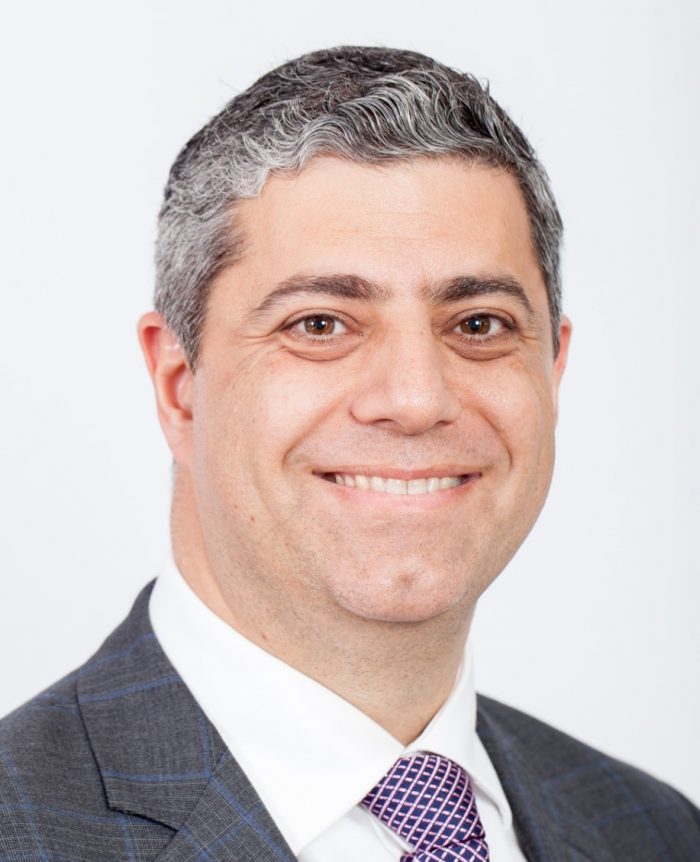 Farah Camile S合作论文数UWA Dental School, University of Western Australia, Nedlands, WA, Australia/Australian Centre for Oral Oncology Research & Education21
 Stephen Porter合作论文数Eastman Dental Institute, University College London;Royal Free Hospital16David Wiesenfeld合作论文数Department of Surgery, Melbourne Medical School, Faculty of Medicine, Dentistry and Health Sciences, The University of Melbourne15
 Reynolds Eric合作论文数Melbourne Dental School, The University of Melbourne15Kendrick Koo合作论文数The Howard Florey Institute, University of Melbourne14
 Mohd Hafiz Arzmi合作论文数Department of Oral Biology, University of Malaya10
 Stuart Dashper合作论文数Melbourne Dental School, Faculty of Medicine, Dentistry and Health Sciences, The University of Melbourne9Angel Christopher M合作论文数9Clemons K V合作论文数9
 Suhail Al-Amad合作论文数College of Dental Medicine, University of Sharjah9
 Ali Alnuaimi合作论文数9
 David A. Stevens合作论文数Division of Infectious Diseases & Geographic Medicine, Department of Medicine, School of Medicine, Stanford University;Maternal & Child Health Research Institute, School of Medicine, Stanford University;Clinical Laboratories, California Institute for Medical Research9Maddalena Manfredi合作论文数Dipartimento di Scienze Otorino-Odonto-Oftalmologiche e Cervico-facciali, Università degli studi di Parma9
 Geraldine Moses合作论文数School of Pharmacy, The University of Queensland8Christine Seers合作论文数Melbourne Dental School, Faculty of Medicine, Dentistry and Health Sciences, The University of Melbourne8
 Rodrigo Marino合作论文数7
 Neil O'Brien-Simpson合作论文数7
 Elizabeth Sari合作论文数7George Dimitroulis合作论文数7Carlos a Angel合作论文数7Z M Al-Karaawi合作论文数6
 Federica Canfora合作论文数Oral Med Unit, Univ Naples Federico II6
 Mathew AWT Lim合作论文数Dental Serv, Alfred Hlth6
 John Henry McCusker合作论文数Department of Molecular Genetics and Microbiology, School of Medicine, Duke University6
 Lorraine O'Reilly合作论文数Walter and Eliza Hall Institute of Medical Research6A Nastri合作论文数5
 Jane Hocking合作论文数Centre for Epidemiology and Biostatistics, Melbourne School of Population and Global Health, Faculty of Medicine, Dentistry and Health Sciences, The University of Melbourne5
 David Manton合作论文数Melbourne Dental School, Faculty of Medicine, Dentistry and Health Sciences, The University of Melbourne5
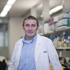 John Silke合作论文数La Trobe University;The Walter and Eliza Hall Institute;WEHI;Faculty of Medicine, Dentistry and Health Sciences, University of Melbourne5Ramdzan Adlin N合作论文数5
 Mohammad Alrashdan合作论文数School of Dentistry, Seoul National University5Paolo Vescovi合作论文数5Nw Savage合作论文数5
 Jacks Jorge合作论文数Piracicaba Dental School, University of Campinas5D Rowler合作论文数4K Stewart合作论文数4Ivy Tan合作论文数4Ali Ibrahim Mohammed合作论文数4
 Suzanne M Garland合作论文数4
 Rishi S. Ramani合作论文数Melbourne Dental School4
 Simone Belobrov合作论文数4
 Shreya Tocaciu合作论文数4J. G. Clement合作论文数4Graham Hepworth合作论文数School of Mathematics and Statistics, Faculty of Science, The University of Melbourne4




