Intracerebral hemorrhage (ICH) is a lethal stroke type; mortality approaches 50%, and current medical therapy against ICH shows only limited effectiveness, so an alternative approach is required, such as stem cell‐based cell therapy. Previously we have shown that intravenously transplanted human neural stem cells (NSCs) selectively migrate to the brain and promote functional recovery in rat ICH model, and others have shown that intracerebral infusion of brain‐derived neurotrophic factor (BDNF) results in improved structural and functional outcome from cerebral ischemia. We postulated that human NSCs overexpressing BDNF transplanted into cerebral cortex overlying ICH lesion could provide improved survival of grafted NSCs and increased angiogenesis and behavioral recovery in mouse ICH model. ICH was induced in adult mice by injection of bacterial collagenase into striatum. The HB1.F3.BDNF (F3.BDNF) human NSC line produces sixfold higher amounts of BDNFF over the parental F3 cell line in vitro, induces behavioral improvement, and produces a threefold increase in cell survival at 2 weeks and 8 weeks posttransplantation. Brain transplantation of human NSCs overexpressing BDNF provided differentiation and survival of grafted human NSCs and renewed angiogenesis of host brain and functional recovery of ICH animals. These results indicate that the F3.BDNF human NSCs should be of great value as a cellular source for experimental studies involving cellular therapy for human neurological disorders, including ICH. © 2010 Wiley‐Liss, Inc.
Background Midkine (MK), a member of the heparin-binding growth factor family, which includes MK and pleiotrophin, is known to possess neurotrophic and neuroprotective properties in the central nervous system. Previous studies have shown that MK is an effective neuroprotective agent in reducing retinal degeneration caused by excessive light and decreasing hippocampal neuronal death in ischemic gerbil brain. The present study was undertaken to investigate whether MK acts as an anticonvulsant in kainic acid (KA)-induced seizure in mouse and blocks KA-mediated neuronal cell death in hippocampus. Results Increased expression of MK was found in hippocampus of mouse following seizures induced by intracerebroventricular injection of KA, and MK expression was found in glial fibrillary acidic protein (GFAP)-positive astrocytes. Concurrent injection of MK and KA attenuated KA-induced seizure activity and cell death of hippocampal neurons including pyramidal cells and glutamic acid decarboxylase 67 (GAD67)-positive GABAergic interneurons in the CA3 and hilar area. Conclusion The results of the present study indicate that MK functions as an anticonvulsant and neuroprotective agent in hippocampus during KA-induced seizures.
We have investigated the effectiveness of transplantation of human neural stem cells into adult rat striatum prior to induction of striatal damage with the mitochondrial toxin 3-nitropropionic acid (3-NP). Systemic 3-NP administration caused widespread neuropathological deficits similar to ones found in Huntington disease (HD) including impairment in motor function (rotarod balance test) and extensive degeneration of neuron-specific nuclear antigen (NeuN)(+) neurons, calbindin(+) neurons and glutamic acid decarboxylase (GAD)(+) striatal neurons. Animals receiving intrastriatal implantation of human neural stem cells (hNSCs) 1 week before 3-NP treatments exhibited significantly improved motor performance and reduced damage to striatal neurons compared with control sham injections. In contrast, transplantation of hNSCs at 12 h after the initial 3-NP administration did not lead to any improvement in motor performance or protect striatal neurons from the 3-NP-induced toxicity. These results indicate that the presence of grafted hNSCs before 3-NP treatment is required for host striatal neuronal protection and enhanced motor function. Immunoreactivity of brain-derived neurotrophic factor (BDNF) was found in vitro in cultured hNSCs and in vivo in grafted NSCs with expression and secretion of BDNF demonstrated by RT-PCR, immunocytochemistry, dot-blot, and ELISA analyses. Thus, protective effects of proactive transplantation of hNSCs may be due, in part, to effects mediated by BDNF. The findings in this work have particular relevance to a rat model of HD in that proactive transplanted hNSCs protect host striatal neurons against neuronal injury and improve motor impairment induced by 3-NP toxicity.
Recent advances in stem cell biology have aroused wide and intense attention by investi gators as well as the general public because of the stem cells’ broad applications in basic biomedical research and transplantation therapies. Stem cells could be used in tissue transplantation therapies for clinical targets, including neurodegenerative diseases, spinal cord injuries, and strokes, among others. To realize the full potential of human multipotent stem cells, however, further research lies ahead of us.
A human hybrid neuronal cell line A1 has been generated by somatic fusion between a human fetal cerebral neuron and a human neuroblastoma cell, and RT-PCR, immunochemical, and electrophysiological studies of the hybrid cells indicated that the cells express faithfully of morphological, immunochemical, physiological, and genetic features of human cerebral neurons. A1 hybrid neurons express neuron-specific markers such as neurofilament-L (NF-L), NF-M, NF-H, MAP-2, and β tubulin III. A1 human hybrid neurons express messages for various cytokines and cytokine receptors which are similar to parental human CNS neurons and different from the other parental cell line, SK-SH-SY5Y neuroblastoma. A1 hybrid neurons also express messages for choline acetyltransferase (ChAT), tyrosine hydroxylase (TH), and glutamic acid decarboxylase (GAD), indicating that they could differentiate into various subsets of neuronal types. Whole-cell patch clamp experiments showed that A1 hybrid neurons expressed Na+ currents, which were completely blocked by tetrodotoxin. In addition, depolarizing and hyperpolarizing voltage clamp steps evoked respective outward and inward K+ currents in these cells. When A1 hybrid neurons were exposed to β amyloid for 72 hr, there was three-fold increase in TUNEL positive cells over controls, indicating that β amyloid is neurotoxic to A1 hybrid neurons. The present study indicates that the A1 human hybrid neuronal cell line should serve as a valuable in vitro model for studies of biology, physiology, and pathology of human neurons in health and disease.
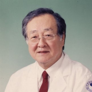 Seung U Kim合作论文数Division of Allergy & Immunology, Department of Medicine, Faculty of Medicine, The University of British Columbia3
 Jae K. Ryu合作论文数Department of Pharmacology and Therapeutics, University of British Columbia3Hong Jun Lee合作论文数Res Inst, Humetacell Inc2Lim In J合作论文数2
 Seung Hyun Kim合作论文数Department of Ophthalmology, College of Medicine, Korea University2
 James Mclarnon合作论文数Department of Anesthesiology, Pharmacology & Therapeutics, Faculty of Medicine, The University of British Columbia2A Nagai合作论文数2Jean Kim合作论文数2Sung-Jun Cho合作论文数1HB Choi合作论文数1Eiji Nakagawa合作论文数1SY Baek合作论文数1B Kim合作论文数1K Hatori合作论文数1
 Atsushi Nagai合作论文数1Ozaki Akihiko合作论文数1Suzuki Yuji合作论文数1Dongsun Park合作论文数1
 Hyun B Choi合作论文数1
 Evan Snyder合作论文数Center for Neurologic Diseases, Sanford Burnham Prebys;Stem Cell Reserch Center, Sanford-Burnham Medical Research Institute1




