Telomeres, complexes of DNA and proteins, protect ends of linear chromosomes. In humans, the two shelterin proteins TRF1 and TIN2, along with cohesin subunit SA1, were proposed to mediate telomere cohesion. Although the ability of the TRF1-TIN2 and TRF1-SA1 systems to compact telomeric DNA by DNA-DNA bridging has been reported, the function of the full ternary TRF1-TIN2-SA1 system has not been explored in detail. Here, we quantify the compaction of nanochannel-stretched DNA by the ternary system, as well as its constituents, and obtain estimates of the relative impact of its constituents and their interactions. We find that TRF1, TIN2, and SA1 work synergistically to cause a compaction of the DNA substrate, and that maximal compaction occurs if all three proteins are present. By altering the sequence with which DNA substrates are exposed to proteins, we establish that compaction by TRF1 and TIN2 can proceed through binding of TRF1 to DNA, followed by compaction as TIN2 recognizes the previously bound TRF1. We further establish that SA1 alone can also lead to a compaction, and that compaction in a combined system of all three proteins can be understood as an additive effect of TRF1-TIN2 and SA1-based compaction. Atomic force microscopy of intermolecular aggregation confirms that a combination of TRF1, TIN2, and SA1 together drive strong intermolecular aggregation as it would be required during chromosome cohesion.
The methyl-CpG-binding domain 2 and 3 proteins (MBD2 and MBD3) provide structural and DNA-binding function for the Nucleosome Remodeling and Deacetylase (NuRD) complex. The two proteins form distinct NuRD complexes and show different binding affinity and selectivity for methylated DNA. Previous studies have shown that MBD2 binds with high affinity and selectivity for a single methylated CpG dinucleotide while MBD3 does not. However, the NuRD complex functions in regions of the genome that contain many CpG dinucleotides (CpG islands). Therefore, in this work, we investigate the binding and diffusion of MBD2 and MBD3 on more biologically relevant DNA templates that contain a large CpG island or limited CpG sites. Using a combination of single-molecule and biophysical analyses, we show that both MBD2 and MBD3 diffuse freely and rapidly across unmethylated CpG-rich DNA. In contrast, we found methylation of large CpG islands traps MBD2 leading to stable and apparently static binding on the CpG island while MBD3 continues to diffuse freely. In addition, we demonstrate both proteins bend DNA, which is augmented by methylation. Together, these studies support a model in which MBD2-NuRD strongly localizes to and compacts methylated CpG islands while MBD3-NuRD can freely mobilize nucleosomes independent of methylation status.
Single-molecule techniques provide important details supplementing the framework obtained from traditional bulk experiments. Many cellular processes such as DNA replication, DNA repair, and telomere maintenance involve interaction among multiple proteins, their co-factors, and DNA. To investigate such interactions and to differentiate the function of each component necessitate a technique that allows the combinatorial exposure of DNA to multiple proteins and co-factors as well as manipulation of the DNA configuration. We present a nanofluidic device with the capability of active combinatorial exchange of up to three buffers in real-time and dynamic manipulation of DNA under physiologically relevant conditions. We demonstrate its utility in monitoring compaction of DNA by telomeric proteins, DNA modification by an endonuclease, and DNA loop extrusion by cohesin.
The telomere specific shelterin complex, which includes TRF1, TRF2, RAP1, TIN2, TPP1 and POT1, prevents spurious recognition of telomeres as double-strand DNA breaks and regulates telomerase and DNA repair activities at telomeres. TIN2 is a key component of the shelterin complex that directly interacts with TRF1, TRF2 and TPP1. In vivo, the large majority of TRF1 and TRF2 are in complex with TIN2 but without TPP1 and POT1. Since knockdown of TIN2 also removes TRF1 and TRF2 from telomeres, previous cell-based assays only provide information on downstream effects after the loss of TRF1/TRF2 and TIN2. Here, we investigated DNA structures promoted by TRF2-TIN2 using single-molecule imaging platforms, including tracking of compaction of long mouse telomeric DNA using fluorescence imaging, atomic force microscopy (AFM) imaging of protein-DNA structures, and monitoring of DNA-DNA and DNA-RNA bridging using the DNA tightrope assay. These techniques enabled us to uncover previously unknown unique activities of TIN2. TIN2S and TIN2L isoforms facilitate TRF2-mediated telomeric DNA compaction (cis-interactions), dsDNA-dsDNA, dsDNA-ssDNA and dsDNA-ssRNA bridging (trans-interactions). Furthermore, TIN2 facilitates TRF2-mediated T-loop formation. We propose a molecular model in which TIN2 functions as an architectural protein to promote TRF2-mediated trans and cis higher-order nucleic acid structures at telomeres.
TIN2 is a core component of the shelterin complex linking double-stranded telomeric DNA-binding proteins (TRF1 and TRF2) and single-strand overhang-binding proteins (TPP1-POT1). In vivo, the large majority of TRF1 and TRF2 exist in complexes containing TIN2 but lacking TPP1/POT1; however, the role of TRF1-TIN2 interactions in mediating interactions with telomeric DNA is unclear. Here, we investigated DNA molecular structures promoted by TRF1-TIN2 interaction using atomic force microscopy (AFM), total internal reflection fluo-rescence microscopy (TIRFM), and the DNA tightrope assay. We demonstrate that the short (TIN2S) and long (TIN2L) isoforms of TIN2 facilitate TRF1-mediated DNA compaction (cis-in-teractions) and DNA-DNA bridging (trans-interactions) in a telomeric sequence-and length-dependent manner. On the short telomeric DNA substrate (six TTAGGG repeats), the ma-jority of TRF1-mediated telomeric DNA-DNA bridging events are transient with a lifetime of similar to 1.95 s. On longer DNA substrates (270 TTAGGG repeats), TIN2 forms multiprotein complexes with TRF1 and stabilizes TRF1-mediated DNA-DNA bridging events that last on the order of minutes. Preincubation of TRF1 with its regulator protein Tankyrase 1 and the cofactor NAD+ significantly reduced TRF1-TIN2 mediated DNA-DNA bridging, whereas TIN2 protected the disassembly of TRF1-TIN2 mediated DNA-DNA bridging upon Tankyrase 1 addition. Furthermore, we showed that TPP1 inhibits TRF1-TIN2L-mediated DNA-DNA bridging. Our study, together with previous findings, supports a molecular model in which protein assemblies at telomeres are heterogeneous with distinct subcomplexes and full shelterin complexes playing distinct roles in telomere protection and elongation.
Cohesin SA1 (STAG1) and SA2 (STAG2) are key components of the cohesin complex. Previous studies have highlighted the unique contributions by SA1 and SA2 to 3D chromatin organization, DNA replication fork progression, and DNA double-strand break (DSB) repair. Recently, we discovered that cohesin SA1 and SA2 are DNA binding proteins. Given the recently discovered link between SA2 and RNA-mediated biological pathways, we investigated whether or not SA1 and SA2 directly bind to RNA using a combination of bulk biochemical assays and single-molecule techniques, including atomic force microscopy (AFM) and the DNA tightrope assay. We discovered that both SA1 and SA2 bind to various RNA containing substrates, including ssRNA, dsRNA, RNA:DNA hybrids, and R-loops. Importantly, both SA1 and SA2 localize to regions on dsDNA that contain RNA. We directly compared the SA1/SA2 binding and R-loops sites extracted from Chromatin Immunoprecipitation sequencing (ChIP-seq) and DNA-RNA Immunoprecipitation sequencing (DRIP-Seq) data sets, respectively. This analysis revealed that SA1 and SA2 binding sites overlap significantly with R-loops. The majority of R-loop-localized SA1 and SA2 are also sites where other subunits of the cohesin complex bind. These results provide a new direction for future investigation of the diverse biological functions of SA1 and SA2.
BACKGROUND:MeCP2 and MBD2 are members of a family of proteins that possess a domain that selectively binds 5-methylcytosine in a CpG context. Members of the family interact with other proteins to modulate DNA packing. Stretching of DNA-protein complexes in nanofluidic channels with a cross-section of a few persistence lengths allows us to probe the degree of compaction by proteins. RESULTS:We demonstrate DNA compaction by MeCP2 while MBD2 does not affect DNA configuration. By using atomic force microscopy (AFM), we determined that the mechanism for compaction by MeCP2 is the formation of bridges between distant DNA stretches and the formation of loops. CONCLUSIONS:Despite sharing a similar specific DNA-binding domain, the impact of full-length 5-methylcytosine-binding proteins can vary drastically between strong compaction of DNA and no discernable large-scale impact of protein binding. We demonstrate that ATTO 565-labeled MBD2 is a good candidate as a staining agent for epigenetic mapping.
Tumor-associated adipocytes promote tumor growth by providing energy and causing chronic inflammation. Here, we have exploited the lipid metabolism to engineer adipocytes that serve as a depot to deliver cancer therapeutics at the tumor site. Rumenic acid (RA), as an anticancer fatty acid, and a doxorubicin prodrug (pDox) with a reactive oxygen species (ROS)-cleavable linker, are encapsulated in adipocytes to deliver therapeutics in a tumor-specific bio-responsive manner. After intratumoral or postsurgical administration, lipolysis releases the RA and pDox that is activated by intracellular ROS-responsive conversion, subsequently promoting antitumor efficacy. Furthermore, downregulation of PD-L1 expression is observed in tumor cells, favoring the emergence of CD4(+) and CD8(+) T cell-mediated immune responses.
 Parminder Kaur合作论文数7
 Hong Wang合作论文数Natl Inst Environm Hlth Sci, Mol Genet Lab, Res Triangle Pk, NC USA7
 Hai Pan合作论文数Nanjing University, Nanjing University7
 Robert Riehn合作论文数Cavendish Laboratory, University of Cambridge7
 Ariana C Detwiler合作论文数4
 Patricia L Opresko合作论文数4
 Jacob Piehler合作论文数Division of Biophysics, Department of Biology/Chemistry, University of Osnabrück4
 Changjiang You合作论文数4Chelsea Mahn合作论文数3Dhruv Bhattaram合作论文数2Miao Jin合作论文数2Saroj Dangi合作论文数Department of Electrical and Computer Engineering, North Carolina State University2
 David Williams合作论文数Department of Pathology and Laboratory Medicine, School of Medicine, The University of North Carolina at Chapel Hil2Barnes Ryan P合作论文数2
 Gage Oliver Leighton合作论文数2
 Yizhi Jane Tao (陶一之)合作论文数Department of Biosciences, Wiess School of Natural Sciences, Rice University2Di Wen合作论文数1Maedeh Roushan合作论文数1Jennifer Soto合作论文数1Yanlin Fan合作论文数1Warren Lu合作论文数1Zach Messenger合作论文数1Edgar M. Faison合作论文数1Pengning Xu合作论文数1
 Quanyin Hu (胡全银)合作论文数Pharmaceutical Sciences Division, School of Pharmacy, University of Wisconsin-Madison;Wisconsin Center for NanoBioSystems, University of Wisconsin-Madison1Elizabeth Marie Irvin合作论文数1
 Ashwin Ghadiyaram合作论文数Phys Dept, North Carolina State Univ1
 Zubair Azad合作论文数Dept Phys, North Carolina State Univ1
 Pengyu Hao合作论文数Physics Department, North Carolina State University1Samantha Sanford合作论文数1
 Zejun Wang合作论文数College of Sciences, Northeastern University1
 George Van Den Driessche合作论文数Dept Chem, North Carolina State Univ1
 Henry Miller合作论文数Univ Texas Hlth San Antonio, Greehey Childrens Canc Res Inst, San Antonio, TX 78229 USA1
 Peter Abdou合作论文数Department of Bioengineering, University of California, Los Angeles1Guozhou Xu合作论文数1
 Alex Bishop合作论文数Univ Texas Hlth Sci Ctr San Antonio, Greehey Childrens Canc Res Inst, San Antonio, TX 78229 USA1
 Hongjun Li (李洪军)合作论文数School of Pharmacy, Zhejiang University;The Second Affiliated Hospital, School of Medicine, Zhejiang University1
 Guojun Chen合作论文数Department of Biomedical Engineering, Faculty of Medicine and Health Sciences, McGill University1
 Jonathan Hall合作论文数Department of Chemistry and Applied Biosciences, Eidgenössische Technische Hochschule Zürich1
 Jinqiang Wang (王金强)合作论文数School of Pharmacy, Zhejiang University1
 Saeid Movahed合作论文数1Bilinovich Stephanie M合作论文数1
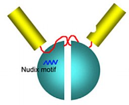 Qingyu Tang合作论文数Shanghai Institute for Biological Sciences Chinese Academy of Sciences, Chinese Academy of Sciences1Countryman Preston合作论文数1
 Yuqi Zhang (张宇琪)合作论文数College of Pharmaceutical Sciences, Zhejiang University1
 Qian Chen (陈倩)合作论文数College of Nano Science & Technology, Soochow University1
 Ohashi Masao合作论文数Graduate School of Medicine, Dentistry and Pharmaceutical Sciences, Okayama University1
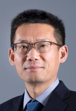 Hongtao Yu (于洪涛)合作论文数School of Life Sciences, Westlake University1
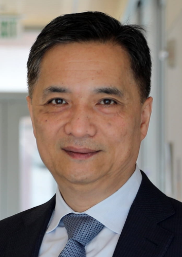 Song Li (李松)合作论文数Department of Bioengineering, Samueli School of Engineering, University of California, Los Angeles;Department of Medicine, Samueli School of Engineering, University of California, Los Angeles1




