Purpose The scapholunate ligament (SLL) is the most frequently injured wrist ligament. The aim of this study was to investigate cellular and extracellular changes within the SLL following injury. Methods Fifteen SLLs were harvested, ranging between 39 days to 20 years from time of injury. These specimens were subject to immunohistochemical analysis to characterize their vascular and collagen constitution. Results Of the 15 ligaments, 4 were harvested <3 months from injury, and 11 harvested >3 months from injury. The mean type I collagen density was 45.6% (25.2% to 55.9%) in all specimens. The mean type III collagen density was 47% (38.2% to 51.8%) of the ligament area in specimens <3 months after injury and 30.6% (13.3% to 44.1%) in those >3 months after injury. Type III collagen density was highest in the volar subunit. Type I collagen decreased only minimally in specimens taken within 2 years of injury. The increase in the type I:III collagen ratio reflected the decline in type III collagen. Blood vessels were found in 13 of 15 specimens. Mean vessel density for all specimens was 1.3% (0% to 7.1%), with the highest density of 1.8% (0% to 10%) in the volar subunit. The vessel density decreased from 2.9% (1.3% to 4.3%) to 1.6% (0% to 10%) in the volar subunit in specimens harvested >3 months after injury. Conclusions Mean type III collagen density decreased with time, most notably within the volar subunit. Mean type I collagen density held comparatively stable in ligaments taken within 2 years from injury. Blood vessels were detected in 87% of specimens, with the highest density in the volar subunit. Clinical relevance The SLL displays a collagen profile similar to other ligaments with favorable healing capacity. The volar subunit possessed a collagen ratio and vessel density that may suggest its acute repair and inclusion in reconstructive techniques has merit.
Organoid transplantation has a promising future in the treatment of liver disease, but a major limitation is the lack of guidance on the most appropriate method for transplantation that maximises organoid survival. Human induced pluripotent stem cell (hiPSC)-derived liver progenitor cell organoids were transplanted into four different transplantation sites in a mouse model of liver disease, using five organoid delivery methods. Organoids were transplanted into the vascularised chamber device established in the groin, or into the liver, spleen, and subcutaneous fat. For organoid transplantations into the liver, organoids were delivered either in Matrigel alone, or in Matrigel and a polyurethane scaffold. At 2 weeks post-transplantation, the vascularised chamber had the highest organoid survival, which was 5.1x higher than the site with second highest survival (p=0.0002), being the intra-hepatic scaffold approach. No organoid survival was observed when delivered into the liver without a scaffold, or when injected into the spleen. Very low survival occurred in transplantations into subcutaneous fat. Animals with the vascularised chamber also had the highest levels of human albumin (0.33 ± 0.09 ng/mL). This study provides strong evidence supporting the use of the vascularised chamber for future liver organoid transplantation studies, including its translation into clinical therapy.
Background & AimsLiver sinusoidal endothelial cells (LSECs) are important in liver development, regeneration and pathophysiology, but the differentiation process underlying their tissue-specific phenotype is poorly understood and difficult to study as primary human cells are scarce. The aim of this study was to use human induced pluripotent stem cell (hiPSC)-derived LSEC-like cells to investigate the differentiation process of LSECs.MethodshiPSC-derived endothelial cells were transplanted into the livers of Fah−/−/Rag2−/−/Il2rg−/− mice and assessed over a 12-week period. Lineage tracing, immunofluorescence, flow cytometry, plasma human factor VIII measurement and bulk and single cell transcriptomic analysis were used to assess the molecular and functional changes that occur with transplantation.ResultsProgressive and long-term repopulation of the liver vasculature occurred as iECs expanded along the sinusoids between hepatocytes and increasingly produced human factor VIII, indicating differentiation into LSEC-like cells. To chart the developmental profile associated with LSEC specification, the bulk transcriptome of transplanted cells between 1- and 12-weeks post-transplantation were compared against primary human adult LSECs. This demonstrated a chronological increase in LSEC markers, LSEC differentiation pathways, and zonation. Bulk transcriptome analysis suggested that the transcription factors NOTCH1, GATA4, and FOS play a central role in LSEC specification, interacting with a network of 27 transcription factors. Novel markers associated with this process include EMCN and CLEC14A. Additionally, single cell transcriptomic analysis demonstrated that transplanted iECs at 4-weeks contain zonal subpopulations with a region-specific phenotype.ConclusionsCollectively, this study confirms that hiPSC can adopt LSEC-like features and provides insight into LSEC specification. This humanised xenograft system can be applied to further interrogate LSEC developmental biology and pathophysiology, bypassing current logistical obstacles associated with primary human LSECs.Impact And ImplicationsLiver sinusoidal endothelial cells (LSECs) are important cells for liver biology, but better model systems are required to study them. We present a pluripotent stem cell xenografting model which produces human LSEC-like cells. A detailed and longitudinal transcriptomic analysis of the development of LSEC-like cells is included, which will guide future studies to interrogate LSEC biology and produce LSEC-like cells which could be used for regenerative medicine.
PURPOSE:Scapholunate ligaments (SLLs) play a well-established role in maintaining carpal alignment and kinematics, and are innervated with sensory mechanoreceptors located within the ligaments. They are involved in the afferent arc of dynamic wrist stability. The aim of this study was to describe the changes in these mechanoreceptor populations in injured SLLs. METHODS:Injured SLLs were collected from human wrists at the time of SLL reconstruction or limited wrist fusion, where the ligament remnants would otherwise be discarded. These specimens were formalin-fixed and paraffin-embedded for immunohistochemical analysis to identify mechanoreceptors, which were then classified by type and location within the ligament. RESULTS:A total of 15 ligaments were collected, with the interval from injury ranging from 39 days-20 years. Eleven ligaments were collected less than one year after injury, and four ligaments were collected two years or more after injury. A total of 66 mechanoreceptors were identified, with 50 mechanoreceptors identified in nine of the 11 specimens collected less than one year after injury. In this group, 54% of the mechanoreceptors resided in the volar subunit, 20% in the dorsal subunit, and 26% in the proximal subunit. Two of the four specimens collected two years or later after injury contained mechanoreceptors, all of which were located in the dorsal subunit. Increasing time from injury demonstrated a decline in mechanoreceptor numbers within the volar subunit. CONCLUSIONS:Mechanoreceptors were consistently located in the SLL, particularly in the volar subunit of specimens collected less than one year after injury. CLINICAL RELEVANCE:Ligament reconstruction techniques aim to primarily reconstitute the biomechanical function of the disrupted SLL; however, re-establishing the afferent proprioceptive capacity of the SLL may be a secondary objective. This suggests the need to consider the reconstruction of its volar subunit particularly in those managed within one year of injury.
This chapter reviews the history and indications of digital replantation and provides a thorough assessment of hand function to guide the surgeon. A technical guide to replantation is provided based on guiding principles of hand surgery and microsurgery. We provide a useful framework for the functional and cosmetic principles of microsurgical thumb reconstruction as well as pollicization and phalangization. We recommend a deep understanding of the hand's function prior to embarking on digital replantation and thumb reconstruction, as well as individualizing reconstruction to patient needs.
Plastic surgery knowledge in the Middle Ages is known to have progressed via the Sushruta Samhita and Pragmateia of Paul of Aegina . Both texts influenced numerous medical authors of the Middle Ages, particularly those from the Islamic Golden Age. Little is known of how this information was transferred to these great writers. This article examines how the Sushruta Samhita and the Pragmateia crossed international borders and contributed to the practice of plastic surgery in the early Middle Ages. A comprehensive review of medical, medical humanities and history databases (PubMed; MEDLINE; Web of Knowledge; Anthropology; JSTOR, Encyclopedia of ancient history), non-digital printed texts and digitised manuscripts (Biblioteca Medicea Laurenziana and BnF Gallica) was conducted using multiple search terms and filters including Middle Ages Surgery; Paul of Aegina; Sushruta; Branca Family; Reconstruction; Plastic Surgery; Islamic Medicine; Nasal reconstruction and Rhinoplasty. The search was restricted to publications which focused on the period between 476 and 1453 AD. A seventh century translator, Hunain ibn Ishaq; a thirteenth century manuscript Par. gr. 2293; the Arab conquest of Sicily; and an eleventh century translator, Constantine the African were identified. From Sushruta and Paul, our speciality proliferated in the Middle Ages due to the Great Translation Movement and the prosperity of the Islamic Golden Age. It influenced several medical authors like Albucasis, resulting in Paul and Sushruta’s techniques crossing international borders. Level of evidence: Not ratable.
A major hurdle in tissue engineering of organs is the incorporation of a functioning blood vessel network integrated throughout the engineered tissue that readily links to the surrounding host blood vessels to provide the oxygen and nutrients required by the engineered construct. In the early years of tissue engineering development, vascularization was not a priority and generally angiogenic ingrowth from neighboring host capillary networks, a process termed extrinsic vascularization was used to vascularize implanted tissue engineering constructs. Extrinsic vascularization takes weeks, and much of the implanted tissue becomes ischemic and dies before capillary ingrowth is complete. In 2000, intrinsic vascularization was devised by Tanaka et al. who isolated a macrovascular pedicle in a plastic chamber which subsequently underwent considerable angiogenic sprouting. A new arteriovenous capillary network was therefore formed within the chamber space which was capable of growing with and supporting the survival of tissue/organ specific cells implanted in the chamber. There was a time lag to development of this pedicle-based angiogenic network, and in recent years a new technique termed pre-vascularization has been developed that involves co-culture of endothelial cells with parenchymal cells or stem cells as they assemble in vitro. Capillary networks are formed throughout the construct, and upon implantation inosculate (functionally join) with host capillaries. Inosculation takes at least 2 days and provides blood flow within this time period within the construct. The most efficient vascularization technique for thick three-dimensional tissue engineering would be the combination of pre-vascularization in vitro with vascularization via angiogenic sprouting of a vascular pedicle, this combination has rarely been successfully utilized.
BACKGROUND:Plastic and Reconstructive Surgery (PRS) can trace its origins as far back as 3000 BC. Despite this, it remained a relatively rare and unestablished branch of surgery until the devastating injuries of the World Wars necessitated reconstruction. Returning wartime surgeons used the skills they had learned on the battlefield to continue PRS in Australia and New Zealand. This article examines the significant contributions of Australian and New Zealand surgeons to the founding of PRS as a global specialty and provides an account of the strenuous dedicated competition that led to the development of microsurgery and advances in reconstruction.METHODS:A comprehensive review of medical, medical humanities, and history databases (PubMed; MEDLINE; Web of Knowledge; Anthropology; Encyclopaedia of ancient history) and non-digital printed texts was conducted using multiple search terms and filters including Reconstruction; Plastic Surgery; Burns; Flaps; and Microsurgery). The search was restricted to publications that focused on the period between 1818 CE to current.RESULTS:Significant contributions of surgeons from the Antipodes occurred during several periods including the Industrial era, World Wars, Post-war and in the modern age.CONCLUSIONS:Stirred by their wartime experience, surgeons from Australia and New Zealand laid the foundations of the global success of Plastic Surgery in the modern age and helped establish it as a specialty in its own right.
The adipose tissue engineering paradigm of in vitro cell-seeded scaffolds subsequently implanted in vivo failed because of inadequate vascularization. Consequently, entirely in vivo models of tissue engineering are being trialled where angiogenic growth is stimulated in unison with expansion of implanted cells and matrices. In animals, impressive amounts of fat and fibroblastic tissue have been grown by matrix induction of preadipocytes or by redirecting a vascular pedicle with fat into a sealed chamber space and proof of principle shown in a human trial. The models are cumbersome limiting clinical translation and currently direct fat transfer by injection is simpler. Unlike true tissue engineering there is, however, no net gain of tissue and even when ‘successful’ it is debated whether the graft survives or is replaced by newly regenerated adipocytes. Research focuses on stem cell differentiation, cell survival, and matrix and biomechanical manipulations.
Though the industrial era (1750 to 1914 AD) and the world wars (1914 to 1945 AD) were the catalysts for the establishment of Plastic and Reconstructive Surgery as a specialty in its own right, a careful examination of ancient texts and literature reveals that those conditions generally attributed to the craft were recognised and treated as early as 3000 BC. This article examines the remarkable medical documents of ancient civilisations as they pertain to our specialty and their legacies in the modern world. A comprehensive review of medical, medical humanities, and history databases (PubMed; MEDLINE; Web of Knowledge; Anthropology; Encyclopedia of ancient history) and non-digital printed texts was conducted using multiple search terms and filters including Reconstruction; Plastic Surgery; Ancient Surgery; Burns; Flaps; Sutures; and Dressings. The search was restricted to publications that focused on the period between 3100 BC and 476 AD. Additional information was sought from an eminent scholar of the Vedic period as well as the chief librarian of the Kaiser Library to aid in the identification of provenance. Five ancient periods and associated manuscripts were identified. Our specialty draws its earliest roots from antiquity. Reconstructive procedures used today were described in ancient eras indicating that the practice of plastic surgery is as old as surgery itself. Level of evidence: Not ratable.
The structural and physiological complexity of currently available liver organoids is limited, thereby reducing their relevance for drug studies, disease modelling, and regenerative therapy. In this study we combined mouse liver progenitor cells (LPCs) with mouse liver sinusoidal endothelial cells (LSECs) to generate hepatobiliary organoids with liver-specific vasculature. Organoids consisting of 5x10(3) cells were created from either LPCs, or a 1:1 combination of LPC/LSECs. LPC organoids demonstrated mild hepatobiliary differentiation in vitro with minimal morphological change; in contrast LPC/LSEC organoids developed clusters of polygonal hepatocyte-like cells and biliary ducts over a 7 day period. Hepatic (albumin, CPS1, CYP3A11) and biliary (GGT1) genes were significantly upregulated in LPC/LSEC organoids compared to LPC organoids over 7 days, as was albumin secretion. LPC/LSEC organoids also had significantly higher in vitro viability compared to LPC organoids. LPC and LPC/LSEC organoids were transplanted into vascularised chambers created in Fah(-/-)/Rag2(-/-)/Il2rg(-/-) mice (50 LPC organoids, containing 2.5x10(5) LPCs, and 100 LPC/LSEC organoids, containing 2.5x10(5) L(PC)s). At 2 weeks, minimal LPCs survived in chambers with LPC organoids, but robust hepatobiliary ductular tissue was present in LPC/LSEC organoids. Morphometric analysis demonstrated a 115-fold increase in HNF4 alpha(+) cells in LPC/LSEC organoid chambers (17.26 +/- 4.34 cells/mm(2) vs 0.15 +/- 0.15 cells/mm(2), p = 0.018), and 42-fold increase in Sox9(+) cells in LPC/LSEC organoid chambers (28.29 +/- 6.05 cells/mm(2) vs 0.67 +/- 0.67 cells/mm(2), p = 0.011). This study presents a novel method to develop vascularised hepatobiliary organoids, with both in vitro and in vivo results confirming that incorporating LSECs with LPCs into organoids significantly increases the differentiation of hepatobiliary tissue within organoids and their survival post-transplantation.
For decades, plastic surgeons have spent considerable effort exploring anatomical regions for free flap design. More recently, tissue-engineering approaches have been utilised in an attempt to grow transplantable tissue flaps in vivo. The aim of this study was to engineer a fat flap with a vascular pedicle by combining autologous fat grafts and a novel acellular hydrogel (Adipogel) in an established tissue-engineering model comprising a chamber and blood vessel loop. An arteriovenous loop was created in the rat groin from the femoral vessels and positioned inside a perforated polycarbonate chamber. In Group 1, the chamber contained minced, centrifuged autologous fat; in Group 2, Adipogel was added to the graft; and in Group 3, Adipogel alone was used. Constructs were histologically examined at 6 and 12 weeks. In all groups, new tissue was generated. Adipocytes, although appearing viable in the graft at the time of insertion, were predominantly nonviable at 6 weeks. However, by 12 weeks, new fat had formed in all groups and was significantly greater in the combined fat/Adipogel group. No significant difference was seen in final construct total volume or construct neovascularisation between the groups. This study demonstrated that a pedicled adipose flap can be generated in rats by combining a blood vessel loop, an adipogenic hydrogel, and a lipoaspirate equivalent. Success appears to be based on adipogenesis rather than on adipocyte survival, and consistent with our previous work, this adipogenesis occurred subsequent to graft death and remodelling. The regenerative process was significantly enhanced in the presence of Adipogel.
Skin grafts are often used to restore skin defects after trauma and cancer. Scarcity of skin and donor site morbidity have led to the development of various bioengineered and synthetic alternatives to promote healing in superficial and deep skin wounds. This chapter gives an updated overview of the different bioengineered and synthetic products that are currently available for treatment of dermal skin defects. An overview is given of scaffolds that substitute the dermal layer or both the epidermal and dermal layer. Results of relevant clinical studies using these products are given. A variety of skin substitutes are on the market today. Some show to be excellent alternatives for autologous skin grafts. Despite this, current challenges still to be addressed are the high cost, the variable graft take in the wound, the altering pigmentation of the newly formed skin, and the often reduced skin mobility and elasticity in the treated area. Using skin scaffolds shows promising clinical results. Future developments in this field are, however, necessary to overcome current disadvantages such as high cost, variable take, uneven pigmentation, or reduced skin mobility and elasticity.
Background: Bacterial infection is a common and serious complication in orthopedic implants following traumatic injury, which is often associated with extensive soft tissue damage and contaminated wounds. Multidrug-resistant bacteria have been found in these infected wounds, especially in patients who have multi trauma and prolonged stay in intensive care units.Purpose: The objective of this study was to develop a coating on orthopedic implants that is effective against drug-resistant bacteria. Methods and results: We applied nanoparticles (30-70nm) of the trace element selenium (Se) as a coating through surface-induced nucleation-deposition on titanium implants and investigated the antimicrobial activity against drug resistant bacteria including Methicillin-resistant Staphylococcus aureus (MRSA) and Methicillin-resistant Staphylococcus epidermidis (MRSE) in vitro and in an infected femur model in rats.The nanoparticles were shown in vitro to have antimicrobial activity at concentrations as low as 0.5ppm. The nanoparticle coatings strongly inhibited biofilm formation on the implants and reduced the number of viable bacteria in the surrounding tissue following inoculation of implants with biofilm forming doses of bacteria. Conclusion: This study shows a proof of concept for a selenium nanoparticle coatings as a potential anti-infective barrier for orthopedic medical devices in the setting of contamination with multi-resistant bacteria. It also represents one of the few (if only) in vivo assessment of selenium nanoparticle coatings on reducing antibiotic-resistant orthopedic implant infections.
Tissue flaps are used to cover large/poorly healing wounds, but involve complex surgery and donor site morbidity. In this study a tissue flap is assembled using the mammalian body as a bioreactor to functionally connect an artery and vein to a human capillary network assembled from induced pluripotent stem cell-derived endothelial cells (hiPSC ECs). In vitro: Porous NovoSorb' scaffolds (3 mm x 1.35 mm) were seeded with 200,000 hiPSC ECs 100,000 human vascular smooth muscle cells (hvSMC), and cultured for 1-3 days, with capillaries formed by 24 h which were CD3r, VE-Cadherin', EphB4., VEGFR2' and Ki67, whilst hvSMCs (calponin+) attached abluminally. In vivo: In SCID mice, bi-lateral epigastric vascular pellicles were isolated in a silicone chamber for a 3 week 'delay period' for pedicle capillary sprouting, then reopened, and two hiPSC EC hvSMCs seeded scaffolds transplanted over the pedicle. The chamber was either resealed (Group 1), or removed and surrounding tissue secured around the pedicle + scaffolds (Group 2), for 1 or 2 weeks. Human capillaries survived in vivo and were CD31 +, VE-Cadherin' and VEGFR2'. Human vSMCs remained attached, and host mesenchymal cells also attached abluminally. Systemically injected FITC-dextran present in human capillary lumens indicated inosculation to host capillaries. Human iPSC EC capillary morphometric parameters at one week in vivo were equal to or higher than the same parameters measured in human abdominal skin. This 'proof of concept' study has demonstrated that bio-engineering an autologous human tissue flap based on hiPSC EC could minimize the use of donor flaps and has potential applications for complex wound coverage. Statement of Significance Tissue flaps, used for surgical reconstruction of wounds, require complex surgery, often associated with morbidity. Bio-engineering a simpler alternative, we assembled a human induced pluripotent stem cell derived endothelial cell (hiPSC ECs) capillary network in a porous scaffold in vitro, which when transplanted over a mouse vascular pedicle in vivo formed a functional tissue flap with mouse blood flow in the human capillaries. Therefore it is feasible to form an autologous tissue flap derived from a hiPSC EC capillary network assembled in vitro, and functionally connect to a vascular pedicle in vivo that could be utilized in complex wound repair for chronic or acute wounds. (C) 2019 Acta Materialia Inc. Published by Elsevier Ltd. All rights reserved.
Vascularisation is key to developing large transplantable tissue constructs capable of providing therapeutic benefits. The vascularised tissue engineering chamber originates from surgical concepts in tissue prefabrication and microsurgery. It serves as an in vivo bioreactor in the form of a closed, protected space surgically created and embedded within the body by fitting a noncollapsible chamber around major blood vessels. This creates a highly angiogenic environment which facilitates the engraftment and survival of transplanted cells and tissue constructs. This article outlines the chamber concept and explores its application in the context of recent advances in biomedical engineering, and how this can play a role in the future of cell therapies and regenerative medicine.
BACKGROUND:Free fat grafting is popular, but it is still unclear how it works. Although focusing on graft survival seems an obvious direction for improving clinical results, the authors' research suggests that long-term volume retention is in part attributable to new fat regeneration. Measures to facilitate adipogenesis may therefore be equally important.METHODS:To investigate the relative roles of survival and regeneration of fat grafts, the authors measured the fate of human lipoaspirate implanted into the scalps of immunodeficient mice, with and without stromal vascular fraction and a porcine extracellular matrix (Adipogel). Specifically, the authors were interested in volume retention, and the composition of implanted or regenerated tissue at 6 and 12 weeks.RESULTS:Free fat grafts exhibited poor volume retention and survival. Almost all of the injected human adipocytes died, but new mouse fat formed peripheral to the encapsulated fat graft. Adipogel and stromal vascular fraction improved proliferation of murine fat and human vasculature. Human CD34 stromal cells were present but only in the periphery, and there was no evidence that these cells differentiated into adipocytes.CONCLUSIONS:In the authors' model, most of the implanted tissue died, but unresorbed dead fat accounted substantially for the long-term, reduced volume. A layer of host-derived, regenerated adipose tissue was present at the periphery. This regeneration may be driven by the presence of dying fat, and it was enhanced by addition of the authors' adipogenic adjuncts. Future research should perhaps focus not only on improving graft survival but also on enhancing the adipogenic environment conducive to fat regeneration.
Vascularization is a major hurdle for growing three-dimensional tissue engineered constructs. This study investigated the mechanisms involved in hypoxic preconditioning of primary rat myoblasts in vitro and their influence on local angiogenesis postimplantation. Primary rat myoblast cultures were exposed to 90min hypoxia at < 1% oxygen followed by normoxia for 24h. Real time (RT) polymerase chain reaction evaluation indicated that 90min hypoxia resulted in significant downregulation of miR-1 and miR-206 (p < 0.05) and angiopoietin-1 (p < 0.05) with upregulation of vascular endothelial growth factor-A (VEGF-A; p < 0.05). The miR-1 and angiopoietin-1 responses remained significantly downregulated after a 24h rest phase. In addition, direct inhibition of miR-206 in L6 myoblasts caused a significant increase in VEGF-A expression (p < 0.05), further establishing that changes in VEGF-A expression are influenced by miR-206. Of the myogenic genes examined, MyoD was significantly upregulated, only after 24h rest (p < 0.05). Preconditioned or control myoblasts were implanted with Matrigel into isolated bilateral tissue engineering chambers incorporating a flow-through epigastric vascular pedicle in severe combined immunodeficiency mice and the chamber tissue harvested 14days later. Chambers implanted with preconditioned myoblasts had a significantly increased percentage volume of blood vessels (p = 0.0325) compared with chambers implanted with control myoblasts. Hypoxic preconditioned myoblasts promote vascularization of constructs via VEGF upregulation and downregulation of angiopoietin-1, miR-1 and miR-206. The relatively simple strategy of hypoxic preconditioning of implanted cells - including non-stem cell types - has broad, future applications in tissue engineering of skeletal muscle and other tissues, as a technique to significantly increase implant site angiogenesis.
 Geraldine M. Mitchell合作论文数Microsurgery Research Centre, St Vincent's Hospital46Kenneth R. Knight合作论文数39Bm Obrien合作论文数36
 Anthony Penington合作论文数St. Vincent's Hospital, University of Melbourne;Department of Paediatrics, Faculty of Medicine, Dentistry and Health Sciences, University of Melbourne;The Royal Children's Hospital Melbourne35Aurora Messina合作论文数Bernard O'Brien Institute of Microsurgery28Keren M. Abberton合作论文数24J V Hurley合作论文数20
 Alastair Stewart合作论文数Division of Medicine, Dentistry and Health Sciences, The University of Melbourne;Department of Biochemsitry and Pharmacology, The University of Melbourne18
 Jason Palmer合作论文数Swartz Center for Computational Neuroscience17
 Michael J. Hickey合作论文数Leukocyte Trafficking Group, Centre for Inflammatory Disease Monash Health, Monash University14
 Rik Thompson合作论文数School of Biomedical Sciences, Faculty of Health, Queensland University of Technology;Institute of Health & Biomedical Innovation, Queensland University of Technology14Rosalind Romeo合作论文数12Am Macleod合作论文数11Xiaolian Han合作论文数11DA LEPORE合作论文数11Keramidaris Effie合作论文数10Daniel M. Crowe合作论文数10
 Geoffrey Stevens合作论文数Department of Chemical and Biomolecular Engineering, The University of Melbourne9
 David Mccombe合作论文数9
 Michael W Findlay合作论文数8
 Andrea O’Connor合作论文数Department of Biomedical Engineering, School of Chemical and Biomedical Engineering, Faculty of Engineering and Information Technology, The University of Melbourne8Pribaz Julian J合作论文数7Webb Angela合作论文数Division of Plastic &Reconstructive Surgery, Peter MacCallum Cancer Centre;Department of Plastic &Reconstructive Surgery, St. Vincent's Hospital Melbourne7
 Heidi Debels合作论文数7
 Juergen H Dolderer合作论文数7P.G. Hayward合作论文数6
 Rodney J Dilley合作论文数Alfred Hospital, Baker Heart Research Institute6
 Filip Stillaert合作论文数6Bruce J. Dowsing合作论文数6Heather Cleland合作论文数Victorian Adult Burns Service, The Alfred Hospital6
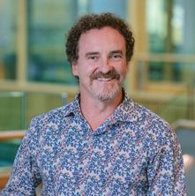 Justin J. Cooper White合作论文数Australian Institute for Bioengineering and Nanotechnology, University of Queensland6
 Zerina Lokmic合作论文数5Alan M. MacLeod合作论文数5
 Michele Riccio合作论文数5Shaun S. Tan合作论文数5
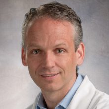 Stefan Hofer合作论文数Department of Surgery, Temerty Faculty of Medicine, University of Toronto;Division of Plastic, Reconstructive & Aesthetic Surgery, University of Toronto;Toronto General Hospital5Yap Kiryu K合作论文数Department of Surgery, St Vincent's Hospital Melbourne;Discipline of Medicine, University of Adelaide and Royal Adelaide Hospital;Mannum District Hospital;Mannum District Hospital, University of Adelaide and Royal Adelaide Hospital5Sinha Shiba合作论文数5Nicholas Trost合作论文数5Randall O. Craft合作论文数5
 George Yeoh合作论文数Australian Centre for RNA Therapeutics in Cancer, School of Human Sciences, University of Western Australia;arry Perkins Institute of Medical Research5
 Milind Wagh合作论文数Department of Plastic and Reconstructive Surgery, Lokmanya Tilak Municipal Medical College and Hospital5Bernard Mcc. O'Brien合作论文数5D.R. Theile合作论文数5A.F. Breidahl合作论文数4Gregory P.L. Thomas合作论文数4A MACLEOD合作论文数4B Zhang合作论文数4
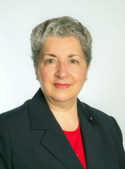 Mary Galea AM合作论文数Department of Medicine, Melbourne Medical School, Faculty of Medicine, Dentistry and Health Sciences, The University of Melbourne4




