PURPOSE:We tested feasibility of burst wave lithotripsy and ultrasonic propulsion to noninvasively fragment and expel small, asymptomatic renal stones in awake participants. MATERIALS AND METHODS:Adult patients suspected of having 2- to 7-mm stones were consented and screened for eligibility. Burst wave lithotripsy and ultrasonic propulsion were applied to up to 3 stones in 1 kidney of qualifying participants for a 30-minute total exposure. Participants completed a CT scan and the Wisconsin Stone Quality-of-Life (WISQOL) questionnaire within 90 days before and 120 days after the procedure. Participants were contacted weekly for 3 weeks after the procedure to assess adverse events (AEs). Outcomes included (1) no fragment > 2 mm, (2) unanticipated health care visits, (3) change in stone volume, (4) reported AEs, and (5) WISQOL score. RESULTS:Forty-one participants were enrolled between April 2023 and October 2024. Twenty-one participants failed screening because no stones were seen, stones were too large or small, stone visibility was too deep or obstructed, or they declined to participate. Twenty participants with 31 stones received the research procedure with 7 undergoing a single repeat procedure. Twenty-two of 31 stones (71%) met the primary effectiveness outcome of no fragment > 2 mm, with 17 of 31 stones (55%) reported as stone free. Median stone volume reduction (IQR) was 100% (88%-100%). No participants returned unexpectedly for care related to the procedure. AEs were all grade I by modified Clavien classification. WISQOL scores improved on 10 of 15 completed questionnaires. CONCLUSIONS:Small, asymptomatic renal stones were effectively and safely removed in awake participants in a clinic setting.
Introduction: Current American Urological Association guidelines recommend that patients with acute obstructive kidney stone requiring continuous anticoagulation/antiplatelet therapy should not be treated by shockwave lithotripsy or percutaneous nephrolithotomy because of the risk of catastrophic renal hemorrhage possible with those techniques. Currently, ureteroscopy is the only recommended surgical treatment. We evaluated if burst wave lithotripsy (BWL) could be used in these cases by treating pigs with BWL while undergoing anticoagulation therapy. Materials and Methods: Six pigs (31-37 kg) were given 200 units/kg porcine heparin, and then the right kidney of each animal was treated with a dose of BWL (18,000 ultrasound pulses at 10 Hz, 20 cycles/pulse, peak positive pressure of 12 MPa) known to fracture kidney stones. The contralateral kidney served as a control. Therapeutic anticoagulation was confirmed by evaluating activated partial thromboplastin time (aPTT). Microhematuria was checked by urine test strips. Kidneys were subsequently evaluated for hemorrhagic injury via MRI. Results: aPTT was significantly elevated (>100 seconds) throughout the entire treatment period (p = <0.001) indicating therapeutic anticoagulation. Gross hematuria was not observed in any of the pigs. After BWL, microhematuria averaged 145.8 red blood cells (RBC)/mu L in the BWL-treated kidney and 48 RBC/mu L in the control kidneys, and there was no statistically significant difference noted in microhematuria rates between the two groups (p = 0.16). Hemorrhagic injury measured only 0.10 +/- 0.02% of the tissue in the BWL-treated kidneys and 0.12 +/- 0.04% in the control side, with a paired difference of -0.03 +/- 0.05%, showing no significant difference (p = 0.58). Conclusions: A typical clinical dose of BWL causes no hemorrhagic injury to the kidney even during therapeutic anticoagulation therapy. This result suggests that BWL should be safe to use in patients with stone undergoing anticoagulation/antiplatelet therapy.
You have accessJournal of UrologyStone Disease: Surgical Therapy (Including ESWL) III (MP29)1 May 2024MP29-11 FEASIBILITY OF BURST WAVE LITHOTRIPSY AND ULTRASONIC PROPULSION TO EXPEL SMALL, ASYMPTOMATIC, RENAL STONES Arturo E. Holmes, Mathew D. Sorensen, Barbrina Dunmire, Jeff Thiel, Barbara H. Burke, Christina Popchoi, John C. Kucewicz, Yak-Nam Wang, Stephanie Totten, Adam D. Maxwell, M. Kennedy Hall, James E. Lingeman, Alana C. Desai, Branda Levchak, Claire C. Yang, Michael R. Bailey, and Jonathan D. Harper Arturo E. HolmesArturo E. Holmes , Mathew D. SorensenMathew D. Sorensen , Barbrina DunmireBarbrina Dunmire , Jeff ThielJeff Thiel , Barbara H. BurkeBarbara H. Burke , Christina PopchoiChristina Popchoi , John C. KucewiczJohn C. Kucewicz , Yak-Nam WangYak-Nam Wang , Stephanie TottenStephanie Totten , Adam D. MaxwellAdam D. Maxwell , M. Kennedy HallM. Kennedy Hall , James E. LingemanJames E. Lingeman , Alana C. DesaiAlana C. Desai , Branda LevchakBranda Levchak , Claire C. YangClaire C. Yang , Michael R. BaileyMichael R. Bailey , and Jonathan D. HarperJonathan D. Harper View All Author Informationhttps://doi.org/10.1097/01.JU.0001008680.67760.37.11AboutPDF ToolsAdd to favoritesDownload CitationsTrack CitationsPermissionsReprints ShareFacebookLinked InTwitterEmail Abstract INTRODUCTION AND OBJECTIVE: The feasibility of burst wave lithotripsy (BWL) and ultrasonic propulsion to noninvasively fragment and expel small, asymptomatic, renal stones in awake subjects is being tested. A previous randomized control trial reported that removal of secondary, small, asymptomatic renal stones during surgery for a primary stone reduced relapse by 82% (Sorensen et al., NEJM, 2022;387:506-13). Our objective was to treat small asymptomatic stones with BWL and ultrasonic propulsion in a clinic-based setting without anesthesia. METHODS: Participants with up to three, 2-7 mm stones in one kidney seen on computerized tomography (CT) within 90 days were consented and screened to assure targetability with the ultrasound device. Untreated infection or inability to hold anticoagulation were exclusions. Transcutaneous ultrasound imaging with BWL therapy to break stones and ultrasonic propulsion to reposition fragments were applied to awake subjects for a 30-minute total exposure under continuous cardiac monitoring. Pain was assessed immediately before and after the procedure. Postoperative urine samples were graded on a published hematuria score (0-10). Participants were asked to strain their urine and they were contacted weekly for 3 weeks to assess for adverse events (AEs) and fragment passage. The primary outcome was stone free on CT 90 days post procedure. Secondary outcomes included change in stone volume, fragment passage, and AEs. RESULTS: Thirteen participants have been enrolled; 4 failed screening because no stones were seen (2), stones were too large (1), and the individual chose bilateral surgery instead (1). Nine participants with 11 stones received the research procedure: all tolerated treatment. Seven participants have received follow-up CT to date, 2 were stone free with 3 stones completely cleared. Mean reduction in stone volume was 70±25%. Video recordings of the research procedures showed stone fragmentation and repositioning. Six of 9 provided photographs of passed fragments. AEs were mild and self-resolving and included: hematuria (6, average score 2.0±1.6), renal colic (1), back pain (3), urinary urgency (1), and change in urinary frequency (1). One participant with a history of urinary tract infections was given antibiotics post procedure despite a negative urinalysis. All pain scores were zero, except one 2, which the participant attributed to positioning. CONCLUSIONS: It is feasible to remove small, asymptomatic, renal stones noninvasively in awake participants with only mild transient AEs. BWL and ultrasonic propulsion may in the not-too-distant future offer a way to prophylactically remove small stones before they require an emergency department visit or surgery. Source of Funding: Work supported by NIH NIDDK P01 DK043881 © 2024 by American Urological Association Education and Research, Inc.FiguresReferencesRelatedDetails Volume 211Issue 5SMay 2024Page: e485 Advertisement Copyright & Permissions© 2024 by American Urological Association Education and Research, Inc.Metrics Author Information Arturo E. Holmes More articles by this author Mathew D. Sorensen More articles by this author Barbrina Dunmire More articles by this author Jeff Thiel More articles by this author Barbara H. Burke More articles by this author Christina Popchoi More articles by this author John C. Kucewicz More articles by this author Yak-Nam Wang More articles by this author Stephanie Totten More articles by this author Adam D. Maxwell More articles by this author M. Kennedy Hall More articles by this author James E. Lingeman More articles by this author Alana C. Desai More articles by this author Branda Levchak More articles by this author Claire C. Yang More articles by this author Michael R. Bailey More articles by this author Jonathan D. Harper More articles by this author Expand All Advertisement PDF downloadLoading ...
PURPOSE:Ultrasonic propulsion is an investigational procedure for awake patients. Our purpose was to evaluate whether ultrasonic propulsion to facilitate residual kidney stone fragment clearance reduced relapse. MATERIALS AND METHODS:This multicenter, prospective, open-label, randomized, controlled trial used single block randomization (1:1) without masking. Adults with residual fragments (individually ≤5 mm) were enrolled. Primary outcome was relapse as measured by stone growth, a stone-related urgent medical visit, or surgery by 5 years or study end. Secondary outcomes were fragment passage within 3 weeks and adverse events within 90 days. Cumulative incidence of relapse was estimated using the Kaplan-Meier method. Log-rank test was used to compare the treatment (ultrasonic propulsion) and control (observation) groups. RESULTS:The trial was conducted from May 9, 2015, through April 6, 2024. Median follow-up (interquartile range) was 3.0 (1.8-3.2) years. The treatment group (n = 40) had longer time to relapse than the control group (n = 42; P < .003). The restricted mean time-to-relapse was 52% longer in the treatment group than in the control group (1530 ± 92 days vs 1009 ± 118 days), and the risk of relapse was lower (hazard ratio 0.30, 95% CI 0.13-0.68) with 8 of 40 and 21 of 42 participants, respectively, experiencing relapse. Omitting 3 participants not asked about passage, 24 treatment (63%) and 2 control (5%) participants passed fragments within 3 weeks of treatment. adverse events were mild, transient, and self-resolving, and were reported in 25 treated participants (63%) and 17 controls (40%). CONCLUSIONS:Ultrasonic propulsion reduced relapse and added minimal risk.
Marine mammals may develop kidney stones, which can be challenging to treat. We describe burst wave lithotripsy (BWL) and ultrasonic propulsion to treat ureteral calculi in a 48-year-old female bottlenose dolphin (Tursiops truncatus) and to reduce renal stone burden in a 23-year-old male harbor seal (Phoca vitulina). BWL and ultrasonic propulsion were delivered transcutaneously in sinusoidal ultrasound bursts to fragment and reposition stones. Targeting and monitoring were performed with real-time imaging integrated within the BWL system. Four dolphin stones were obtained and fragmented ex vivo. The dolphin case received a 10-min and a 20-min BWL treatment conducted approximately 24 h apart to treat two 8–10 mm partially obstructing right mid-ureteral stones, using oral sedation alone. For the harbor seal, while under general anesthesia, retrograde ureteroscopy attempts were unsuccessful because of ureteral tortuosity, and a 30-min BWL treatment was targeted on one 10-mm right kidney stone cluster. All 4 stones fragmented completely to < 2-mm fragments in < 20 min ex vivo. In the dolphin case, the ureteral stones appeared to fragment, spread apart, and move with ultrasonic propulsion. On post-treatment day 1, the ureteral calculi fragments shifted caudally reaching the ureteral orifice on day 9. On day 10, the calculi fragments passed, and the hydroureter resolved. In the harbor seal, the stone cluster was observed to fragment and was not visible on the post-operative computed tomography scan. The seal had gross hematuria and a day of behavior indicating stone passage but overall, an uneventful recovery. BWL and ultrasonic propulsion successfully relieved ureteral stone obstruction in a geriatric dolphin and reduced renal stone burden in a geriatric harbor seal.
You have accessJournal of UrologyStone Disease: Surgical Therapy (Including ESWL) III (MP29)1 May 2024MP29-10 OFFICE-BASED REMOVAL OF SMALL RESIDUAL KIDNEY STONE FRAGMENTS REDUCES RELAPSE RATES Mathew D. Sorensen, Barbrina Dunmire, Jeff Thiel, Bryan W. Cunitz, Barbara H. Burke, Branda J. Levchak, Christina Popchoi, Arturo E. Holmes, John C. Kucewicz, M. Kennedy Hall, Manjiri Dighe, Jessica C. Dai, Fionnuala C. Cormack, Ziyue Liu, Michael R. Bailey, Michael P. Porter, and Jonathan D. Harper Mathew D. SorensenMathew D. Sorensen , Barbrina DunmireBarbrina Dunmire , Jeff ThielJeff Thiel , Bryan W. CunitzBryan W. Cunitz , Barbara H. BurkeBarbara H. Burke , Branda J. LevchakBranda J. Levchak , Christina PopchoiChristina Popchoi , Arturo E. HolmesArturo E. Holmes , John C. KucewiczJohn C. Kucewicz , M. Kennedy HallM. Kennedy Hall , Manjiri DigheManjiri Dighe , Jessica C. DaiJessica C. Dai , Fionnuala C. CormackFionnuala C. Cormack , Ziyue LiuZiyue Liu , Michael R. BaileyMichael R. Bailey , Michael P. PorterMichael P. Porter , and Jonathan D. HarperJonathan D. Harper View All Author Informationhttps://doi.org/10.1097/01.JU.0001008680.67760.37.10AboutPDF ToolsAdd to favoritesDownload CitationsTrack CitationsPermissionsReprints ShareFacebookLinked InTwitterEmail Abstract INTRODUCTION AND OBJECTIVE: The benefit of removing residual kidney stone fragments is unknown. U.S. urology guidelines recommend imaging after surgery to detect fragments and offering a second endoscopic surgery to remove fragments as patients with fragments frequently require additional clinical care. Human feasibility studies and randomized clinical trials have shown fragments may be removed by nonsurgical, noninvasive technologies. METHODS: In a multi-center, randomized, controlled trial, 82 adults with residual kidney stone fragments (individually≤5 mm) were randomized to receive a noninvasive, nonsurgical investigative procedure to attempt to facilitate fragment clearance or no procedure and observation of their fragments (ClinicalTrials.gov number, NCT02028559). The primary outcome was relapse as measured by future symptomatic, unscheduled medical visits, surgeries, or stone growth measured on annual CT exams. RESULTS: Demographic and clinical characteristics were similar (Table 1). After a median follow-up of 2.4 years, the risk of relapse was 68% lower in the treatment group than the control group (odds ratio 0.32, 95% confidence interval 0.14-0.73) with absolute difference in relapse of 20% vs. 48% (treatment versus control). The treatment group had 51% longer time-to-relapse than controls (restricted mean of 1505±99 days for treatment vs 995±120 days for control, p<0.004, log-rank test) (Figure 1). Excluding 3 participants not asked about passage, asymptomatic passage of fragments within the first 3 weeks was over 12-fold higher in the treatment group (n=24, 63%) compared to control (n=2, 5%). After 3 weeks, asymptomatic passage was similar between groups (n=10 treatment vs 12 control subjects). Average number of treatments was 1.35. Adverse events were mild and transient and included 1 report of a bruise and 11 cases of mild discomfort. CONCLUSIONS: Removal of residual fragments by ultrasonic propulsion added minimal risk and reduced relapse. Download PPT Source of Funding: Funding provided by the NIH NIDDK P01 DK043881 and the Puget Sound Veterans Affairs © 2024 by American Urological Association Education and Research, Inc.FiguresReferencesRelatedDetails Volume 211Issue 5SMay 2024Page: e484 Advertisement Copyright & Permissions© 2024 by American Urological Association Education and Research, Inc.Metrics Author Information Mathew D. Sorensen More articles by this author Barbrina Dunmire More articles by this author Jeff Thiel More articles by this author Bryan W. Cunitz More articles by this author Barbara H. Burke More articles by this author Branda J. Levchak More articles by this author Christina Popchoi More articles by this author Arturo E. Holmes More articles by this author John C. Kucewicz More articles by this author M. Kennedy Hall More articles by this author Manjiri Dighe More articles by this author Jessica C. Dai More articles by this author Fionnuala C. Cormack More articles by this author Ziyue Liu More articles by this author Michael R. Bailey More articles by this author Michael P. Porter More articles by this author Jonathan D. Harper More articles by this author Expand All Advertisement PDF downloadLoading ...
You have accessJournal of UrologyParadigm-shifting, Practice-changing Clinical Trials in Urology (P2)1 May 2024P2-11 OFFICE-BASED REMOVAL OF SMALL URINARY STONES Mathew D. Sorensen, Barbrina Dunmire, Jeff Thiel, Bryan W. Cunitz, Barbara H. Burke, Branda J. Levchak, Christina Popchoi, Arturo E. Holmes, John C. Kucewicz, M. Kennedy Hall, Manjiri Dighe, Jessica C. Dai, Fionnuala C. Cormack, Ziyue Liu, Michael R. Bailey, Michael P. Porter, and Jonathan D. Harper Mathew D. SorensenMathew D. Sorensen , Barbrina DunmireBarbrina Dunmire , Jeff ThielJeff Thiel , Bryan W. CunitzBryan W. Cunitz , Barbara H. BurkeBarbara H. Burke , Branda J. LevchakBranda J. Levchak , Christina PopchoiChristina Popchoi , Arturo E. HolmesArturo E. Holmes , John C. KucewiczJohn C. Kucewicz , M. Kennedy HallM. Kennedy Hall , Manjiri DigheManjiri Dighe , Jessica C. DaiJessica C. Dai , Fionnuala C. CormackFionnuala C. Cormack , Ziyue LiuZiyue Liu , Michael R. BaileyMichael R. Bailey , Michael P. PorterMichael P. Porter , and Jonathan D. HarperJonathan D. Harper View All Author Informationhttps://doi.org/10.1097/01.JU.0001015816.87470.c9.11AboutPDF ToolsAdd to favoritesDownload CitationsTrack CitationsPermissionsReprints ShareFacebookLinked InTwitterEmail Abstract INTRODUCTION AND OBJECTIVES: We investigate changing practice to treat small stones and fragments. We published that removing by ureteroscopy small, asymptomatic renal stones during surgery for ureteral or contralateral renal stones reduced relapse by 82% in 4-year follow-up [NEJM 2022;387:506-513]. Here we report relapse following ultrasonic propulsion - noninvasive, nonsurgical treatment - to reposition small, asymptomatic residual fragments to facilitate clearance. We have begun studies adding burst wave lithotripsy to ultrasonic propulsion in the same device to break ureteral and renal stones and reposition fragments to facilitate clearance in awake patients. METHODS: In a multi-center, randomized, controlled trial, 82 adults with residual kidney stone fragments (individually ≤5 mm) were randomized to receive ultrasonic propulsion to attempt to facilitate fragment clearance or no procedure and observation of their fragments (ClinicalTrials.gov NCT02028559). The primary outcome was relapse as measured by future symptomatic, unscheduled medical visits, surgeries, or stone growth measured on annual CT exams. RESULTS: Demographic and clinical characteristics were similar (Table 1). After a mean follow-up of 2.6 years, the risk of relapse was 70% lower in the treatment group than the control group (hazard ratio 0.30, 95% confidence interval 0.13-0.68) with absolute difference in relapse of 20% vs. 50% (treatment vs. control). The treatment group had 52% longer time-to-relapse than controls (restricted mean of 1530 ± 92 days for treatment vs. 1009 ± 118 days for control, p<0.003, log-rank test) (Fig. 1). Excluding 3 participants not asked about passage, passage of fragments within the first 3 weeks was over 12-fold higher in the treatment group (24 of 38, 63%) compared to control (2 of 41, 5%). Adverse events were mild and transient and were reported in 25 (63%) of 40 patients in the treatment group and 17 (40%) of 42 patients in the control group. CONCLUSIONS: Removal of residual fragments by ultrasonic propulsion added minimal risk and reduced relapse. Download PPT Source of Funding: Funding provided by the NIH NIDDK P01 DK043881 and the Puget Sound Veterans Affairs © 2024 by American Urological Association Education and Research, Inc.FiguresReferencesRelatedDetails Volume 211Issue 5S2May 2024Page: e7 Advertisement Copyright & Permissions© 2024 by American Urological Association Education and Research, Inc.Metrics Author Information Mathew D. Sorensen More articles by this author Barbrina Dunmire More articles by this author Jeff Thiel More articles by this author Bryan W. Cunitz More articles by this author Barbara H. Burke More articles by this author Branda J. Levchak More articles by this author Christina Popchoi More articles by this author Arturo E. Holmes More articles by this author John C. Kucewicz More articles by this author M. Kennedy Hall More articles by this author Manjiri Dighe More articles by this author Jessica C. Dai More articles by this author Fionnuala C. Cormack More articles by this author Ziyue Liu More articles by this author Michael R. Bailey More articles by this author Michael P. Porter More articles by this author Jonathan D. Harper More articles by this author Expand All Advertisement PDF downloadLoading ...
Despite the growing incidence of urinary stones, the fundamental interventions for urinary stones have remained the same for several decades. However, technical innovations in ultrasound have enabled new strategies for stone management. Our team aims to employ these methods to establish a new paradigm to treat stones in an office or clinic rather than a surgical suite. Technologies include new imaging methods to detect and characterize stones, burst wave lithotripsy to fragment stones using focused ultrasound rather than shock waves, and ultrasonic propulsion to promote clearance of fragments, all using a single ultrasound platform. This presentation will describe the principles and development of these technologies, as well as the fruitful partnerships between teams of basic scientists, engineers, physicians, and industry that have enabled rapid development of these concepts to successful clinical trials. [Work supported by NIDDK P01 DK043881 and the Focused Ultrasound Foundation.]
You have accessJournal of UrologyCME1 Apr 2023MP23-05 NONINVASIVE ACOUSTIC FORCEPS USING ULTRASOUND TO MANIPULATE OBJECTS IN THE URINARY TRACT Mohamed Ghanem, Arturo Holmes, Adam Maxwell, Michael Bailey, Mathew Sorensen, and Jonathan Harper Mohamed GhanemMohamed Ghanem More articles by this author , Arturo HolmesArturo Holmes More articles by this author , Adam MaxwellAdam Maxwell More articles by this author , Michael BaileyMichael Bailey More articles by this author , Mathew SorensenMathew Sorensen More articles by this author , and Jonathan HarperJonathan Harper More articles by this author View All Author Informationhttps://doi.org/10.1097/JU.0000000000003248.05AboutPDF ToolsAdd to favoritesDownload CitationsTrack CitationsPermissionsReprints ShareFacebookLinked InTwitterEmail Abstract INTRODUCTION AND OBJECTIVE: Ultrasonic propulsion has been demonstrated in clinical trials to noninvasively reposition renal stones along the direction of the probe. However, extracorporeal manipulation of stones causing ureteral stone obstruction requires controlled movement along a complex path in the urinary tract. Here, we evaluate the safety and utility of a method for controlled manipulation of stone models in vivo in a complex three-dimensional (3D) path using focused ultrasound beams. METHODS: A multi-element ultrasound array transducer with a 12-cm focus was used to produce an acoustic trap around a stone model (3 mm glass sphere) in the bladder of three pigs under anesthesia. The array was coupled to the pig by a water bath and synchronized with a P6-3 ultrasound imaging probe for targeting. A cystoscope was inserted into the bladder to visualize the motion. The sphere was acoustically trapped and steered along three different paths (Figure 1a-c). Deviation of the measured from the intended path was calculated to quantify the accuracy. The total energy exposure to manipulate the stone was low; at 1.5 MHz, the maximum exposure had a maximum peak pressure of 1.4 MPa and a pulse average intensity of 133 W/cm2. The bladder was then evaluated histologically. RESULTS: Spheres were levitated and moved in the three paths in all pigs for a total of 26 manipulations. The spheres were lifted and moved 1) laterally 3 mm and back down, 2) in a horizontal, and 3) in a vertical loop of 3 mm radius. The average discrepancy between the desired path and that observed on ultrasound was 0.24±0.07 mm, and by the cystoscope was 0.17±0.09 mm. The sphere was dragged for 5 cm along the bladder wall by moving the array mechanically (Figure 1d). The skin to stone depth was 2±0.5 cm. Histology showed no signs of injury to the bladder wall. CONCLUSIONS: This work demonstrated the controlled, noninvasive manipulation of stone models in vivo at safe ultrasound power levels. This work potentiates the use of noninvasive ultrasound tweezers to facilitate movement of stones in any direction which could expand the capabilities of ultrasonic propulsion alone. Source of Funding: Work supported by NIDDK P01 DK043881, K25 DK132416 © 2023 by American Urological Association Education and Research, Inc.FiguresReferencesRelatedDetails Volume 209Issue Supplement 4April 2023Page: e307 Advertisement Copyright & Permissions© 2023 by American Urological Association Education and Research, Inc.MetricsAuthor Information Mohamed Ghanem More articles by this author Arturo Holmes More articles by this author Adam Maxwell More articles by this author Michael Bailey More articles by this author Mathew Sorensen More articles by this author Jonathan Harper More articles by this author Expand All Advertisement PDF downloadLoading ...
Acoustic radiation forces can surround and trap objects to manipulate them in three dimensions. It has been demonstrated that the radiation forces can maneuver solid heavy objects similar to kidney stones in live animals under safe acoustic exposure levels. However, initial experiments identified limitations on the steering range of objects because of the array design, and tissue aberrations. Here, we present a method to design an in-house 256 multi-element array to improve the steering range of 2–5 mm stones. We modeled the acoustic field for various geometrical parameters including the element size, transducer focusing, and F-number at a frequency range from 0.5 to 1 MHz. The parameters were then used to simulate the trapping radiation forces on stone models to finalize the design of the array. The array was fabricated with a center frequency of 950 kHz and a focal distance located at 120 mm and an F-number of 1.5. A holography scan was performed to characterize the transducer output. Various trapping beams were measured and compared to simulations. The resulting radiation forces produced were on the order of the weight of the trapped 2-5 mm stone models. [Work supported by NIH P01-DK043881, K25-132416, and Applied Physics Laboratory SEED fellowship.]
Acoustic radiation forces can remotely manipulate particles. Forces from a standing wave field align microscale particles along the nodal or anti-nodal locations of the field to form three-dimensional (3D) patterns. These patterns can be used to form 3D microstructures for tissue engineering applications. However, standing wave generation requires more than one transducer or a reflector, which is challenging to implement in vivo. Here, a method is developed and validated to manipulate microspheres using a travelling wave from a single transducer. Diffraction theory and an iterative angular spectrum approach are employed to design phase holograms to shape the acoustic field. The field replicates a standing wave and aligns polyethylene microspheres in water, which are analogous to cells in vivo, at pressure nodes. Using Gor’kov potential to calculate the radiation forces on the microspheres, axial forces are minimized, and transverse forces are maximized to create stable particle patterns. Pressure fields from the phase holograms and resulting particle aggregation patterns match predictions with a feature similarity index > 0.92, where 1 is a perfect match. The resulting radiation forces are comparable to those produced from a standing wave, which suggests opportunities for in vivo implementation of cell patterning toward tissue engineering applications.
Acoustic fields can generate radiation forces that can align particles in patterns. Forces from standing waves pattern particles in three dimensions (3-D) at either nodal or anti-nodal regions. These patterns can be utilized to form 3-D microstructures for applications in tissue engineering or fabrication of layered materials. However, standing waves require more than one transducer or a reflector, which can complicate implementation. Here, we developed a method to suspend and align microspheres using a traveling wave from a single transducer. Acoustic fields were shaped to align polyethylene microspheres mimicking tissue cells in parallel planes along the axis of the transducer. A Bessel-like beam was developed using diffraction theory and an iterative angular spectrum approach were used to design phase holograms to shape pressure fields. Gor’kov potential was used to calculate radiation forces while minimizing the axial forces to create a stable trap. The resulting pressure fields and particle patterns matched predictions with a similarity index >0.92, where 1 is a perfect match. The transverse radiation force was ten times each microsphere’s weight and comparable to the standing wave radiation forces. Next steps are in vivo implementation of cell patterning for tissue engineering. [Work supported by NIH K25-DK132416 and P01-DK043881.]
You have accessJournal of UrologyCME1 Apr 2023MP23-11 REMOVAL OF SMALL ASYMPTOMATIC RENAL STONES TO REDUCE RELAPSE Mathew D. Sorensen, Jonathan D. Harper, Michael S. Borofsky, Tariq A. Hameed, Kimberly J. Smoot, Barbara H. Burke, Branda J. Levchak, James C. Williams, Ziyue Liu, Michael R. Bailey, and James E. Lingeman Mathew D. SorensenMathew D. Sorensen More articles by this author , Jonathan D. HarperJonathan D. Harper More articles by this author , Michael S. BorofskyMichael S. Borofsky More articles by this author , Tariq A. HameedTariq A. Hameed More articles by this author , Kimberly J. SmootKimberly J. Smoot More articles by this author , Barbara H. BurkeBarbara H. Burke More articles by this author , Branda J. LevchakBranda J. Levchak More articles by this author , James C. WilliamsJames C. Williams More articles by this author , Ziyue LiuZiyue Liu More articles by this author , Michael R. BaileyMichael R. Bailey More articles by this author , and James E. LingemanJames E. Lingeman More articles by this author View All Author Informationhttps://doi.org/10.1097/JU.0000000000003248.11AboutPDF ToolsAdd to favoritesDownload CitationsTrack CitationsPermissionsReprints ShareFacebookLinked InTwitterEmail Abstract INTRODUCTION AND OBJECTIVE: The benefit of endoscopically removing small (≤6 mm) asymptomatic renal stones is unknown. AUA guidelines leave the decision to the urologist and patient. A prospective study with shock wave lithotripsy and some retrospective studies favor observation. However, several studies quoted in AUA guidelines show about half of small renal stones cause a stone event within 5 years. We performed a randomized, controlled trial (ClinicalTrials.gov number, NCT02210650) of removing small asymptomatic renal stones at the time of ureteral or contralateral stone surgery and hope to discuss the results with the AUA community. METHODS: In a multi-center, randomized, controlled trial, 38 patients had their small asymptomatic renal stones removed, and 35 patients (Control group) did not, during endoscopic removal of ureteral or contralateral renal stones. The primary outcome was relapse measured by future emergency department (ED) visits, surgeries, and stone growth. RESULTS: After a mean follow up duration of 4.2 years, the Treatment group had longer relapse time than the Control group (p<0.0001, logrank test). The restricted mean time-to-relapse was 75% longer in the Treatment group (1631.6 (standard error 72.8) days vs. 934.2 (121.8)). Relapse risk was 82% lower in the Treatment group (hazard ratio 0.18, 95% confidence interval 0.07-0.44) with 16% vs. 63% of subjects relapsing. Treatment added a median of 25.6 (interquartile range 18.5-35.2) minutes to surgery. ED visits within 2 weeks of surgery were 5 in the Treatment group and 4 in the Control group. Eight Treatment and 10 Control subjects reported passing stones. CONCLUSIONS: Removal of small, asymptomatic renal stones during surgery for ureteral or contralateral renal stones reduced relapse. Treatment increased surgery duration but did not increase surgical returns to the ED or passing stones. As surgical tools and techniques continue to improve, the balance may further tilt toward early intervention. Source of Funding: Supported by NIH NIDDK grant P01 DK043881 and from resources through the Veterans Affairs Puget Sound Health Care System © 2023 by American Urological Association Education and Research, Inc.FiguresReferencesRelatedDetails Volume 209Issue Supplement 4April 2023Page: e310 Advertisement Copyright & Permissions© 2023 by American Urological Association Education and Research, Inc.MetricsAuthor Information Mathew D. Sorensen More articles by this author Jonathan D. Harper More articles by this author Michael S. Borofsky More articles by this author Tariq A. Hameed More articles by this author Kimberly J. Smoot More articles by this author Barbara H. Burke More articles by this author Branda J. Levchak More articles by this author James C. Williams More articles by this author Ziyue Liu More articles by this author Michael R. Bailey More articles by this author James E. Lingeman More articles by this author Expand All Advertisement PDF downloadLoading ...
You have accessJournal of UrologyCME1 Apr 2023PD28-04 ADVANCED BURST WAVE LITHOTRIPSY FOR HUMAN TRIALS Arturo Holmes, Ekaterina Kuznetsova, Shivani Ramesh, Adam D. Maxwell, Ga Won Kim, John Kucewicz, Bryan W. Cunitz, Wayne Kreider, Barbrina Dunmire, Michael R. Bailey, Mathew D. Sorensen, and Jonathan D. Harper Arturo HolmesArturo Holmes More articles by this author , Ekaterina KuznetsovaEkaterina Kuznetsova More articles by this author , Shivani RameshShivani Ramesh More articles by this author , Adam D. MaxwellAdam D. Maxwell More articles by this author , Ga Won KimGa Won Kim More articles by this author , John KucewiczJohn Kucewicz More articles by this author , Bryan W. CunitzBryan W. Cunitz More articles by this author , Wayne KreiderWayne Kreider More articles by this author , Barbrina DunmireBarbrina Dunmire More articles by this author , Michael R. BaileyMichael R. Bailey More articles by this author , Mathew D. SorensenMathew D. Sorensen More articles by this author , and Jonathan D. HarperJonathan D. Harper More articles by this author View All Author Informationhttps://doi.org/10.1097/JU.0000000000003313.04AboutPDF ToolsAdd to favoritesDownload CitationsTrack CitationsPermissionsReprints ShareFacebookLinked InTwitterEmail Abstract INTRODUCTION AND OBJECTIVE: The first in human trials of burst wave lithotripsy (BWL) demonstrated consistent fragmentation of upper tract stones with no or mild hematuria. However, for a randomized, controlled trial of treating 2-7 mm stones we wanted improved image resolution to not miss any fragments and ability to break stones to<1 mm at skin to stone depths of 4-10 cm on ultrasound. The system was redesigned and tested to add these improvements. METHODS: A new ultrasound platform for imaging, ultrasonic propulsion, and BWL was designed and constructed (‘Propulse2’). The imaging employed high-bandwidth phased array probes and Verasonics Vantage imaging engine. Resolution changes were compared against existing devices in a standard imaging phantom. Longer focus therapy transducers were designed. Additionally, the transducers operate at frequencies between 350-800 kHz. A high-power pulse generator was developed to achieve pressure at greater depths, produce longer duration pulses, and enable interleaving BWL with ultrasonic propulsion pulses and accelerate fragmentation. To test the therapeutic capabilities, rehydrated 2-7 mm calcium oxalate monohydrate stones were sonicated with each transducer and frequency in a calyx phantom in 2.5-minute intervals for 20 minutes. After each interval, the fragments were weighed and sieved to 1 and 2 mm, then returned to the phantom. Burst cycle durations between 5 to 160 cycles were tested with and without interleaved pulses and depths 4-10 cm all at focal peak negative pressures of 7 MPa. RESULTS: Imaging contrast (signal to noise ratio) with the old and new systems was 1.62 and 3.45 at 6 cm depth and 1.07 and 1.75 at 10 cm depth. Lateral resolution was improved by a factor of 2.1 at 6 cm and 2.5 at 10 cm. Targeting accuracy was measured to be within 1 mm for all transducers. On average for all stones and conditions, 81% and 75% of the stone mass was comminuted to<2 mm and < 1mm fragments respectively in 20 minutes (Figure 1A). Subanalysis of fragmentation to<2 mm fragments showed fewer than 20 cycles were less effective even with increased pulse repetition rate to equalize energy (Figure 1B). CONCLUSIONS: The system demonstrated over twice the imaging contrast and resolution and capability to fragment stones more finely over a 4-10 cm depth. Propulse2 has been approved for human trials (ClinicalTrials.gov number, NCT04796792). Source of Funding: Work supported by NIDDK P01 DK043881. © 2023 by American Urological Association Education and Research, Inc.FiguresReferencesRelatedDetails Volume 209Issue Supplement 4April 2023Page: e819 Advertisement Copyright & Permissions© 2023 by American Urological Association Education and Research, Inc.MetricsAuthor Information Arturo Holmes More articles by this author Ekaterina Kuznetsova More articles by this author Shivani Ramesh More articles by this author Adam D. Maxwell More articles by this author Ga Won Kim More articles by this author John Kucewicz More articles by this author Bryan W. Cunitz More articles by this author Wayne Kreider More articles by this author Barbrina Dunmire More articles by this author Michael R. Bailey More articles by this author Mathew D. Sorensen More articles by this author Jonathan D. Harper More articles by this author Expand All Advertisement PDF downloadLoading ...
Upper urinary tract stones are increasingly prevalent in pet cats and are difficult to manage. Surgical procedures to address obstructing ureteroliths have short- and long-term complications, and medical therapies (e.g., fluid diuresis and smooth muscle relaxants) are infrequently effective. Burst wave lithotripsy is a non-invasive, ultrasound-guided, handheld focused ultrasound technology to disintegrate urinary stones, which is now undergoing human clinical trials in awake unanesthetized subjects. In this study, we designed and performed in vitro testing of a modified burst wave lithotripsy system to noninvasively fragment stones in cats. The design accounted for differences in anatomic scale, acoustic window, skin-to-stone depth, and stone size. Prototypes were fabricated and tested in a benchtop model using 35 natural calcium oxalate monohydrate stones from cats. In an initial experiment, burst wave lithotripsy was performed using peak ultrasound pressures of 7.3 (n = 10), 8.0 (n = 5), or 8.9 MPa (n = 10) for up to 30 min. Fourteen of 25 stones fragmented to < 1 mm within the 30 min. In a second experiment, burst wave lithotripsy was performed using a second transducer and peak ultrasound pressure of 8.0 MPa (n = 10) for up to 50 min. In the second experiment, 9 of 10 stones fragmented to < 1 mm within the 50 min. Across both experiments, an average of 73–97
Objectives The number and distribution of lung ultrasound (LUS) imaging artifacts termed B‐lines correlate with the presence of acute lung disease such as infection, acute respiratory distress syndrome (ARDS), and pulmonary edema. Detection and interpretation of B‐lines require dedicated training and is machine and operator‐dependent. The goal of this study was to identify radio frequency (RF) signal features associated with B‐lines in a cohort of patients with cardiogenic pulmonary edema. A quantitative signal indicator could then be used in a single‐element, non‐imaging, wearable, automated lung ultrasound sensor (LUSS) for continuous hands‐free monitoring of lung fluid. Methods In this prospective study a 10‐zone LUS exam was performed in 16 participants, including 12 patients admitted with acute cardiogenic pulmonary edema (mean age 60 ± 12 years) and 4 healthy controls (mean age 44 ± 21). Overall,160 individual LUS video clips were recorded. The LUS exams were performed with a phased array probe driven by an open‐platform ultrasound system with simultaneous RF signal collection. RF data were analyzed offline for candidate B‐line indicators based on signal amplitude, temporal variability, and frequency spectrum; blinded independent review of LUS images for the presence or absence of B‐lines served as ground truth. Predictive performance of the signal indicators was determined with receiving operator characteristic (ROC) analysis with k‐fold cross‐validation. Results Two RF signal features—temporal variability of signal amplitude at large depths and at the pleural line—were strongly associated with B‐line presence. The sensitivity and specificity of a combinatorial indicator were 93.2 and 58.5%, respectively, with cross‐validated area under the ROC curve (AUC) of 0.91 (95% CI = 0.80–0.94). Conclusion A combinatorial signal indicator for use with single‐element non‐imaging LUSS was developed to facilitate continuous monitoring of lung fluid in patients with respiratory illness.
 Oleg A. Sapozhnikov合作论文数201
 Lawrence Crum合作论文数Department of Bioengineering, College of Engineering and UW Medicine, University of Washington;Applied Physics Laboratory, University of Washington155
 Adam D. Maxwell合作论文数Department of Urology, University of Washington School of Medicine124
 Bryan W. Cunitz合作论文数122
 Wayne Kreider合作论文数115
 Vera A. Khokhlova合作论文数Physics Faculty, Moscow State University96
 Jonathan D Harper合作论文数93
 Barbrina Dunmire合作论文数83Mathew Sorensen合作论文数70James A. Mcateer合作论文数59
 Julianna C Simon合作论文数56
 James C. Williams合作论文数Department of Anatomy and Cell Biology, Indiana University School of Medicine53
 Peter J. Kaczkowski合作论文数Center for Industrial and Medical Ultrasound, University of Washington50
 Andrew P. Evan合作论文数Emeritus of Anatomy and Cell Biology Indiana University Purdue University Indianapolis School of Medicine Department of Anatomy and Cell Biology48
 Jeff Thiel合作论文数41
 Khokhlova Tatiana D合作论文数41
 Michael Canney合作论文数Carthera38
 Robin Cleveland合作论文数Department of Engineering Science, Magdalen College, University of Oxford34Marla Paun合作论文数32
 Ryan Hsi合作论文数31La Crum合作论文数30
 Liu Ziyue合作论文数28
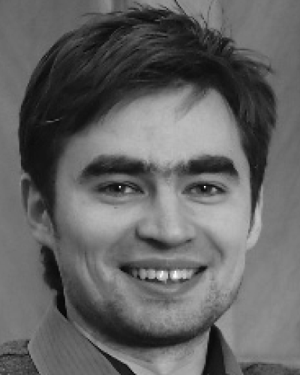 P. V. Yuldashev合作论文数28
 Yuri Pishchalnikov合作论文数Applaud Medical25
 James E. Lingeman合作论文数Indiana Clinic Urology Methodist Professional Center25Vera Khokhlova合作论文数24Frank Starr合作论文数24
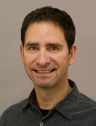 Thomas J. Matula合作论文数Department of Bioengineering, University of Washington24
 Yak-Nam Wang合作论文数24
 John C. Kucewicz合作论文数24
 Neil R. Owen合作论文数23
 Franklin Lee合作论文数21
 Tim Colonius合作论文数Andrew and Peggy Cherng Department of Medical Engineering, Division of Engineering and Applied Science, California Institute of Technology;Department of Mechanical and Civil Engineering, Division of Engineering and Applied Science, California Institute of Technology19Brian MacConaghy合作论文数18
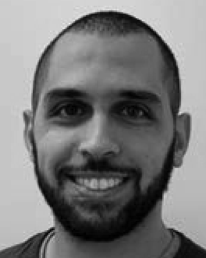 Mohamed Ghanem合作论文数17
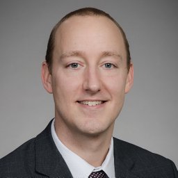 Schade George R合作论文数17Wei Lu合作论文数17Ro Cleveland合作论文数16Anup Shah合作论文数16
 Shahram Vaezy合作论文数University of Washington15
 M. Kennedy Hall合作论文数Department of Emergency Medicine, University of Washington14
 Partanen Ari合作论文数12
 Farr Navid合作论文数Department of Bioengineering, University of Washington12
 Hong Chen (陈红)合作论文数Department of Biomedical Engineering, McKelvey School of Engineering, Washington University in St. Louis12
 Joo Ha Hwang合作论文数Division of General Surgery, Stanford Medicine, Stanford University;Division of Gastroenterology and Hepatology, Department of Medicine, Stanford Medicine, Stanford University12
 Jessica Dai合作论文数11Bret A. Connors合作论文数11




