Fresnel zone Fresnel zone plate plates are the most commonly used optic in x-ray microscopes. Following a short discussion of historical developments, the properties of zone plates are outlined, along with the microscope systems that employ them. A number of applications of x-ray microscopes are then surveyed, including in biology, environmental science, and materials science.
Using an example of Kirkpatrick-Baez mirrors for the micro-diffraction beamline 12.3.2, we present the recent development of bendable x-ray optics and experimental techniques used for focusing of beams of soft and hard x-rays at the Advanced Light Source (ALS). We briefly review the nature of the bending and analyze a generalized solution of the bending equation for a side-profiled elliptically bent mirror substrate. A scaling rule is derived to understand a range of reliable tunability of the bendable optics for different applications (e.g., different focal distances and grazing incidence angles). Original design approaches are developed for assembly of bendable mirrors with minimal spurious stress and misalignment and for final precision compensation of the residual stress, substrate twist, and roll-off misalignment of the mirrors. Procedures used for optimal shaping and alignment of bendable optics at the ALS optical metrology laboratory are also briefly reviewed.
A new facility is described that is being constructed for coherent scattering and microscopy at the Advanced Light Source. The undulator beamline delivers maximum coherent flux to two experiment programs. One is for soft x-ray coherent scattering and correlation studies, the other is for soft x-ray diffractive imaging using ptychographic reconstruction techniques. The energy resolution is sufficient for NEXAFS spectroscopy with full polarization control, from below the carbon edge to 2500eV. New instrumentation is planned for wavelength limited 2D imaging and 3D imaging at spatial resolution better than 10nm and for coherent scattering studies with a variety of sample environments including strong magnetic fields and cryogenic temperatures. High speed CCD detectors will make optimum use of the available flux on dedicated data transmission networks.
In this paper the choice between bending magnets and insertion devices as sample illuminators for a hard X-ray full-field microscope is investigated. An optimized bending-magnet beamline design is presented. Its imaging speed is very competitive with the performance of similar microscopes installed currently at insertion-device beamlines. The fact that imaging X-ray microscopes can accept a large phase space makes them very well suited to the output characteristics of bending magnets which are often a plentiful and paid-for resource. There exist opportunities at all synchrotron light sources to take advantage of this finding to build bending-magnet beamlines that are dedicated to transmission X-ray microscope facilities. It is expected that demand for such facilities will increase as three-dimensional tomography becomes routine and advanced techniques such as mosaic tomography and XANES tomography (taking three-dimensional tomograms at different energies to highlight elemental and chemical differences) become more widespread.
Convenience and cost often lead to synchrotron beamlines where the final bendable Kirkpatrick-Baez focusing pair must relay the final image to different samples at different image distances e.g., [Proc. FEL2009, 246-249 (2009)] either for different experimental chambers, or diagnostics. We present an initial analytical approach, starting from, and extending the work of Howells et al. [OE 39(10), 2748-62 (2000)] to analyze the trade-offs between choice of mirror, bending couples and the given, shaped sagittal width of the optic. Both experimentally and in simulation, we have found that after an appropriate re-bending, sagittally shaped optics can perform with high quality at significantly different incidence angles and conjugate distances. We present one successful demonstration from the ALS Optical Metrology Beamline 5.3.1, and review some new closed form analytical solutions with a view towards understanding our results.
In this paper is demonstrated a complete algorithm for determining the electron density of an individual particle from diffraction patterns of many particles, randomly oriented about a single axis. The algorithm operates on angular correlations among the measured intensity distributions. We also demonstrate the ability to recover the angular correlation functions of a single particle from measured diffraction patterns.
We propose a method for determining the projected electron density of an individual molecule from diffraction patterns of many such simultaneously illuminated molecules, randomly oriented about an axis parallel to an incident x-ray beam. We illustrate the idea with a simulation of a structure determination of a K-channel membrane protein in situ.
X-ray diffraction microscopy (XDM) is a new form of X-ray imaging that is being practiced at several third-generation synchrotron-radiation X-ray facilities. Nine years have elapsed since the technique was first introduced and it has made rapid progress in demonstrating high-resolution three-dimensional imaging and promises few-nanometer resolution with much larger samples than can be imaged in the transmission electron microscope. Both life- and materials-science applications of XDM are intended, and it is expected that the principal limitation to resolution will be radiation damage for life science and the coherent power of available X-ray sources for material science. In this paper we address the question of the role of radiation damage. We use a statistical analysis based on the so-called “dose fractionation theorem” of Hegerl and Hoppe to calculate the dose needed to make an image of a single life-science sample by XDM with a given resolution. We find that the needed dose scales with the inverse fourth power of the resolution and present experimental evidence to support this finding. To determine the maximum tolerable dose we have assembled a number of data taken from the literature plus some measurements of our own which cover ranges of resolution that are not well covered otherwise. The conclusion of this study is that, based on the natural contrast between protein and water and “Rose-criterion” image quality, one should be able to image a frozen-hydrated biological sample using XDM at a resolution of about 10nm.
Symptoms of radiation damage are reviewed, followed by a brief description of the three main damage mechanisms: knock-on displacement (predominant in electrically conducting specimens), ionization damage (radiolysis), and electrostatic charging effects in poorly conducting specimens. Measurements of characteristic dose and damage cross section are considered, together with direct and inverse dose-rate effects. Dose limited resolution is defined in terms of a characteristic dose and instrumental parameters. Damage control is discussed in terms of low-dose technique, choice of imaging mode, specimen temperature, specimen environment and TEM accelerating voltage. We examine the possibility of performing electron cryomicroscopy in STEM mode, with a judicious choice of probe current and probe diameter.
Modern polishing methods of ion-beam milling, and single atom removal techniques are beginning to allow the fabrication of arbitrary surface shapes for reflecting grazing incidence optics. Moreover, the total expense of fabrication, coating, measuring, mounting, aligning, cooling, and surrounding the optic with vacuum make the reduction of optical part count attractive for the latest generation x-ray sources, not even considering potential effects on the scattering and reflective losses of the radiation. These two developments converge to effectively suggest the question of what surface would be the optimally de-magnifying surface to replace a toroid illuminated by a wave cylindrical in the sagittal direction if the sag of the single surface were determined by a function, and not constrained to be a typical optical shape. To address this we derive a simplified case of the formalism of Chrisp, using the classical optical path function approach of Fermat to give a power series calculation of this best surface. This surface, the "diaboloid," would in principle earn its name by its, at least ab initio, consideration of being very difficult to manufacture. We show an example of improvement this surface would provide.
The MISTRAL beamline is one of the seven phase-I beamlines at the ALBA synchrotron light source (Barcelona, Spain) that will be opened to users at the end of 2010. MISTRAL will be devoted to cryotomography in the water window and multi-keV spectral regions for biological applications. The optics design consists of a plane-grating monochromator that has been implemented using variable-line-spacing gratings to fulfil the requirements of X-ray microscopy using a reflective condenser. For instance, a fixed-focus condition independent of the included angle, constant magnification as well as coma and spherical aberration corrections are achieved with this system. The reported design is of wider use.
Ultralow density polymers, metals, and ceramic nanofoams are valued for their high strength-to-weight ratio, high surface area, and insulating properties ascribed to their structural geometry. We obtain the labrynthine internal structure of a tantalum oxide nanofoam by x-ray diffractive imaging. Finite-element analysis from the structure reveals mechanical properties consistent with bulk samples and with a diffusion-limited cluster aggregation model, while excess mass on the nodes discounts the dangling fragments hypothesis of percolation theory.
Integration of the Two-Dimensional Power Spectral Density into Specifications for the X-ray Domain—Problems and Opportunities W. R. McKinney, M. R. Howells, V. V. Yashchuk Lawrence Berkeley National Laboratory, Berkeley, California 94720 An implementation of the two-dimensional statistical scattering theory of Church and Takacs for the prediction of scattering from x-ray mirrors is presented with a graphical user interface. The process of this development has clarified several problems which are of significant interest to the synchrotron community. These problems have been addressed to some extent, for example, for large astronomical telescopes, and at the National Ignition Facility for normal incidence optics, but not in the synchrotron community for grazing incidence optics. Since it is based on the Power Spectral Density (PSD) to provide a description of the deviations from ideal shape of the surface, accurate prediction of the scattering requires an accurate estimation of the PSD. Specifically, the spatial frequency range of measurement must be the correct one for the geometry of use of the optic—including grazing incidence and coherence effects, and the modifications to the PSD of the Optical Transfer Functions (OTF) of the measuring instruments must be removed. A solution for removal of OTF effects has been presented previously, the Binary Pseudo-Random Grating. Typically, the frequency range of a single instrument does not cover the range of interest, requiring the stitching together of PSD estimations. difficulties in two dimensions. This combination generates its own set of Fitting smooth functions to two dimensional PSDs, particularly in the case of spatial non-isotropy of the surface, which is often the case for optics in synchrotron beam lines, can be difficult. The convenient, and physically accurate fractal for one dimension does not readily transfer to two dimensions. Finally, a completely statistical description of scattering must be integrated with a deterministic low spatial frequency component in order to completely model the intensity near the image. An outline for approaching these problems, and our proposed experimental program is given.
Advances in the development of free-electron lasers offer the realistic prospect of nanoscale imaging on the timescale of atomic motions. We identify X-ray Fourier-transform holography 1 , 2 , 3 as a promising but, so far, inefficient scheme to do this. We show that a uniformly redundant array 4 placed next to the sample, multiplies the efficiency of X-ray Fourier transform holography by more than three orders of magnitude, approaching that of a perfect lens, and provides holographic images with both amplitude- and phase-contrast information. The experiments reported here demonstrate this concept by imaging a nano-fabricated object at a synchrotron source, and a bacterial cell with a soft-X-ray free-electron laser, where illumination by a single 15-fs pulse was successfully used in producing the holographic image. As X-ray lasers move to shorter wavelengths we expect to obtain higher spatial resolution ultrafast movies of transient states of matter.
A method is proposed for obtaining three simultaneous projections of a target from a single radiation pulse, which also allows the relative orientation of successive targets to be determined. The method has application to femtosecond x-ray diffraction, and does not require solution of the phase problem. We show that the principal axes of a compact charge-density distribution can be obtained from projections of its autocorrelation function, which is directly accessible in diffraction experiments. The results may have more general application to time resolved tomographic pump-probe experiments and time-series imaging.
An ultra-high resolution beamline is being constructed at the Advanced Light Source (ALS) for the study of low energy excitations in strongly correlated systems with the use of high-resolution inelastic scattering and angle-resolved photoemission. This new beamline, given the acronym Merlin (for meV resolution line), will cover the energy range 10-150 eV. The monochromator has fixed entrance and exit slits and a plane mirror that can illuminate a spherical grating at the required angle of incidence (as in the SX-700 mechanism). The monochromator can be operated in two different modes. In the highest resolution mode, the energy scanning requires translating the monochromator chamber (total travel 1.1 m) as well as rotating the grating and the plane mirror in front of the grating. The resolution in this mode is practically determined by the slits width. In the second mode, the scanning requires rotating the grating and the plane mirror. This mode can be used to scan a few eV without a significant resolution loss. The source for the beamline is a 1.9 in long, 90 nun period quasi periodic EPU. The expected flux at the sample is higher than 10(11) photons/s at a resolving power of 5x10(4) in the energy range 16-130 eV. A second set of gratings can be used to obtain higher flux at the expense of resolution.
The resolution of X-ray diffraction microscopy is limited by the maximum dose that can be delivered prior to sample damage. In the proposed serial crystallography method, the damage problem is addressed by distributing the total dose over many identical hydrated macromolecules running continuously in a single-file train across a continuous X-ray beam, and resolution is then limited only by the available molecular and X-ray fluxes and molecular alignment. Orientation of the diffracting molecules is achieved by laser alignment. The incident X-ray fluence (energy/area) is evaluated that is required to obtain a given resolution from (i) an analytical model, giving the count rate at the maximum scattering angle for a model protein, (ii) explicit simulation of diffraction patterns for a GroEL-GroES protein complex, and (iii) the spatial frequency cut-off of the transfer function following iterative solution of the phase problem, and reconstruction of an electron density map in the projection approximation. These calculations include counting shot noise and multiple starts of the phasing algorithm. The results indicate counting time and the number of proteins needed within the beam at any instant for a given resolution and X-ray flux. An inverse fourth-power dependence of exposure time on resolution is confirmed, with important implications for all coherent X-ray imaging. It is found that multiple single-file protein beams will be needed for sub-nanometer resolution on current third-generation synchrotrons, but not on fourth-generation designs, where reconstruction of secondary protein structure at a resolution of 7 A should be possible with relatively short exposures.
 Chris Jacobsen合作论文数Department of Physics & Astronomy, Weinberg College of Arts & Sciences, Northwestern University50
 Janos Kirz合作论文数Physics Department, SUNY40
 J. C. H. Spence合作论文数Physics Department,Arizona State University27
 Henry Chapman合作论文数Coherent X-Ray Imaging Division, Center for Free-Electron Laser Science, Deutsches Elektronen-Synchrotron;University of Hamburg25
 Stefano Marchesini合作论文数SLAC National Accelerator Laboratory, Stanford University24
 Uwe Weierstall合作论文数Department of Physics, Arizona State University24
 Howard Padmore合作论文数Lawrence Berkeley National Laboratory21
 Ian Mcnulty合作论文数Argonne National Laboratory17
 Wayne Mckinney合作论文数Advanced Light Source, Lawrence Berkeley National Laboratory17
 S. Lindaas合作论文数16H. Rarback合作论文数15H. He合作论文数13David Sayre合作论文数12
 Zahid Hussain合作论文数Department of Material Physics, Materials Sciences Division, Lawrence Berkeley National Laboratory11
 Edward J. Moler合作论文数Tethys BioScience, Inc11Tony Warwick合作论文数11
 Harald Ade合作论文数Department of Physics and Astronomy, College of Sciences, North Carolina State University10
 Alastair MacDowell合作论文数Lawrence Berkeley National Laboratory10
 Chris Buckley合作论文数Ge Healthcare UK Limited;GE Healthcare9D. Shapiro合作论文数8JCH Spence合作论文数Arizona State Univ, Dept Phys, Tempe, AZ 85287 USA8
 Peter Z. Takacs合作论文数Brookhaven National Laboratory8
 Alex Noy合作论文数Lawrence Livermore National Laboratory;School of Natural Sciences, University of California Merced8
 Anton Barty合作论文数Center for Data and Computing in Natural Sciences, Universitat Hamburg8C. Cui合作论文数7
 Wenbing Yun合作论文数Sigray, Inc.7
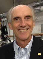 Gwyn P. Williams合作论文数Triseka7
 Dong Shu (舒东)合作论文数School of Chemistry, South China Normal University7Stefan P. Hau-Riege合作论文数7Tobias Beetz合作论文数Department of Physics and Astronomy;SUNY Stony Brook;Department of Physics and Astronomy, SUNY Stony Brook7
 Stephen Rothman合作论文数7r digennaro合作论文数6
 David A. Shirley合作论文数Pennsylvania State University6
 Erik H. Anderson合作论文数6
 Kevin Schmidt合作论文数Department of Physics, Arizona State University6Sue Wirick合作论文数6
 Valeriy V. Yashchuk合作论文数Lawrence Berkeley National Laboratory6H. Hogrefe合作论文数6S. Irick合作论文数6Steven C. Irick合作论文数6S. P. Hau-Riege合作论文数5E. Lima合作论文数5Steven L Hulbert合作论文数5Dmitri Starodub合作论文数Department of Physics;Arizona State University5Robert Duarte合作论文数5S. Williams合作论文数4RM Duarte合作论文数4j chin合作论文数4




