In eukaryotes, the separation of transcription from translation enables extensive mRNA processing (capping, splicing, and polyadenylation) before translation. This review focuses on recent work that provides considerable insight into how mRNAs navigate these processes in which a spectrum of RNA-binding proteins (RBPs) coordinate different processing steps and couple them to nuclear export. Although the principal components in these pathways have been identified, the precise way in which RBPs bind to mRNAs, some aspects of how their binding and release are mediated by DEAD-box ATPases, and the complete structures of some messenger ribonucleoprotein complexes (mRNPs) remain unclear. Moreover, the checkpoints that recognize both completion of mRNA processing and the generation of mature mRNPs, as well as how they are coordinated, are only partially characterized.
The rabies virus (RABV) phosphoprotein (P protein) has multiple functions, including acting as the essential non-catalytic cofactor of the viral polymerase (L protein) for genome replication and transcription; the principal viral antagonist of the interferon (IFN)-mediated innate immune response; and the chaperone for the viral nucleoprotein (N protein). Although P protein is known to undergo phosphorylation by cellular kinases, the location and functions of the phosphorylation sites remains poorly defined. Here, we report the identification by mass-spectrometry (MS) of residues of P protein that are modified by phosphorylation in mammalian cells, including several novel sites. Analysis of P protein with phospho-mimetic and phospho-inhibitory mutations of three novel residues/clusters that were commonly identified by MS (Ser48, Ser183/187, Ser217/219/220) indicate that phosphorylation at each of these sites does not have a major influence on nuclear trafficking or antagonistic functions toward IFN signalling pathways. However, phosphorylation of Ser48 in the N-terminus of P protein impaired function in transcription/replication and in the formation of replication structures that contain complexes of P and N proteins, suggestive of altered interactions of these proteins. The crystal structure of P protein containing the S48E phospho-mimetic mutation indicates that Ser48 phosphorylation facilitates the binding of residues 41–52 of P protein into the RNA-binding groove of non-RNA-bound N protein (N0), primarily through the formation of a salt bridge with Arg434 of N protein. These data indicate that Ser48 modification regulates the cycling of P-N0 chaperone complexes that deliver N protein to RNA to enable transcription/replication, such that enhanced interaction due to S48E phospho-mimetic mutation reduces N protein delivery to the RNA, inhibiting subsequent transcription/replication processes. These data are, to our knowledge, the first to implicate phosphorylation of RABV P protein in conserved replication functions of the P gene.
Andrew McLachlan enjoyed a wide-ranging career, starting as a theoretical chemist and ending as a molecular biologist. After reading physics as an undergraduate at Trinity College, Cambridge, he joined the Theoretical Chemistry Section of the Chemistry Department to study for a PhD. His work on electron spin resonance then led to his election as a research fellow at Trinity. Following postdoctoral work in the United States, he returned to Cambridge as a college lecturer in physics at Trinity and was eventually appointed a university lecturer in chemistry. Despite a productive research programme in various aspects of theoretical chemistry, he decided that a move into the newly developing subject of molecular biology would provide an exciting opportunity. Accordingly, in 1967 he accepted an offer to join the Medical Research Council Laboratory of Molecular Biology. The initial plan was to work on the mechanism of oxygen uptake by haemoglobin, but he quickly became interested in wider problems of protein structure. In particular, he developed methods for aligning the sequences of different proteins and for detecting repeated sequences and motifs, leading to an interest in the evolution of protein sequences. In collaboration with others, he applied his methods to a wide range of proteins, providing important understanding of their structures and relationships.
Australian bat lyssavirus (ABLV) shows similar clinical symptoms as rabies, but there are currently no protein structures available for ABLV proteins. In lyssaviruses, the interaction between nucleoprotein (N) and phosphoprotein (N) in the absence of RNA generates a complex (N0P) that is crucial for viral assembly, and understanding the interface between these two proteins has the potential to provide insight into a key feature: the viral lifecycle. In this study, we used recombinant chimeric protein expression and X-ray crystallography to determine the structure of ABLV nucleoprotein bound to residues 1–40 of its phosphoprotein chaperone. Comparison of our results with the recently generated structure of RABV CVS-11 N0P demonstrated a highly conserved interface in this complex. Because the N0P interface is conserved in the lyssaviruses of phylogroup I, it is an attractive therapeutic target for multiple rabies-causing viral species.
Although the separation of transcription and translation, mediated by the nuclear envelope, is the defining characteristic of Eukaryotes, the barrier between the nuclear and cytoplasmic compartments needs to be semipermeable to enable material to be moved between them. Moreover, each compartment needs to have a distinctive complement of macromolecules to mediate specific functions and so movement between them needs to be controlled. This is achieved through the selective active transport of macromolecules through the nuclear pores that stud the nuclear envelope, and which serve as a conduit between these compartments. Nuclear pores are huge cylindrical macromolecular assemblies and are constructed from the order of 30 different proteins called nucleoporins. Nuclear pores have a central transport channel that is filled with a dense network of natively unfolded portions of many different nuclear pore proteins (nucleoporins or nups). This network generates a barrier that impedes, but does not entirely prevent, the diffusion of many macromolecules through the pores. The rapid movement of a range of proteins and RNAs through the pores is mediated by a range of transport factors that bind their cargo in one compartment and release it in the other. However, although as their size increases the diffusion of macromolecules through nuclear pores is progressively impaired, additional mechanisms, including the binding of some macromolecules to immobile components of each compartment and also the active removal of macromolecules from the inappropriate compartment, are needed to fully maintain the distinctive compositions of each compartment.
Human respiratory syncytial virus (RSV) causes severe respiratory illness in children and the elderly. Treatments for RSV disease are however limited and efforts to produce an effective vaccine have so far been unsuccessful. Understanding RSV virion structure is an important prerequisite for developing interventions to treat or prevent infection but has been challenging because of the fragility of virions propagated in cell culture. Here we show, using cryogenic electron microscopy (cryoEM) and cryogenic electron tomography (cryoET) of RSV particles cultivated directly on transmission electron microscopy (TEM) grids, that there is extensive helical symmetry in RSV filamentous virions. We have calculated a 16 Å resolution three-dimensional reconstruction of the viral envelope, targeting the matrix protein (M) that forms an endoskeleton below the viral membrane. These data define a helical lattice of M proteins, showing how M is oriented relative to the viral envelope and that helical ordering of viral glycoproteins that stud the viral envelope is coordinated by the M layer. Moreover, the helically ordered viral glycoproteins in RSV filamentous virions cluster in pairs, which may have implications for the conformation of fusion (F) glycoprotein epitopes that are the principal target for vaccine and monoclonal antibody development. We also report the presence, in authentic virus infections, of N-RNA rings packaged within RSV filamentous virions. Overall, the structural data obtained provides molecular insight into the organization of the virion and the mechanism of its assembly.
The MERS coronavirus (MERS-CoV) is a highly pathogenic, emerging virus that produces accessory proteins to antagonize the host innate immune response. The MERS-CoV ORF4b protein has been shown to bind preferentially to the nuclear import adapter IMPα3 in infected cells, thereby inhibiting NF-κB-dependent innate immune responses. Here, we report high-resolution structures of ORF4b bound to two distinct IMPα family members. Each exhibit highly similar binding mechanisms that, in both cases, lack a prototypical Lys bound at their P2 site. Mutations within the NLS region dramatically alter the mechanism of binding, which reverts to the canonical P2 Lys binding mechanism. Mutational studies confirm that the novel binding mechanism is important for its nuclear import, IMPα interaction, and inhibition of innate immune signaling pathways. In parallel, we determined structures of the nuclear binding domain of NF-κB component p50 bound to both IMPα2 and α3, demonstrating that p50 overlaps with the ORF4b binding sites, suggesting a basis for inhibition. Our results provide a detailed structural basis that explains how a virus can target the IMPα nuclear import adapter to impair immunity, and illustrate how small mutations in ORF4b, like those found in closely related coronaviruses such as HKU5, change the IMPα binding mechanism.
Human respiratory syncytial virus (RSV) causes severe respiratory illness in children and the elderly. Here, using cryogenic electron microscopy and tomography combined with computational image analysis and three‐dimensional reconstruction, we show that there is extensive helical ordering of the envelope‐associated proteins and glycoproteins of RSV filamentous virions. We calculated a 16 Å resolution sub‐tomogram average of the matrix protein (M) layer that forms an endoskeleton below the viral envelope. These data define a helical lattice of M‐dimers, showing how M is oriented relative to the viral envelope. Glycoproteins that stud the viral envelope were also found to be helically ordered, a property that was coordinated by the M‐layer. Furthermore, envelope glycoproteins clustered in pairs, a feature that may have implications for the conformation of fusion (F) glycoprotein epitopes that are the principal target for vaccine and monoclonal antibody development. We also report the presence, in authentic virus infections, of N‐RNA rings packaged within RSV virions. These data provide molecular insight into the organisation of the virion and the mechanism of its assembly. CryoEM and tomography analysis of the human pathogen respiratory syncytial virus shows the formation of filamentous virions that exhibit helical ordering of the envelope associated matrix protein layer, which in turn coordinates helical ordering and clustering of viral glycoproteins. CryoEM and tomography analysis of the human pathogen RSV shows the formation of filamentous virions that exhibit helical ordering of the envelope associated matrix protein layer that in turn coordinates helical ordering and clustering of viral glycoproteins.
DNA nanotechnology provides a versatile and powerful tool to dissect the structure-function relationship of biomolecular machines like the nuclear pore complex (NPC), an enormous protein assembly that controls molecular traffic between the nucleus and cytoplasm. To understand how the intrinsically disordered, Phe-Gly-rich nucleoporins (FG-nups) within the NPC's central transport channel impede the diffusion of macromolecules, we built a DNA-origami NanoTrap. The NanoTrap comprises precisely arranged FG-nups in an NPC-like channel, which sits on a baseplate that captures macromolecules that pass through the FG network. Using this biomimetic construct, we determined that the FG-motif type, grafting density and spatial arrangement are critical determinants of an effective diffusion barrier. Further, we observe that diffusion barriers formed with cohesive FG-interactions dominate in mixed-FG-nup scenarios. Our DNA-origami platform thus sheds light on how NPCs sieve inert macromolecules and will provide a valuable tool for studying nuclear transport.
SOX (SRY-related HMG-box) transcription factors perform critical functions in development and cell differentiation. These roles depend on precise nuclear trafficking, with mutations in the nuclear targeting regions causing developmental diseases and a range of cancers. SOX protein nuclear localization is proposed to be mediated by two nuclear localization signals (NLSs) positioned within the extremities of the DNA-binding HMG-box domain and, although mutations within either cause disease, the mechanistic basis has remained unclear. Unexpectedly, we find here that these two distantly positioned NLSs of SOX2 contribute to a contiguous interface spanning 9 of the 10 ARM domains on the nuclear import adapter IMPα3. We identify key binding determinants and show this interface is critical for neural stem cell maintenance and for Drosophila development. Moreover, we identify a structural basis for the preference of SOX2 binding to IMPα3. In addition to defining the structural basis for SOX protein localization, these results provide a platform for understanding how mutations and post-translational modifications within these regions may modulate nuclear localization and result in clinical disease, and also how other proteins containing multiple NLSs may bind IMPα through an extended recognition interface.
The importin α/β transport machinery mediates the nuclear import of cargo proteins that bear a classical nuclear localization sequence (cNLS). These cargo proteins are linked to the major nuclear protein import factor, importin-β, by the importin-α adapter, after which cargo/carrier complexes enter the nucleus through nuclear pores. In the nucleus, cargo is released by the action of RanGTP and the nuclear pore protein Nup2, after which the importins are recycled to the cytoplasm for further transport cycles. The nuclear export of importin-α is mediated by Cse1/CAS. Here, we exploit structures of functionally important complexes to identify residues that are critical for these interactions and provide insight into how cycles of protein import and recycling of importin-α occur in vivo using a Saccharomyces cerevisiae model. We examine how these molecular interactions impact protein localization, cargo import, function and complex formation. We show that reversing the charge of key residues in importin-α (Arg44) or Cse1 (Asp220) results in loss of function of the respective proteins and impairs complex formation both in vitro and in vivo. To extend these results, we show that basic residues in the Nup2 N-terminus are required for both Nup2 interaction with importin-α and Nup2 function. These results provide a more comprehensive mechanistic model of how Cse1, RanGTP and Nup2 function in concert to mediate cNLS-cargo release in the nucleus.
Defects in the mRNA export scaffold protein GANP, encoded by the MCM3AP gene, cause autosomal recessive early-onset peripheral neuropathy with or without intellectual disability. We extend here the phenotypic range associated with MCM3AP variants, by describing a severely hypotonic child and a sibling pair with a progressive encephalopathic syndrome. In addition, our analysis of skin fibroblasts from affected individuals from seven unrelated families indicates that disease variants result in depletion of GANP except when they alter critical residues in the Sac3 mRNA binding domain. GANP depletion was associated with more severe phenotypes compared with the Sac3 variants. Patient fibroblasts showed transcriptome alterations that suggested intron content-dependent regulation of gene expression. For example, all differentially expressed intronless genes were downregulated, including ATXN7L3B, which couples mRNA export to transcription activation by association with the TREX-2 and SAGA complexes. Our results provide insight into the molecular basis behind genotype-phenotype correlations in MCM3AP-associated disease and suggest mechanisms by which GANP defects might alter RNA metabolism.
The poly(A) RNA binding Zn finger ribonucleoprotein Nab2 functions to control the length of 3' poly(A) tails in Saccharomyces cerevisiae as well as contributing to the integration of the nuclear export of mature mRNA with preceding steps in the nuclear phase of the gene expression pathway. Nab2 is constructed from an N-terminal PWI-fold domain, followed by QQQP and RGG motifs and then seven CCCH Zn fingers. The nuclear pore-associated proteins Gfd1 and Mlp1 bind to opposite sides of the Nab2 N-terminal domain and function in the nuclear export of mRNA, whereas the Zn fingers, especially fingers 5-7, bind to A-rich regions of mature transcripts and function to regulate poly(A) tail length as well as mRNA compaction prior to nuclear export. Nab2 Zn fingers 5-7 have a defined spatial arrangement, with fingers 5 and 7 arranged on one side of the cluster and finger 6 on the other side. This spatial arrangement facilitates the dimerization of Nab2 when bound to adenine-rich RNAs and regulates both the termination of 3' polyadenylation and transcript compaction. Nab2 also functions to coordinate steps in the nuclear phase of the gene expression pathway, such as splicing and polyadenylation, with the generation of mature mRNA and its nuclear export. Nab2 orthologues in higher Eukaryotes have similar domain structures and play roles associated with the regulation of splicing and polyadenylation. Importantly, mutations in the gene encoding the human Nab2 orthologue ZC3H14 and cause intellectual disability.
In eukaryotes, the separation of translation from transcription by the nuclear envelope enables mRNA modifications such as capping, splicing, and polyadenylation. These modifications are mediated by a spectrum of ribonuclear proteins that associate with preRNA transcripts, coordinating the different steps and coupling them to nuclear export, ensuring that only mature transcripts reach the cytoplasmic translation machinery. Although the components of this machinery have been identified and considerable functional insight has been achieved, a number of questions remain outstanding about mRNA nuclear export and how it is integrated into the nuclear phase of the gene expression pathway. Nuclear export factors mediate mRNA transit through nuclear pores to the cytoplasm, after which these factors are removed from the mRNA, preventing transcripts from returning to the nucleus. However, as outlined in this review, several aspects of the mechanism by which transport factor binding and release are mediated remain unclear, as are the roles of accessory nuclear components in these processes. Moreover, the mechanisms by which completion of mRNA splicing and polyadenylation are recognized, together with how they are coordinated with nuclear export, also remain only partially characterized. One attractive hypothesis is that dissociating poly(A) polymerase from the cleavage and polyadenylation machinery could signal completion of mRNA maturation and thereby provide a mechanism for initiating nuclear export. The impressive array of genetic, molecular, cellular, and structural data that has been generated about these systems now provides many of the tools needed to define the precise mechanisms involved in these processes and how they are integrated.
The Three prime repair exonuclease 2 (TREX-2) complex functions as a platform to which many of the components of the nuclear mRNA processing machinery bind, facilitating integration of this phase of the gene expression pathway, as well as mediating the re-positioning of highly regulated actively transcribing genes (such as GAL1) to nuclear pores (NPCs) to accelerate their activation. In Saccharomyces cerevisiae the TREX-2 complex is based on a Sac3 scaffold to which Thp1, Sem1, Cdc31 and two Sus1 chains are bound. A combination of X-ray crystallography and electron microscopy studies have established the structure of two major regions of this complex: the M-region that functions to bind nucleic acids and the CID region that functions to link the complex to nuclear pores. These structures have facilitated the engineering of mutants that have been used to define the contributions made by the TREX-2 complex to locating high-expressed genes to nuclear pores and the contributions made to mRNA nuclear export.
 Anita Corbett合作论文数Department of Biology, College of Arts and Sciences, Emory University27
 Roberts Thomas M合作论文数27Helen M. Kent合作论文数22
 Airlie Mccoy合作论文数Cambridge Institute for Medical Research, University of Cambridge14David Neuhaus合作论文数13Rw Kensler合作论文数12
 Eugene Valkov合作论文数Centre for Infectious Disease Research, University of Queensland11Andrew D Mclachlan合作论文数10
 Richard Bayliss合作论文数MRC Laboratory of Molecular Biology, Hills Rd, Cambridge CB2 2QH, England UK10LL LeClaire合作论文数9
 Ed Hurt合作论文数Biochemistry Center, Heidelberg University9
 Shintaro Aibara合作论文数Stockholm Univ, Dept Biochem & Biophys, Sci Life Lab, Solna, Sweden9
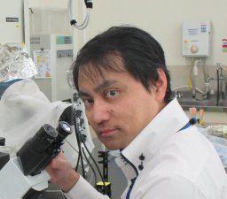 Katsuya Shimabukuro合作论文数Department of Biological Science, Florida State University9Angelika A. Noegel合作论文数8Richard P. Grant合作论文数MRC Laboratory of Molecular Biology, Hills Rd., Cambridge CB2 2QH, UK8WD Clarkson合作论文数7
 B. Booth Quimby合作论文数7Mt Harreman合作论文数7
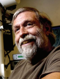 Terrance J. Beveridge合作论文数Department of Molecular and Cellular Biology, College of Biological Science, University of Guelph7
 Jade Forwood合作论文数Gulbali Institute, Charles Sturt University7Noda Naoki合作论文数7
 Fasken Milo B合作论文数Department of Biochemistry, Emory University School of Medicine7
 Roy Quinlan合作论文数Department of Biosciences, The University of Durham7KL KING合作论文数6IK BUCKLEY合作论文数6Je Italiano合作论文数6Jani Divyang合作论文数6Masasuke Yoshida合作论文数Kyoto Sangyo University6
 Paola Fucini合作论文数6
 Trichur R Raju合作论文数Department of Neurophysiology, National Institute of Mental Health and Neurosciences6S WHYTOCK合作论文数6Tl Bullock合作论文数5RD Moir合作论文数5Rjc Levine合作论文数5AME Baker合作论文数5Michael Schleicher合作论文数5
 Takeyuki Wakabayashi合作论文数5Sj Atkinson合作论文数4
 Ellisdon Andrew M合作论文数Monash Biomedicine Discovery Institute, Monash University4
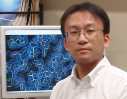 Matsuura Yoshiyuki合作论文数名古屋大学大学院4Neil J. Marshall合作论文数4
 Stephen H McLaughlin合作论文数School of Biological Sciences, University of Manchester4L. S. Miao合作论文数4Alwin Köhler合作论文数4
 Catherine Dargemont合作论文数4
 Trevor Littlewood合作论文数Department of Biochemistry, University of Cambridge4
 Allison Lange合作论文数4
 Avril V. Somlyo合作论文数Department of Molecular Physiology and Biological Physics, School of Medicine, University of Virginia4A.M.E. Baker合作论文数3




