BACKGROUND:A severely subluxated crystalline lens refers to significant displacement of the eye's natural lens from its normal position, often due to zonular weakness or rupture.[1] This condition can result from trauma, connective tissue disorders like Marfan syndrome, or advanced pseudoexfoliation.[2,3] Visual symptoms include blurred vision, monocular diplopia, and glare. On examination, lens edge may be visible within the pupil, and phacodonesis (lens wobbling) may be noted. Severe subluxation increases the risk of complications such as angle-closure glaucoma, lens-induced uveitis, or retinal detachment. Management typically involves surgical removal of the lens, with or without intraocular lens implantation, depending on zonular support.[4] Sinha et al.[5] had described the intralenticular bimanual irrigation and aspiration in 2005 for a sublaxated lens in Marfan syndrome. Khokhar et al.[6] described the modified technique of endocapsular lens aspiration for severely subluxated lenses in 2018. In 2022, Urkude et al.[7] described the "Kissing microvitreoretinal (MVR) blade technique" for microspherophakia. The surgical challenges associated with such clinical conditions are as follows:[4,8] Poor capsular support makes the capsulorhexis difficult. Lens instability: The lens-bag complex is mobile and making the surgical process of lens aspiration extremely hard. There is always a possibility of lenticular matter or the capsular bag falling into the vitreous cavity. PURPOSE:The authors describe "Best Pick and Hold" with "Wrap and Roll" technique for extreme subluxated lens. These novel techniques can be used to perform lens aspiration with ease and negate the possibility of previously described complications. SYNOPSIS:The procedure starts with inserting two MVR blades through clear corneal incisions into the anterior chamber. Clear corneal incisions are preferred as technical challenges are anticipated in pediatric eyes, which have lower scleral rigidity, smaller axial lengths, thinner sclera, and more elastic tissues, making scleral tunnel construction and wound closure more difficult compared to adults. The first MVR blade is precisely advanced to enter the lens capsule and pick the subluxated lens to the central visual axis, followed by the entry of the second MVR blade at the opposite points, creating stable access. The surgeon then introduces a vitrectomy cutter and irrigation cannula, sequentially exchanging them for the MVRs. This strategic maneuver, referred to as the "Best Pick and Hold" technique, allows secure stabilization and controlled intralenticular lens aspiration while minimizing early stress to residual zonules. An MVR entry can also stabilize the lens; however, there is a risk of extension and cut-through caused by the sharp edge of the MVR. Hand-to-hand holding with the vitrectomy cutter and irrigation cannula provides better hold and stabilization of the lens. In the second phase, the capsule is wrapped over the irrigation cannula by using a vitrectomy probe, and the vitrectomy cutter is employed to excise it, a technique called "Wrap and Roll." This procedure helps to reduce capsular bag remnants falling into the vitreous cavity. HIGHLIGHTS:"Best Pick and Hold" with "Wrap and Roll" can be used in the management of severely sublaxated crystalline lens. VIDEO LINK:https://youtu.be/G0LgweQT0d0.
Purpose: Comparative evaluation of the corneal biomechanical parameters by the dynamic corneal response imaging with CORVIS-ST ST in normal and post-keratoplasty eyes. Methods: Cross-sectional observational study of CORVIS-ST imaging of 150 normal corneas and 150 post-keratoplasty corneas was done to evaluate corneal biomechanics of normal Asian-Indian corneas and post-keratoplasty corneas. Data on demographic details, postoperative period, visual acuity (UCVA, BCVA) (logmar units), refraction, axial length (D), corneal topography Scheimpflug’s imaging with Pentacam [Anterior-K1(AK1), Anterior-K2 (AK2), Posterior-K1(PK1), PosteriorK2(PK2), Mean keratometry (Kmean), Keratometry maximum (Kmax), Q-value, D-value, Thinnest pachymetry (TP), Corneal elevation [anterior (CE ant) and posterior (CE post)], CORVIS-ST imaging (bIOP, 1st and 2nd Appl time HCT, 1st Appl length, DA, TBI, CBI, HC rad, Vel in and out, DA ratio, IR, Inv CR, SSI, SPA1, ARTh) were recorded. Results: Comparative evaluation of CORVIS-ST in 150 normal corneas [mean age 32.54 ± 10 (9–68) years] and 150 post-keratoplasty corneas [mean age 35.14 ± 16.96 (8–72) years; PK (58), DSAEK (29), DALK (58), HALK (8), and SALK (3)] noted 18 parameters (bIOP, 1st and 2nd Appl time HCT, 1st Appl length, DA, TBI, CBI, HC rad, Vel in and out, DA ratio, IR, Inv CR, SSI, SPA1, ARTh) and showed that 12 of these were statistically different in post-keratoplasty corneas. Parameters of DA, TBI, CBI, IR, and Inv CR were higher, whereas HC rad, SSI, SPA1, and ARTh showed significantly lesser values in post-keratoplasty corneas as compared to normal corneas. Subgroup analysis (PK, DSAEK, DALK, HALK) showed eyes that underwent DSAEK had biomechanical values closest to that of normal corneas. Post-DALK in comparison to post-PK corneas showed significantly higher values in CBI and IR, and lower values in HC rad, SSI, and ARTh (0.87 ± 0.17, 9.9 ± 3.5, 0.85 ± 0.25, 190.92 ± 119.06 and 0.71 ± 0.25, 8.19 ± 1.92, 1.02 ± 0.38, and 340.90 ± 178.37, respectively). Correlational analysis showed significant mild positive correlation of CBI with corneal curvature in normal and post-PK corneas (P = 0.004 and 0.040, respectively). Conclusion: Post-keratoplasty corneas were seen to have lesser biomechanical strength as compared to normal corneas. Post-DSAEK corneas had a better biomechanical strength than other groups and closest biomechanics to normal corneas. Post-DALK in comparison to post-PK corneas showed a lower biomechanical strength in our study.
BACKGROUND:Chemical injuries to the eye are a significant cause of vision impairment worldwide. These injuries demand immediate and appropriate intervention due to their potential for causing visual morbidity and long-term sequelae. The severity and managmenet is influenced by factors such as the chemical type, concentration, duration of exposure, and the extent of ocular surface involvement. Chemical injuries to the eye are a significant cause of vision impairment worldwide. These injuries demand immediate and appropriate intervention due to their potential for causing visual morbidity and long-term sequelae. The severity and managmenet is influenced by factors such as the chemical type, concentration, duration of exposure, and the extent of ocular surface involvement. PURPOSE:To provide a comprehensive overview of current perspectives in the classification and management of ocular chemical injuries, with emphasis on recent advances in treatment protocols during both the acute and chronic phases. METHODS:After thorough literature search in PubMed and MEDLINE 72 studies with maximum relevance that were published as systematic reviews, as well as randomized and non-randomized comparative studies (cohort or case series) on the topic of chemical injuries classification and management were finally selected for this article. RESULTS:Early management is centered on immediate irrigation, neutralization of the chemical agent, and prevention of further tissue damage through clinical assessment and medical therapy. In severe cases, surgical intervention may be necessary to restore ocular integrity. In the chronic phase, once the ocular surface is stabilized, visual rehabilitation becomes the focus, involving a multifactorial decision-making approach tailored to individual patient needs. CONCLUSION:Effective management of ocular chemical injuries requires a dynamic, phased approach, integrating timely acute intervention with personalized long-term rehabilitation strategies. Recent advancements in therapeutic techniques have improved outcomes, but ongoing research and clinical vigilance remain essential for optimizing care.
PURPOSE:The aim of this study was to evaluate the role of topical human IgG eye drops (4 mg/mL) as adjunct therapy in eyes with moderate to severe refractory dry eye disease (DED). METHODOLOGY:A prospective, longitudinal, open label, dual cohort study of patients with chronic DED of moderate to severe grade comprise of OSIG therapy group cohort on topical immunoglobulin IgG 0.4% four times daily, as adjunctive therapy for one year, to study the role of OSIG therapy in comparison with conventional treatment. Patients above 18 years of age with chronic moderate to severe grade DED with tear break-up time (TBUT) ≤7 seconds, Schirmer test ≤9 mm/5 minutes, ocular surface disease index (OSDI) score ≥13, National Eye Institute (NEI) corneal staining score >3, and conjunctival staining score >3 were recruited into the study. Study participants in the conventional treatment cohort group were on topical lubricants four hourly (carboxymethyl cellulose 0.5% and lubricant ointment at bedtime), topical steroids (prednisolone 1%/fluorometholone 0.1%/loteprednol 0.5%) therapy as per the severity of the DED requirement and topical immunomodulators (cyclosporine A 0.1% eye drops or tacrolimus 0.1% eye ointment) twice daily. Study participants in the OSIG treatment cohort group were started on topical human immunoglobulin IgG 0.4% four times daily as adjunct therapy in addition to the all the above-mentioned medications for a period of 12 months. Study parameters included DED severity level assessment, NIH score, ocular surface evaluation tests, Schirmer's I test, TBUT, corneal and conjunctival staining score, OSDI score, and conjunctival hyperemia score. Ocular surface imaging using LipiView and ocular surface analyzer imaging for non-invasive TBUT (NITBUT), meibomian gland, and tear imaging was performed for the OSIG treatment cohort. Study characteristics were evaluated at time of recruitment into the study (baseline visit) and at serial follow-up of months 3, 6, and 12 in OSIG treatment group and follow-up at month 6 in conventional treatment group. Treatment grading score was devised in accordance to the frequency of application of lubricants and steroids and potency of steroids. RESULTS:Our study of 71 eyes of 36 patients [mean age 39 ± 11.80 years] in OSIG treatment cohort group and 64 eyes of 32 patients [mean age 40 ± 11.53 years] in conventional treatment cohort group observed a significant reduction in the objective ocular surface symptom assessment parameters of OSDI scores, and dry eye severity level with improvement in clinical assessment parameters of TBUT, ocular surface staining scores, and conjunctival hyperemia grades in eyes on adjunct OSIG treatment cohort as compared to the conventional therapy cohort. The frequency of topical lubricants and steroids along with the potency of steroids used showed a decreasing trend with 45.28% noted to have become free of steroid dependence at the end of 12 months of OSIG therapy. Adjunct topical IgG therapy also enabled the shifting of 37.14% of eyes from higher to lower potency steroids by the end of 12 months of treatment with OSIG therapy. The frequency of administration of lubricants decreased in 83.33% of eyes 12 months follow-up. The success rate determined based on improvement in ocular surface evaluation was 75.76%, treatment scores improved in 74.98%, and symptoms evaluation (OSDI) was 75.76% in the OSIG therapy cohort. CONCLUSION:Topical immunoglobulin IgG 0.4% as adjunct therapy seems to have a definitive role in reducing topical steroid and lubricant dependency in moderate to severe chronic DED compared to conventional therapy.
PURPOSE:To compare the clinical response of patients with moderate-to-severe dry eye disease to treatment with 0.05% cyclosporine eye drops as an aqueous solution or oil emulsion. STUDY DESIGN:Prospective randomized clinical trial. METHODS:An institutional study where 88 patients with moderate-to-severe dry eye was enrolled after written informed consent and randomized to receive either aqueous solution (Group 1) or oil emulsion (Group 2) 0.05% cyclosporine eye drops in twice daily dosing in addition to lubricant eyedrop 0.5% carboxymethylcellulose six times/day in both eyes. Comprehensive eye examination and baseline parameters were recorded, and clinical assessment was repeated at 4, 8, and 12 weeks. Parameters evaluated included ocular surface disease index score (OSDI), Schirmer test, tear break-up time (TBUT), corneal fluorescein staining - National Eye Institute (NEI) scoring, lipid layer thickness (LLT), tear meniscus height (TMH), non-invasive tear break-up time (NIBUT), percentage loss of meibomian glands, and impression cytology (number of goblet cells/hpf). RESULTS:The mean age was 39 ± 15.6 years and 42 ± 17.7 years, and the M:F ratio was 26:19 and 20:24 in Groups 1 and 2, respectively. Both the groups showed comparable values at baseline. After treatment, there was statistically significant improvement over baseline values in both groups at 4, 8, and 12 weeks. At 12 weeks for the following parameters, there was no significant difference between the groups, though there was an improvement over baseline in Groups 1 and 2, respectively, as follows: OSDI score - 30.89, 33.28 (P < 0.001), Schirmer test - 5 mm, 4 mm (P < 0.001), TBUT - 2.65 sec, 3.07 sec (P < 0.001), NEI score - 2, 2 (P < 0.001) and the number of goblet cells/hpf - 1.5,9 (P = 0016, P = 0.001). A higher number of patients, by a value of 9, in Group 2 showed an increase in the number of goblet cells/hpf. The NIBUT value showed statistically significant improvement in Group 2 compared to Group 1 (P = 0.011). Group 2 also showed statistically significant improvement in TMH by the 8th week (P = 0.015) and in LLT by the 12th week (P < 0.001). Group 1 comparatively showed earlier improvement in LLT by the 4th week (P = 0.027). CONCLUSION:Both aqueous solution and oil emulsion 0.05% cyclosporine formulations appear to be equally effective in the management of dry eye disease with a comparatively better response with oil emulsion formulations.
Refractive kerato-lenticule extraction surgery (KLex), also known as "small incision lenticule extraction (SMILE)," or "smooth incision keratomileusis" (SILK), represents a significant advancement in refractive surgery with its minimally invasive approach and impressive success rates. Despite these advancements, postoperative infectious keratitis, though rare, poses a critical challenge and profoundly impacts visual outcomes. Unlike infections following surface ablation procedures, which benefit from direct drug penetration into the site of infection, KLex and LASIK face hurdles due to the depth of infection location. In laser-assisted in situ keratomileusis (LASIK), infections begin at the interface between the flap and the stromal bed, whereas in KLex, they occur within the stromal bed. This position of the infiltrate poses the challenge of inadequate drug penetration, making management more complex. This review explores the nuances of post-KLex infectious keratitis, delving into the incidence, predisposing factors, and pathophysiology. It also covers the common organisms causing the infection, clinical manifestations, and management strategies. By offering a comprehensive guide, this paper aims to furnish clinicians with the knowledge necessary for vigilant monitoring and timely intervention, thereby enhancing patient outcomes following KLex procedures.
PURPOSE:The purpose is to compare the utilization rate and corneal graft outcomes from multiorgan donors (MDs) versus those from cadaveric donors (CDs). METHODS:In this ambispective and observational study, 36 corneas obtained from MDs and 72 corneas from their age-matched and sex-matched voluntary donors under the Hospital Cornea Retrieval Program (HCRP) from an eye bank were assessed clinically using slit lamp and under a specular microscope for their suitability for utilization. The percent utilization for keratoplasties was noted and compared between the two donor sources as the primary outcome. The transplanted corneas were followed up for 18 months for the assessment of graft survival and other outcome parameters. RESULTS:The utilization rate of corneas obtained from MDs (80.6%) was significantly higher than that of corneas obtained from CDs (68.1%). The mean endothelial cell density (ECD) was significantly higher in corneas from MD as compared to those from CD ( P = 0.037) at 18 months. The Kaplan-Meier survival analysis revealed a cumulative graft failure rate over 1.5 years as 10.3% for corneas obtained from MD and 16.3% for corneas obtained from CD. The microbiological analysis of the corneoscleral rim sent intraoperatively revealed three culture-positive specimens, two from CD, and one from MD. CONCLUSION:The donor source was a significant factor in determining the suitability of corneas for keratoplasty. Death preservation interval, cause of death, ECD, and processing had a modest influence on tissue suitability or quality. This data may help eye banks formulate policies that can maximize the procurement of corneas from MDs under HCRP.
Topical biological agents represent a significant advancement in the treatment of ocular surface diseases, offering a regenerative and therapeutic approach beyond conventional therapies. These agents are derived from serum (autologous or allogeneic), platelets, amniotic membrane, and pooled intravenous immunoglobulin. Their efficacy stems from their rich composition of growth factors, cytokines, and anti-inflammatory molecules that promote tissue healing, reduce inflammation, and enhance corneal regeneration. Autologous serum eye drops, closely mimicking natural tears, have been widely utilized for conditions such as dry eye disease, neurotrophic keratopathy, and persistent epithelial defects. Similarly, platelet derivatives, including platelet-rich plasma (PRP) and platelet lysate (PL), have demonstrated accelerated wound healing and nerve regeneration benefits. Amniotic membrane extracts and human amniotic fluid eye drops share the anti-inflammatory and regenerative properties of the human amniotic membrane. Recent advancements have introduced the use of topical IVIG, which modulates immune responses in severe inflammatory dry eye conditions, such as ocular graft-versus-host disease. Despite these promising applications, challenges such as variability in preparation, storage limitations, and cost remain. Nevertheless, the future of topical biological agents is promising, with emerging recombinant therapies and personalized treatment approaches shaping modern ophthalmologic care. As research continues to expand, these agents are poised to become integral components in managing ocular surface disorders, improving patient outcomes, and reducing dependence on traditional therapies.
This case report details the clinical presentation, diagnosis, and management of an eight-week pregnant 32-year-old female from urban North India with acanthamoeba keratitis (AK). The patient, a contact lens user with proper lens hygiene, presented with redness, irritation, and decreased vision in her left eye. Initial examination revealed perineural keratoneuritis and mild corneal edema. Strong clinical suspicion of AK was confirmed by corneal scraping and confocal microscopy. The patient was treated with anti-acanthamoeba drugs in consultation with her obstetrician. Due to concerns about teratogenic effects, manual punctal occlusion was advised. Despite initial challenges, the patient showed improvement with complete resolution of corneal activity. No antenatal adverse effects were noted, and the baby was delivered healthy at term. This case highlights the complexities of managing AK in pregnancy and suggests that mechanical punctal occlusion may mitigate systemic drug absorption, reducing fetal risk. Even in pregnant patients, prompt recognition and treatment of AK can lead to favorable outcomes.
PURPOSE:Irreversible corneal blindness disproportionately affects low- and middle-income countries where there remains a critical demand for surgeons skilled in corneal transplantation. Penetrating keratoplasty (PK) is a critical skill for ophthalmologists, especially as it remains the most widely performed type of corneal transplant in these parts of the world. Tools to teach and assess trainees in corneal transplantation are needed to help standardize training internationally. Here, we present an Ophthalmology Surgical Competency Assessment Rubric (OSCAR) aimed at assessing the competence and progress of residents in PK. METHODS:A team of cornea specialists developed a rubric for PK using previously published OSCARs as a template. The draft included 11 key steps of PK and six global indices, with a grading scale based on a modified Dreyfus model. The rubric was then reviewed for face and content validity by a panel of eight international experts using an iterative feedback process. The draft underwent multiple cycles of feedback and revisions until a consensus was reached among the authors and the international expert panel. RESULTS:The rubric contains 11 essential steps and six global indices with descriptions of expected functioning at four levels of competency (novice, beginner, advanced beginner, and competent). CONCLUSIONS:This OSCAR for PK contributes to the global standardization of ophthalmology training, helping to meet the increasing demand for corneal transplant services with high-quality care for all.
SETTINGS:Identification of the prevalent species and report clinical features, predisposing factors, and visual outcomes of ocular infections caused by Scedosporium/Lomentospora in a tertiary care eye centre North India. PATIENT/STUDY POPULATION:During a study period of 9 months from December 2022 to August 2023, eight cases of culture positive Scedosporium/Lomentospora ocular infections diagnosed were included in the study. OBSERVATION PROCEDURE:Definitive diagnosis was established on clinical suggestion of fungal infection along with microscopic findings and culture isolation of the fungal pathogen from clinical specimens. The main parameters assessed included predisposing (risk) factors, clinical characteristics, treatment modality used, and visual sequel of individual patients. Time to healing was stated as the time interval from beginning of clinical symptoms to the follow up visit when antifungals were stopped (absolute scarring of infiltrate). OUTCOME MEASURES:Of included eight cases, Scedosporium apiospermum was identified in five cases (including teleomorph state Pseudallescheria boydii and Graphium form) and Lomentospora prolificans was identified in three cases. The mean time to presentation at our hospital from the commencement of clinical disease was 19.2 days (range 1-30 day). History of trauma was present in five (83.3%) of six patients. Different clinical presentations predominantly included keratitis followed by endophthalmitis. Two cases (culture positive Lomentospora prolificans) with provisional diagnosis of fungal keratitis presented with complete corneal melt. Medical management alone with natamycin was successful in two cases, while other patients required surgical intervention in addition to medical management. Scleral involvement and vitreous involvement were associated with poor prognosis. The average time to healing in noted in our series was ~ 90 days. Thus, prompt diagnosis is critical for commencing targeted antifungal therapy as these fungi are resistant to many antifungal agents. Oral and topical antifungals along with surgical management (therapeutic penetrating keratoplasty) were successful for the treatment of S. apiospermum/L. prolificans oculomycosis involving the posterior segment of the eye.
Secondary intraocular lens (IOL) implantation is commonly required in post-traumatic scenarios, complicated cataract surgeries, and in congenital lenticular anomalies. In these scenarios, the capsular bag integrity is compromised, leading to the need of an alternative for IOL fixation.[1] Surgical interventions for secondary IOL implantation in recent times have been enhanced by the availability of a variety of devices and IOL placement strategies. These may be categorized in accordance with the position of the placement as anterior or posterior chamber secondary IOLs. Anterior chamber IOLs may be either angle-supported or iris-fixated anterior chamber IOLs, while posterior chamber ones can be retro iris-fixated or scleral-fixated posterior chamber IOLs. Meticulous preoperative evaluation inclusive of corneal endothelial status, anterior chamber iridocorneal angle, integrity of the iris and capsule, presence of vitreous in the anterior chamber, intraocular pressure, peripheral retinal evaluation, and the health of the macula and optic nerve needs to be done. Compromised corneal endothelial status, shallow anterior chamber angle, poor iris tissue support, and glaucoma preclude the choice of secondary angle-supported anterior chamber IOL implantation. Damaged iris tissue will also not favor anterior or retro-iris claw IOLs. Need for peripheral iridectomy in angle-supported IOLs is to be noted. Scleral-fixated sulcus placement of IOLs is not suitable in case of scleral abnormalities such as Marfan's syndrome, scleral thinning, and in young children. There seemed to be little difference between the complication rates, visual acuity, and refractive outcomes of angle-fixated, iris-fixated, and transscleral-fixated IOLs.[2,3] Refractive surprises, residual cylindrical refractive errors, cystoid macular edema, glaucoma, endothelial decompensation and lens decentration, tilt, and dislocation remain significant concerns in secondary IOL implantations in the absence of adequate capsular support. A recent novel four-point, iris-fixated IOL claims to be successful in addressing aphakia and prevent angle closure glaucoma in eyes requiring secondary IOL implantation.[4] The planar configuration of single-piece IOLs causes a larger contact between the iris and IOL optics, while the thicker haptics of single-piece IOLs cause chronic chaffing of the posterior iris, resulting in uveitis–glaucoma–hyphema syndrome. Hence, single-piece IOLs are not to be placed in the sulcus as secondary implants in cases with favorable residual capsular support.[5,6] Various techniques have been described for transscleral fixation of posterior chamber secondary IOL implantation aided with either sutures (polypropylene or Gore-Tex sutures) or fibrin glue support.[7–9] Haptic extrusion, dislodgement, breakage, inadequate scleral tucking, and infection of sclerotomy site can complicate the surgery. Yamane technique[10,11] and its various modifications that utilize 27-gauge trocars instead of a needle to externalize the haptics and other methods show the popularity of sutureless transscleral fixation of secondary IOLs. The long-term follow-up results of these techniques will help us to understand the refractive outcome and complications associated with these modifications.[12] Careful patient selection, preoperative evaluation, surgical technique and its learning curve, and postoperative outcome and complications need to be revisited to analyze the risk–benefit relationship in these procedures.
 Radhika Tandon合作论文数Dr. Rajendra Prasad Centre for Ophthalmic Sciences, All India Institute of Medical Sciences105
 Gupta Noopur合作论文数Cornea &Dr. Rajendra Prasad Centre for Ophthalmic Sciences, All India Institute of Medical Sciences;Dr. Rajendra Prasad Centre for Ophthalmic Sciences, All India Institute of Medical Sciences47
 Seema Sen合作论文数Rajendra Prasad Centre for Ophthalmic Sciences, All India Institute of Medical Sciences25
 Seema Kashyap合作论文数19
 Anita Panda合作论文数18
 Rasik Vajpayee合作论文数New18Tanuj Dada合作论文数New18
 Tushar Agarwal合作论文数17
 Namrata Sharma合作论文数Dr. Rajendra Prasad Centre for Ophthalmic Sciences, All India Institute Of Medical Sciences, New Delhi16
 Jeewan S Titiyal合作论文数New16
 Sudarshan Khokhar合作论文数New15Neiwete Lomi合作论文数Dr RP Ctr, AIIMS13Ganger Anita合作论文数10
 Sridevi Nair合作论文数9
 Rebika Dhiman合作论文数9
 Neelam Pushker合作论文数9Ahmed Nishat Hussain合作论文数Department of Laboratory Medicine, Delhi State Cancer Institute9
 Ritika Mukhija合作论文数8
 Nimmy Raj合作论文数8
 Sheetal Chauhan合作论文数Dr Rajendra Prasad Centre for Ophthalmic Sciences, All India Institute of Medical Sciences8Gita Satpathy合作论文数8
 Viney Gupta合作论文数All India Institute of Medical Sciences Rajendra Prasad Center for Ophthalmic Sciences8
 Alisha Kishore合作论文数7Archita Singh合作论文数Dr RP Centre for Ophthalmic Sciences, All India Institute of Medical Sciences (AIIMS),7
 Venugopal Anitha合作论文数7Sharma Anjana合作论文数7Rajesh Sinha合作论文数New7
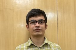 Abhijeet Beniwal合作论文数Dr Rajendra Prasad Centre for Ophthalmic Sciences, AIIMS6
 Velpandian Thirumurthy合作论文数All India Inst Med Sci, Dr Rajendra Prasad Ctr Ophthalm Sci6Rachna Meel合作论文数6Saranya Devi合作论文数5
 Navneet Sidhu合作论文数5
 Rashmi Singh合作论文数5Manu Saini合作论文数Dept Ophthalmol, Post Grad Inst Med Educ & Res5
 Pooja Kumari合作论文数5
 Sujata Mohanty合作论文数Stem Cell Facility, All India Institute of Medical Sciences5Sujith Vengayil合作论文数5
 Parijat Chandra合作论文数New5T Dada合作论文数4Sanjay Kai合作论文数4N Sharma合作论文数4
 Saumya Yadav合作论文数4
 Shikha Gupta合作论文数4A Panda合作论文数4
 Rakhi Kusumesh合作论文数Regional Institute of Ophthalmology, Indira Gandhi Institute of Medical Sciences4
 Manoranjan Mahapatra合作论文数Department of Hematology, All India Institute of Medical Sciences4
 Divya Singh合作论文数CSIR, Central Drug Research Institute4
 Shyam Chauhan合作论文数4
 Yogita Gupta合作论文数Dr Rajendra Prasad Ctr Ophthalm Sci, All India Inst Med Sci3




