Supplementary Figure 4 from Chronic Administration of Valproic Acid Inhibits Prostate Cancer Cell Growth <i>In vitro</i> and <i>In vivo</i>
Supplementary Figure 4 from Chronic Administration of Valproic Acid Inhibits Prostate Cancer Cell Growth <i>In vitro</i> and <i>In vivo</i>
Supplementary Figure 3 from Chronic Administration of Valproic Acid Inhibits Prostate Cancer Cell Growth <i>In vitro</i> and <i>In vivo</i>
Supplementary Figure 1 from Chronic Administration of Valproic Acid Inhibits Prostate Cancer Cell Growth <i>In vitro</i> and <i>In vivo</i>
This paper describes an inclusive biophysical study elucidating the interaction of therapeutic drug azithromycin (Azith) with hen egg white lysozyme (HEWL). Spectroscopic and computational tools have been employed to study the interaction of Azith with HEWL at pH 7.4. The fluorescence quenching constant values (Ksv) exhibited a decrease with the increase in temperature which revealed the occurrence of static quenching mechanism between Azith and HEWL. The thermodynamic data demonstrated that hydrophobic interactions were predominantly involved in the Azith-HEWL interaction. The negative value of standard Gibbs free energy (ΔG°) stated that the Azith-HEWL complex formed via spontaneous molecular interactions. The effect of sodium dodecyl sulfate (SDS) surfactant monomers on the binding propensity of Azith with HEWL was insignificant at lower concentrations however the binding significantly decreased at increased concentrations of the former. Far-UV CD data revealed alteration in the secondary structure of HEWL in the presence of Azith and the overall HEWL conformation changed. Molecular docking results revealed that the binding of Azith with HEWL takes place through hydrophobic interactions and hydrogen bonds.
Supplementary Figure 2 from Chronic Administration of Valproic Acid Inhibits Prostate Cancer Cell Growth In vitro and In vivo
Pancreatic ductal adenocarcinoma (PDAC) is the most common neoplastic disease of the pancreas, accounting for more than 90% of all pancreatic malignancies. As a highly lethal malignancy, PDAC is the fourth leading cause of cancer-related deaths worldwide with a 5-year overall survival of less than 8%. The efficacy and outcome of PDAC treatment largely depend on the stage of disease at the time of diagnosis. Surgical resection followed by adjuvant chemotherapy remains the only possibly curative therapy, yet 80%-90% of PDAC patients present with nonresectable PDAC stages at the time of clinical presentation. Despite our advancing knowledge of PDAC, the prognosis remains strikingly poor, which is primarily due to the difficulty of diagnosing PDAC at the early stages. Recent advances in glycoproteomics and glycomics based on mass spectrometry have shown that aberrations in protein glycosylation plays a critical role in carcinogenesis, tumor progression, metastasis, chemoresistance, and immuno-response of PDAC and other types of cancers. A growing interest has thus been placed upon protein glycosylation as a potential early detection biomarker for PDAC. We herein take stock of the advancements in the early detection of PDAC that were carried out with mass spectrometry, with special focus on protein glycosylation.
Urinary glycoproteins associated with aggressive prostate cancer (AG-PCa) were previously reported using post-digital rectal examination (DRE) urine specimens. To explore the potential of using pre-DRE urine specimens for detecting AG-PCa, we compared glycoproteins between pre- and post-DRE urine specimens, verified the previously identified post-DRE AG-PCa-associated urinary glycoproteins in pre-DRE urine specimens, and explored potential new glycoproteins for AG-PCa detection in pre-DRE urine specimens. Quantitative glycoproteomic data were acquired for 154 pre-DRE urine specimens from 41 patients with no cancer at biopsy, 48 patients with non-AG-PCa (Gleason score = 6), and 65 patients with AG-PCa (Gleason score 7 or above). Compared to glycopeptides from the post-DRE urine data, humoral immunity-related proteins were enriched in pre-DRE urine samples, whereas cell mediated immune response proteins were enriched in post-DRE urine samples. Analyses of AG-PCa-associated glycoproteins from pre-DRE urine revealed that the three urinary glycoproteins, prostate-specific antigen (PSA), prostatic acid phosphatase (ACPP), and CD97 antigen (CD97) that were previously identified in post-DRE urine samples, were also observed as AG-PCa associated glycoproteins in pre-DRE urine. In addition, we identified three new glycoproteins, fibrillin 1 (FBN1), vitronectin (VTN), and hemicentin 2 (HMCN2), to be potentially associated with AG-PCa in pre-DRE urine specimens. In summary, glycoprotein profiles differ between pre- and post-DRE urine specimens. The identified AG-PCa-associated glycoproteins may be further evaluated in large cohort of pre-DRE urine specimens for detecting clinically significant PCa.
Supplementary Figure 2 from Chronic Administration of Valproic Acid Inhibits Prostate Cancer Cell Growth In vitro and In vivo
Serum PSA, together with digital rectal examination and imaging of the prostate gland, have remained the gold standard in urological practices for the management of and intervention for prostate cancer. Based on these adopted practices, the limitations of serum PSA in identifying aggressive prostate cancer has led us to evaluate whether urinary PSA levels might have any clinical utility in prostate cancer diagnosis. Utilizing the Access Hybritech PSA assay, we evaluated a total of n = 437 urine specimens from post-DRE prostate cancer patients. In our initial cohort, PSA tests from a total of one hundred and forty-six (n = 146) urine specimens were obtained from patients with aggressive (Gleason Score ≥ 8, n = 76) and non-aggressive (Gleason Score = 6, n = 70) prostate cancer. A second cohort, with a larger set of n = 291 urine samples from patients with aggressive (GS ≥ 7, n = 168) and non-aggressive (GS = 6, n = 123) prostate cancer, was also utilized in our study. Our data demonstrated that patients with aggressive disease had lower levels of urinary PSA compared to the non-aggressive patients, while the serum PSA levels were higher in patients with aggressive prostate disease. The discordance between serum and urine PSA levels was further validated by immuno-histochemistry (IHC) assay in biopsied tumors and in metastatic lesions (n = 62). Our data demonstrated that aggressive prostate cancer was negatively correlated with the PSA in prostate cancer tissues, and, unlike serum PSA, urinary PSA might serve a better surrogate for capitulating tissue milieus to detect aggressive prostate cancer. We further explored the utility of urine PSA as a cancer biomarker, either alone and in combination with serum PSA, and their ratio (serum to urine PSA) to predict disease status. Comparing the AUCs for the urine and serum PSA alone, we found that urinary PSA had a higher predictive power (AUC= 0.732) in detecting aggressive disease. Furthermore, combining the ratios between serum to urine PSA with urine and serum assay enhanced the performance (AUC = 0.811) in predicting aggressive prostate disease. These studies support the role of urinary PSA in combination with serum for detecting aggressive prostate cancer.
Cells perform various functions by proteins via protein complexes. Characterization of protein complexes is critical to understanding their biological and clinical significance and has been one of the major efforts of functional proteomics. To date, most protein complexes are characterized by the in vitro system from protein extracts after the cells or tissues are lysed, and it has been challenging to determine which of these protein complexes are formed in intact cells. Herein, we report an approach to preserve protein complexes using in vivo cross-linking, followed by size exclusion chromatography and data-independent acquisition mass spectrometry. This approach enables the characterization of in vivo protein complexes from cells or tissues, which allows the determination of protein complexes in clinical research. More importantly, the described approach can identify protein complexes that are not detected by the in vitro system, which provide unique protein function information.
Abstract Objectives Gene therapy using adenoviruses has shown tremendous promise in animal and human models in the past. The tumor responses achieved by these viruses were either because of their oncolytic properties or therapeutic genes expressed from the genome of the virus. We have previously shown enhanced viral replication in cell models that have lost or lower expression of the cyclin dependent kinases inhibitor (p21/Waf-1). Beside the early onset of many viral genes in the p21/Waf-1 knock out cell model, we observed a significantly higher copy number for the viral VA1 but non VA2 non-coding RNA transcripts. In this study we investigated the effect of adenoviral VA1 non-coding RNA on the landscape of cellular proteins. Methods Using a DU145 cell line as a model that was transiently transfected with a plasmid carrying the adenoviral VA1 non-coding RNA, we were able to study changes in the proteome and the cellular cascade of the cells. Using state of the art global proteomics analyses of the differentially expressed proteins between the VA1 overexpressed and control cells demonstrated how the early onset of VA1 transcript affected cellular machinery. Results Using a 1.5 fold cut off between the down-regulated or overexpressing proteins, we were able to demonstrate how the non-coding VA1 gene might be responsible for suppressing the proteasome degradation, the endocytic and lysosomal pathways. Similarly, overexpression of the non-coding VA1 transcript was responsible for the upregulation (1.5 fold) of approximately 40 different proteins in the spliceosome machinery. Conclusions These data demonstrated how the non-coding VA1 RNA functioned independently of any other viral proteins in modulating the cell signaling cascades to promote viral propagation. To our knowledge this is the first report to investigate the function of VA1 non-coding RNA on the cellular proteome.
Prostate cancer (PCa) is a heterogeneous group of tumors, including non aggressive (NAG) and aggressive (AG) cancer, with variable clinical outcomes. Clinically, in order to assess the aggressiveness of a PCa, a core needle biopsy of a tumor is usually obtained to evaluate the Gleason pattern and score of the tumor. However, it may be difficult to assign on a small biopsy sample using histology. Therefore, additional tool is needed to aid in the assessment. We studied the diagnostic utility of 12 protein markers to identify AG tumors using immunohistochemistry (IHC) and tumor tissue microarray (TMA), including 215 cores of PCa and 111 cores of tumor-matched normal adjacent tissue (NAT). Protein markers were evaluated for their potential utility as single or combined panels for identification of AG. Of 12 proteins, PSMA, phospho-EGFR, AR and P16 were over-expressed in AG. Galectin-3, DPP4 and MAN1B1 revealed stronger staining patterns in NAG. The sensitivity and specificity of individual marker varied widely. Based on AUC values of individual marker, we constructed two- and three-marker panels. In two-marker panels, especially in the panel of DPP4 and PSMA, the AUC value reached 0.83 (rangingfrom 0.76 to 0.83). In three-marker panels, containing both DPP4 and PSMA with either Galectin-3 or phospho-EGFR, the AUC value reached 0.86 (ranging from 0.83 to 0.86). The specificities at 95% sensitivity of three-marker panels were also significantly improved. In addition to Gleason score, our IHC panels provide a practical tool to assess the aggressiveness of PCa.
Background: Current PSA-based tests used to detect prostate cancer (PCa) lack sufficient specificity, leading to significant overdetection and overtreatment. Our previous studies showed that serum fucosylated PSA (Fuc-PSA) and soluble TEK receptor tyrosine kinase (Tie-2) had the ability to predict aggressive (AG) PCa. Additional biomarkers are needed to address this significant clinical problem. Methods: A comprehensive Pubmed search followed by multiplex immunoassays identified candidate biomarkers associated with AG PCa. Subsequently, multiplex and lectin-based immunoassays were applied to a case-control set of sera from subjects with AG PCa, low risk PCa, and non-PCa (biopsy negative). These candidate biomarkers were further evaluated for their ability as panels to complement the prostate health index (phi) in detecting AG PCa. Results: When combined through logistic regression, two panel of biomarkers achieved the best performance: 1) phi, Fuc-PSA, SDC1, and GDF-15 for the detection of AG from low risk PCa and 2) phi, Fuc-PSA, SDC1, and Tie-2 for the detection of AG from low risk PCa and non-PCa, with noticeable improvements in ROC analysis over phi alone (AUCs: 0.942 vs 0.872, and 0.934 vs 0.898, respectively). At a fixed sensitivity of 95%, the panels improved specificity with statistical significance in detecting AG from low risk PCa (76.0% vs 56%, p=0.029), and from low risk PCa and non-PCa (78.2% vs 65.5%, p=0.010). Conclusions: Multivariate panels of serum biomarkers identified in this study demonstrated clinically meaningful improvement over the performance of phi, and warrant further clinical validation, which may contribute to the management of PCa.
Recently, we have found that two urinary glycoproteins, prostatic acid phosphatase (ACPP) and clusterin (CLU), combined with serum prostate-specific antigen (PSA) can serve as a three-signature panel for detecting aggressive prostate cancer (PCa) based on a quantitative glycoproteomic study. To facilitate the translation of candidates into clinically applicable tests, robust and accurate targeted parallel reaction monitoring (PRM) assays that can be widely adopted in multiple labs were developed in this study. The developed PRM assays for the urinary glycopeptides, FLN*ESYK from ACPP and EDALN*ETR from CLU, demonstrated good repeatability and a sufficient working range covering three to four orders of magnitude, and their performance in differentiating aggressive PCa was assessed by the quantitative analysis of urine specimens collected from 69 nonaggressive (Gleason score = 6) and 73 aggressive (Gleason ≥ 8) PCa patients. When ACPP combined with CLU, the discrimination power was improved from an area under a curve (AUC) of 0.66 to 0.78. By combining ACPP, CLU, and serum PSA to form a three-signature panel, the AUC was further improved to 0.83 (sensitivity: 84.9%, specificity: 66.7%). Since the serum PSA test alone had an AUC of 0.68, our results demonstrated that the new urinary glycopeptide PRM assays can serve as an adjunct to the serum PSA test to achieve better predictive power toward aggressive PCa. In summary, our developed PRM assays for urinary glycopeptides were successfully applied to clinical PCa urine samples with a promising performance in aggressive PCa detection.
Background Prostate cancer (PCa) is a heterogeneous group of tumors, including non-aggressive (NAG) and aggressive (AG) subtypes, with variable clinical outcomes. We assessed the diagnostic utility of selected protein markers to identify AG tumors. Methods The TMA was constructed, including NAG and AG. 12 protein markers were evaluated using the TMA by IHC stains. The makers were also evaluated for their potential utility as single or panels for distinguishing AG from NAG tumors. Results The higher expressions of four protein markers, including prostate specific membrane antigen (PSMA), phospho-EGFR, androgen receptor (AR), and P16, were identified in AG tumors of Gleason score 4 and 5. In contrast, Galectin-3, DPP4 and MAN1B1 revealed stronger staining patterns in NAG tumors. Sensitivity and specificity of individual marker varied widely. In tow-marker panels, especially in the panel of DPP4 and PSMA, the specificity was 38.46% at 95% sensitivity. To further improve the detection ability, we combined DPP4 and PSMA with either Galectin-3 or phospho-EGFR into three-marker panels. The specificity achieved >46% at 95% sensitivity and AUC was >0.85. Conclusions Our panels can be used to improve the separation of AG from NAG tumor and to add in the optimization of the treatment strategy for patients.
Prostate cancer (PCa) is a heterogeneous group of tumors with variable clinical courses. In order to improve patient outcomes, it is critical to clinically separate aggressive PCa (AG) from non-aggressive PCa (NAG). Although recent genomic studies have identified a spectrum of molecular abnormalities associated with aggressive PCa, it is still challenging to separate AG from NAG. To better understand the functional consequences of PCa progression and the unique features of the AG subtype, we studied the proteomic signatures of primary AG, NAG and metastatic PCa. 39 PCa and 10 benign prostate controls in a discovery cohort and 57 PCa in a validation cohort were analyzed using a data-independent acquisition (DIA) SWATH–MS platform. Proteins with the highest variances (top 500 proteins) were annotated for the pathway enrichment analysis. Functional analysis of differentially expressed proteins in NAG and AG was performed. Data was further validated using a validation cohort; and was also compared with a TCGA mRNA expression dataset and confirmed by immunohistochemistry (IHC) using PCa tissue microarray (TMA). 4,415 proteins were identified in the tumor and benign control tissues, including 158 up-regulated and 116 down-regulated proteins in AG tumors. A functional analysis of tumor-associated proteins revealed reduced expressions of several proteinases, including dipeptidyl peptidase 4 (DPP4), carboxypeptidase E (CPE) and prostate specific antigen (KLK3) in AG and metastatic PCa. A targeted analysis further identified that the reduced expression of DPP4 was associated with the accumulation of DPP4 substrates and the reduced ratio of DPP4 cleaved peptide to intact substrate peptide. Findings were further validated using an independently-collected tumor cohort, correlated with a TCGA mRNA dataset, and confirmed by immunohistochemical stains of PCa tumor microarray (TMA). Our study is the first large-scale proteomics analysis of PCa tissue using a DIA SWATH-MS platform. It provides not only an interrogative proteomic signature of PCa subtypes, but also indicates the critical roles played by certain proteinases during tumor progression. The spectrum map and protein profile generated in the study can be used to investigate potential biological mechanisms involved in PCa and for the development of a clinical assay to distinguish aggressive from indolent PCa.
The emergence of castration-resistance is one of the major challenges in the management of patients with advanced prostate cancer. Although the spectrum of systemic therapies that are available for use alongside androgen deprivation for treatment of castration-resistant prostate cancer (CRPC) is expanding, none of these regimens are curative. Therefore, it is imperative to apply systems approaches to identify and understand the mechanisms that contribute to the development of CRPC. Using comprehensive proteomic approaches, we show that a glycosylation-related enzyme, alpha (1,6) fucosyltransferase (FUT8), which is upregulated in CRPC, might be responsible for resistance to androgen deprivation. Mechanistically, we demonstrated that overexpression of FUT8 resulted in upregulation of the cell surface epidermal growth factor receptor (EGFR) and corresponding downstream signaling, leading to increased cell survival in androgen-depleted conditions. We studied the coregulatory mechanisms of EGFR and FUT8 expression in CRPC xenograft models and found that castration induced FUT8 overexpression associated with increased expression of EGFR. Taken together, our findings suggest a crucial role played by FUT8 as a mediator in switching prostate cancer cells from nuclear receptor signaling (androgen receptor) to the cell surface receptor (EGFR) mechanisms in escaping castration-induced cell death. These findings have clinical implication in understanding the role of FUT8 as a master regulator of cell surface receptors in cancer-resistant phenotypes.
Background: Bronchoalveolar lavage (BAL) is a specific type of air-way fluid. It is a commonly used clinical specimen for the diagnosis of benign diseases and cancers of the lung. Although previous studies have identified several disease-associated proteins in the BAL, the potential utility of BAL in lung cancer is still not well-studied. Based upon the fact that the majority of secreted proteins are glycoproteins, we have profiled N-glycoproteins in BAL collected from lung cancers, and investigated the expression of glycoproteins such as the matrix N-glycoprotein, periostin, in lung cancers. Methods: BAL specimens (n = 16) were collected from lung cancer patients, and analyzed using mass spectrometry-based quantitative N-glycoproteomic technique. Additional BAL specimens (n = 39) were independently collected to further evaluate the expression of periostin by using an enzyme-linked immunosorbent assay (ELISA). Results: A total of 462 glycoproteins were identified in BAL samples using N-glycoproteomic technique, including 290 in lung adenocarcinoma (ADC, n = 5), 376 in squamous cell carcinoma (SQCC, n = 4), 309 in small cell lung carcinoma (SCLC, n = 4), and 316 in benign lung disease (n = 3). The expressions of several glycoproteins were elevated, including 8 in ADC, 12 in SQCC, and 17 in SCLC, compared to benign BALs. The expression of periostin was detected in all subtypes of lung cancers. To further investigate the expression of periostin, an ELISA assay was performed using additional independently collected BALs (n = 39) The normalized levels of periostin in benign disease, ADC, SQCC, and SCLC were 255 ± 104 (mean ± SE) and 4,002 ± 2,181, 3,496 ± 1,765, and 1,772 ± 1,119 ng/mg of total BAL proteins. Conclusion: Our findings demonstrate that proteomic analysis of BAL can be used for the study of cancer-associated extracellular proteins in air-way fluid from lung cancer patients.
Additional file 6: Table S5. Cancer-associated proteins related to Fig. 4, Fig. 5, Figure S4, and Figure S5.
 Hui Zhang合作论文数Department of Otolaryngology-Head and Neck Surgery, School of Medicine, Johns Hopkins University30
 Ronald Rodriguez合作论文数13
 Wasim H Chowdhury合作论文数12
 Tung-Shing Mamie Lih合作论文数Sch Med, Johns Hopkins Univ11
 Yingwei Hu合作论文数11
 Qing Kay Li合作论文数Department of Otolaryngology-Head and Neck Surgery, School of Medicine, Johns Hopkins University10Shabana Shabbeer合作论文数9
 Yangying Zhou合作论文数Xiangya Hosp, Cent South Univ9
 Daniel Wanyui Chan合作论文数Clinical Chemistry Division, Department of Pathology, The Johns Hopkins University School of Medicine;Pathology Core Laboratory, Department of Pathology, The Johns Hopkins University School of Medicine;Center for Biomarker Discovery & Translation, The Johns Hopkins University School of Medicine9Shah Punit P合作论文数Coll Pharm & Pharmaceut Sci, Florida A&M Univ8Mustafa Sadaf合作论文数8
 Chien-Lun Chen合作论文数College of Medicine, Graduate Institute of Clinical Medical Sciences8QingHua Xia (夏庆华)合作论文数7
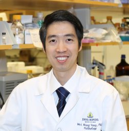 Weiming Yang合作论文数School of Life Sciences, University of Warwick7
 Shuang Yang (杨霜)合作论文数Center for Clinical Mass Spectrometry, College of Pharmaceutical Sciences, Soochow University7
 Jennifer Sung合作论文数Novartis Pharmaceuticals Corporation7Lijun Chen合作论文数6
 Shao-Yung Chen合作论文数Department of Chemical and Biomolecular Engineering, Johns Hopkins University6Michael Carducci合作论文数6
 Zhen Zhang合作论文数Center for Biomarker Discovery and Translation, Johns Hopkins University;School of Medicine, Johns Hopkins University6
 Irfan Shaukat合作论文数Dept Biochem, Univ Narowal5
 Jianbo Pan合作论文数Xiamen University, Xiamen University5Gabrielson Edward合作论文数5
 Mingming Dong (董铭铭)合作论文数School of Bioengineering, Dalian University of Technology5
 Yuefan Wang合作论文数Peking University, Peking University5
 Shisheng Sun (孙士生)合作论文数College of Life Sciences, Northwestern University5Rodrigo Vargas Eguez合作论文数Dept Pathol, Johns Hopkins Univ4Lori J. Sokoll合作论文数4
 Shawn Lupold合作论文数Baltimore, MD4Munazza Khan合作论文数3
 Alan W. Partin合作论文数Department of Urology, Johns Hopkins Medicine3
 Michael A. Carducci合作论文数School of Medicine, Johns Hopkins University;Johns Hopkins Kimmel Cancer Center;Skip Viragh Outpatient Cancer Center;Sibley Memorial Hospital3
 Stefani N Thomas合作论文数Department of Pharmaceutical Sciences, University of Southern California3Sami Siraj合作论文数Institute of Basic Medical Sciences, Khyber Medical University3
 Ganglong Yang (杨刚龙)合作论文数2Tamara Johnson合作论文数2nadine rosmus合作论文数2Xiangchun Wang合作论文数2Jer-Tsong Hsieh合作论文数2Sayf Yassin合作论文数2Johan W R Nortier合作论文数Leiden Univ, Med Ctr, Dept Clin Oncol, Leiden, Netherlands2
 Michael C Haffner合作论文数Johns Hopkins Univ, Sidney Kimmel Comprehens Canc Ctr, Baltimore, MD USA2
 Mohammad T Hedayati合作论文数2
 Liwei Cao (曹丽伟)合作论文数Department of Biochemistry, School of Medicine, Southern University of Science and Technology2Minghui Ao合作论文数Johns Hopkins Univ, Sch Med, Dept Pathol, Baltimore, MD 21205 USA2
 Jing Chen合作论文数NCI, National Institutes of Health;the Early Detection Research Network (EDRN, U01CA152813);the Clinical Proteomic Tumor Analysis Consortium (CPTAC, U24CA160036);NHLBI, National Institutes of Health;Johns Hopkins Proteomics Center;Programs of Excellence in Glycosicences (PEG, P01HL107153);the Swiss National Science Foundation;SystemsX.ch, PhosphoNetPPM;the European Research Council;and PRIME-XS (EC).;National Institutes of Health, National Institutes of Health2
 Luigi Marchionni合作论文数Pathology and Laboratory Medicine, Weill Cornell Medicine, Cornell University2Deangelis Justin合作论文数2Wissing Michel D合作论文数2




