Epilepsy impacts cognition during wakefulness. As epileptic activity is present and even augmented during sleep, epilepsy could also influence sleep-related cognitive processes. However, whether epilepsy modulates sleep-related experiences like dreaming remains poorly known. Here, we prospectively investigated the characteristics and determinants of dreaming in patients with epilepsy. Consecutive adult patients with epilepsy and no major cognitive deficit were recruited in an epilepsy outpatient clinic. They completed a questionnaire about their dreams, sleep and epilepsy over the past year. Medical data on epilepsy characteristics were gathered from the medical file. A generalised linear model was used to explore the determinants of dream recall frequency (DRF). We included 300 patients, with a mean (standard deviation [SD]) age of 40.4 (13.4) years and 51.3% female; 28.6% had more than one seizure/month, and 34.7% already had seizures during sleep. Patients recalled dreams on an average of 1.6 (1.5) days/week and 11% had one or more nightmare/week. Younger age, higher number of nocturnal awakenings, and lower seizures frequency predicted a higher DRF. In patients with focal epilepsy (65.3%), the localisation of the epileptic focus in the parieto-occipital area was negatively associated with DRF. Regarding dream content, 34.0% of patients reported having already dreamt about epilepsy. Dreams of seizures were associated with sleep-related seizures (p = 0.034) and dreams of epilepsy were associated with nightmare frequency (p = 0.004). Our results show that patients with epilepsy share several determinants of DRF (age, awakenings, role of the parieto-occipital area) with healthy subjects. In addition, epilepsy-related factors (seizure frequency, focus localisation) also impact DRF. Investigating dreams in patients with epilepsy can provide information on their epilepsy and their sleep.
ABSTRACT:Considerable progress has been made over the past decade in the use, practicality, and clinical relevance of electroencephalography (EEG) and magnetoencephalography (MEG)-derived data, improving our understanding of cortical nociceptive processing, the transition from nociception to conscious pain, and its cognitive-emotional modulations. Multimodal responses to heat, cold, and tactile/electrical stimuli, combined with autonomic reactions, provide objective sensory tests that bypass the limitations of verbal responses, while emerging approaches to spontaneous EEG/MEG analysis are changing the game in the still elusive area of ongoing pain. We can expect that all departments involved in the assessment of chronic pain will gradually be able to access these powerful and flexible techniques.
Background/Objectives: Degenerative cervical myelopathy (DCM) is the leading cause of functional disabilities of spinal origin in people over 50 years old. The objective of the present study was to establish a multi-parametric weighted scoring system that is easy to use in daily practice, based on the most significant MRI signs and correlated as strongly as possible with the clinical presentation (mJOA)-we call this system the SIMS or Severity on Imaging Myelopathy Score. Methods: Ninety-nine patients who underwent clinical and radiological evaluation by mJOA and MRI between January 2015 and March 2021 were retrospectively included. The variables included in the score were the Fujiwara ratio, the T2-weighted intramedullary hyperintensity, the aspect of the peri-medullary fluid cisterns, the Torg-Pavlov ratio, the local kyphosis and the number of stenotic levels. Each variable was first correlated to the mJOA score for each patient, making it possible to construct the final SIMS at the end, and validate it by comparison with mJOA scores. Results: The variables that were significantly correlated with one another were the T2-weighted intramedullary hyperintensity, the reduction in peri-medullary fluid spaces and the number of stenotic levels (p < 0.05). Then, points were assigned to each variable according to their relative importance and made it possible to construct the definitive SIMS. The final Spearman correlation coefficient between the SIMS and the mJOA score was -0.747. Conclusions: This work showed that this new multi-parametric MRI-based scoring system represents a consistent means to characterize the degree of severity of degenerative cervical myelopathy.
Objectif L’activité épileptique étant activée par le sommeil, elle pourrait influencer les processus cognitifs associés au sommeil, comme le rêve. L’objectif de cette étude prospective était de décrire les caractéristiques des rêves des personnes souffrant d’épilepsie, et d’investiguer les facteurs, associés au sommeil et à l’épilepsie, modulant la fréquence de rappel de rêve (FRR) et le contenu des rêves. Méthodes Des patients de 18 à 65 ans souffrant d’épilepsie et sans déficit cognitif majeur ont été recrutés consécutivement à leur consultation de suivi épileptologique. Ils ont rempli un questionnaire sur leurs rêves, leur sommeil, et leur épilepsie au cours de l’année passée. Les informations concernant l’épilepsie ont été recueillies dans leur dossier médical. Un modèle linéaire généralisé a été utilisé pour explorer les déterminants de la FRR. Résultats Trois cent patients (40,4±13,4 ans, 51,3 % de femmes, 66,4 % avec une épilepsie focale, 48,3 % pharmacorésistants) ont été inclus. Parmi eux, 28,6 % faisaient plus d’une crise épileptique par mois, et 34,7 % avaient déjà présenté des crises pendant le sommeil. La FRR moyenne observée était de 1,6±1,5 rêves/semaine et 11 % des patients faisaient au moins un cauchemar/semaine. Un âge plus jeune (p<0,001), davantage de réveils nocturnes (p=0,019), et une fréquence de crises épileptique moindre (p=0,035) étaient associés à une FRR plus élevée. Chez les patients souffrant d’épilepsie focale, la localisation pariéto-occipitale du foyer épileptique était négativement associé avec la FRR (p=0,034). Concernant le contenu des rêves, 34,0 % des patients rapportaient avoir déjà rêvé d’épilepsie (crises/symptômes de crise/épilepsie en général), et les rêves de crise étaient associés au fait de faire des crises pendant le sommeil (p=0,034). Conclusion Les facteurs prédictifs positifs de la FRR tels qu’un jeune âge et des réveils nocturnes sont similaires à ceux observés dans la population générale. La FRR plus faible associée aux épilepsies pariéto-occipitales est cohérente avec le rôle critique de cette région dans la formation et/ou le rappel des rêves. De plus, la fréquence des crises d’épilepsie impacte négativement la FRR, et la thématique épileptique est incorporée dans le contenu des rêves de nombreux patients, ce qui suggère que l’épilepsie influence la cognition pendant le sommeil.
OBJECTIVE:Acute symptomatic seizures (ASyS) occur in up to 30% of patients with intracerebral hemorrhage (ICH) when continuous electroencephalography (cEEG) is used, potentially worsening outcomes. Identification of early EEG biomarkers of ASyS may help guide personalized antiseizure medication (ASM) prophylaxis. Here, we aimed to describe early interictal EEG patterns, their dynamics, and their association with seizure risk, considering the effect of prophylactic levetiracetam. METHODS:This prospective analysis used data from the PEACH phase 3 trial (2017-2020), which enrolled adults with acute spontaneous supratentorial ICH, randomized to receive levetiracetam or placebo. Patients underwent systematic 48-h cEEG within 48 h of symptom onset. Electrographic seizures and interictal EEG patterns were analyzed using standardized terminology of the American Clinical Neurophysiology Society. Associations between rhythmic and periodic patterns (RPPs) and seizures with clinical and radiological variables were assessed using univariate analyses. We also conducted exploratory testing of the CAV (cortical involvement, age < 65 years, volume > 10 mL) score for predicting ASyS, incorporating RPPs and ASM exposure. RESULTS:Forty-two patients were included (median [Q1-Q3] age = 72 [60-79] years, 29% women), 19 in the levetiracetam group. Interictal EEG abnormalities were common and not influenced by ASM, including background asymmetry (73%), sporadic epileptiform discharges (62%), and RPPs (52%). RPPs were associated with ICH volume (p = .039) and cortical involvement (p = .003). Among patients with RPPs, 50% developed ASyS (20% in those treated with ASM vs. 75% in untreated patients, p = .030). Most patients (91.7%) with seizures had RPPs that preceded seizures, in >90% cases by 12 (Q1-Q3 = 4-25) h. Integrating RPPs into the CAV model led to an improvement of ASyS prediction (area under the curve = .949 vs. .918, p = .53) that was statistically nonsignificant. SIGNIFICANCE:RPPs are strong markers of ictogenesis in acute ICH and precede ASyS, thus offering a potential therapeutic window. These findings support the use of early cEEG for risk stratification and personalized ASM prophylaxis.
The end-stage of amyotrophic lateral sclerosis (ALS) is commonly regarded as a complete Locked-In Syndrome (cLIS). Shifting the perspective from cLIS (assumed consciousness) to Cognitive Motor Dissociation (potentially demonstrable consciousness), we aimed to assess the preservation of covert awareness (internally preserved but externally inaccessible) using a multimodal battery. We evaluate two end-stage ALS patients using neurophysiological testing, passive and active auditory oddball paradigms, an auditory Brain-Computer Interface (BCI), functional activation-task imaging, long-term EEG, brain morphology, and resting-state metabolism to characterize underlying brain function. Patient 1 initially follows simple commands but fails twice at BCI control. At follow-up, command following is no longer observed and his oddball cognitive responses disappear. Patient 2, at a single evaluation, is unable to follow commands or control the BCI. Both patients exhibit altered wakefulness, brain atrophy, and a global cortico–subcortical hypometabolism pattern consistent with a disorder of consciousness, regarded as an extreme manifestation of ALS-associated fronto-temporal dementia. Although it is not possible to firmly prove the absence of awareness, each independent measure concurred with suggesting that a “degenerative disorder of consciousness” rather than a cLIS may constitute the final stage of ALS. This condition appears pathophysiologically distinct from typical tetraplegia and anarthria, in which behavioural communication and BCI use persist to enhance quality of life. Identifying the neuroimaging signatures of this condition represents a substantial milestone in understanding end-stage ALS. Large-scale longitudinal investigations are warranted to determine the prevalence of this profile among patients whose communication appears impossible. Some patients with amyotrophic lateral sclerosis become progressively paralyzed to the extent that they can no longer communicate. This raises a critical question: does consciousness remain despite a complete loss of interaction? To address this issue, we analysed brain activity in two patients at the late stage of this disease. Our findings indicate that no reliable signs of awareness could be detected, even when employing advanced methods to assess brain function, including approaches designed to enable communication without movement. Two hypotheses may account for this outcome: either prolonged paralysis progressively leads to the disappearance of consciousness, or the degeneration of brain function itself directly underlies its loss. However, our conclusions ultimately depend on the reliability of consciousness assessment tools, even when used in combination. Gobert et al. investigate whether consciousness persists in end-stage Amyotrophic Lateral Sclerosis, traditionally assumed as being fully aware but unable to communicate. Multimodal assessments, including Brain-Computer Interface, suggest some individuals may show degenerative disorder of consciousness rather than a complete Locked-In Syndrome.
The continuous monitoring of physiological parameters is now considered as a standard of care in intensive care units (ICU). While multiple techniques are available to guide hemodynamic or respiratory management, the monitoring of neurological function in unconscious patients is usually limited to discontinuous bedside neurological examination or morphological brain imaging. However, cortical activity is accessible at the bedside with electroencephalography (EEG), electrocorticography (ECoG) or evoked potentials. The analysis of the unprocessed signal requires a trained neurophysiologist and could be time consuming. During the past decades, advances in neurophysiological signal acquisition make it possible to calculate quantified EEG parameters in real-time. New monitors also provide ICU friendly display for a dynamic and live assessment of neurological function changes. In this review, we will describe the technical aspects of EEG, ECoG and evoked potentials required for a good signal quality before interpretation. We will discuss how to use those electrophysiological techniques in the ICU to assess neurological function in comatose patients at the acute phase of brain injuries such as traumatic brain injuries, haemorrhagic or ischemic stroke. We will discuss, which quantitative EEG or evoked potentials monitoring parameters can be used at the bedside to guide sedation, evaluate neurological function during awaking and look for new neurological (encephalic or brainstem) injuries. We will present the state of the art and discuss some analyses, which may develop shortly.
Surgery for spinal cord tumors poses a significant challenge due to the inherent risk of neurological deterioration. Despite being performed at numerous centers, there is an ongoing debate regarding the efficacy of pre- and intraoperative neurophysiological investigations in detecting and preventing neurological lesions. This study begins by providing a comprehensive review of the neurophysiological techniques commonly employed in this context. Subsequently, we present findings from a cohort of 67 patients who underwent surgery for intradural tumors. These patients underwent preoperative and intraoperative multimodal somatosensory evoked potentials (SSEPs) and motor evoked potentials (MEPs), with clinical evaluation conducted three months postoperatively. The study aimed to evaluate the neurophysiological, clinical, and radiological factors associated with neurological outcomes. In univariate analysis, preoperative and intraoperative potential alterations, tumor size, and ependymoma-type histology were linked to the risk of worsening neurological condition. In multivariate analysis, only preoperative and intraoperative neurophysiological abnormalities remained significantly associated with such neurological deterioration. Interestingly, transient alterations in intraoperative MEPs and SSEPs did not pose a risk of neurological deterioration. The machine learning model we utilized demonstrated the possibility of predicting clinical outcome, achieving 84% accuracy.
The continuous monitoring of physiological parameters is now considered as a standard of care in intensive care units (ICU). While multiple techniques are available to guide hemodynamic or respiratory management, the monitoring of neurological function in unconscious patients is usually limited to discontinuous bedside neurological examination or morphological brain imaging. However, cortical activity is accessible at the bedside with electroencephalography (EEG), electrocorticography (ECoG) or evoked potentials. The analysis of the unprocessed signal requires a trained neurophysiologist and could be time consuming. During the past decades, advances in neurophysiological signal acquisition make it possible to calculate quantified EEG parameters in real-time. New monitors also provide ICU friendly display for a dynamic and live assessment of neurological function changes. In this review, we will describe the technical aspects of EEG, ECoG and evoked potentials required for a good signal quality before interpretation. We will discuss how to use those electrophysiological techniques in the ICU to assess neurological function in comatose patients at the acute phase of brain injuries such as traumatic brain injuries, haemorrhagic or ischemic stroke. We will discuss, which quantitative EEG or evoked potentials monitoring parameters can be used at the bedside to guide sedation, evaluate neurological function during awaking and look for new neurological (encephalic or brainstem) injuries. We will present the state of the art and discuss some analyses, which may develop shortly.
This pilot study aimed to investigate the interest of high angular resolution diffusion imaging (HARDI) and tractography of the spinal cord (SC) in the management of patients with intramedullary tumors by providing predictive elements for tumor resection. Eight patients were included in a prospective study. HARDI images of the SC were acquired using a 3T MRI scanner with a reduced field of view. Opposed phase-encoding directions allowed distortion corrections. SC fiber tracking was performed using a deterministic approach, with extraction of tensor metrics. Then, regions of interest were drawn to track the spinal pathways of interest. HARDI and tractography added value by providing characteristics about the microstructural organization of the spinal white fibers. In patients with SC tumors, tensor metrics demonstrated significant changes in microstructural architecture, axonal density, and myelinated fibers (all, p < 0.0001) of the spinal white matter. Tractography aided in the differentiation of tumor histological types (SC-invaded vs. pushed back by the tumor), and differentiation of the spinal tracts enabled the determination of precise anatomical relationships between the tumor and the SC, defining the tumor resectability. This study underlines the value of using HARDI and tractography in patients with intramedullary tumors, to show alterations in SC microarchitecture and to differentiate spinal tracts to establish predictive factors for tumor resectability.
The end-stage of amyotrophic lateral sclerosis [ALS] is presumed to be a complete Locked-In Syndrome [cLIS], assuming an internally preserved consciousness that would not be accessible anymore from the outside. However, whether consciousness persists at this stage of ALS remains to be demonstrated. Shifting the perspective from cLIS (presupposed consciousness) to Cognitive Motor Dissociation (to-be-demonstrated consciousness), we attempted to demonstrate consciousness and communication with two cLIS-ALS patients using a multimodal awareness assessment battery. It involved complete neurophysiological assessments, passive and active auditory oddball paradigm (Subject-Own-Name/P300), an auditory-based Brain-Computer-Interface [BCI] and activation-task imaging using functional MRI or [15O]H2O PET. Wakefulness (long-term EEG), brain morphology (CT or MRI scans) and resting brain metabolism ([18F]fluoro-deoxy-glucose PET) were used to describe the underlying cLIS brain function. While Patient 1 could initially follow simple commands, he failed twice to control the BCI. At follow-up, he showed no more evidence of command following and his oddball (Own Name - P300) cognitive responses has disappeared. At his unique evaluation, Patient 2 was neither able to follow simple commands nor to control the BCI. Both patients had altered wakefulness, brain atrophy, and a global cortico-sub-cortical hypometabolism pattern compatible with a disorder of consciousness, regarded as an extreme form of an ALS-associated fronto-temporal dementia. While it is not possible to firmly demonstrate the absence of awareness, each independent measure concurred with suggesting that a degenerative disorder of consciousness rather than a cLIS might be the final stage of ALS. In future cass, this dramatic cognitive decline should be anticipated before communication disappears to enable precise advance directives regarding end-of-life issues in case complete, and neurophysiologically confirmed, unresponsiveness occurs. Altogether, the neuroimaging features distinguishing the mechanisms in this rare condition is a significant milestone to understand end-stage ALS. The present clinical study calls for further exploration of this terminal stage to determine the prevalence of this profile in whom communication seems hopeless. ### Competing Interest Statement The authors have declared no competing interest. ### Funding Statement AO was funded by the Fondation pour la Recherche Medicale (FRM, ING20121226307). PS, JM, EM were funded by one grant from the Fondation pour la Recherche Medicale (FRM, FDM201906008524). JM, EM and PS were funded by ANR-17-CE40-0005, MindMadeClear & ANR-20-CE17-0023, ANR HiFi. PS was funded by Perce-Neige Fondation. The teams of the Lyon Neurocience Research Center are funded by the Labex cortex. ### Author Declarations I confirm all relevant ethical guidelines have been followed, and any necessary IRB and/or ethics committee approvals have been obtained. Yes The details of the IRB/oversight body that provided approval or exemption for the research described are given below: IRB of Hospices Civils de LYON CSE-HCL - IRB 00013204; Pr Cyrille Confavreux gave ethical approval for this work (approval N. 24-310). I confirm that all necessary patient/participant consent has been obtained and the appropriate institutional forms have been archived, and that any patient/participant/sample identifiers included were not known to anyone (e.g., hospital staff, patients or participants themselves) outside the research group so cannot be used to identify individuals. Yes I understand that all clinical trials and any other prospective interventional studies must be registered with an ICMJE-approved registry, such as ClinicalTrials.gov. I confirm that any such study reported in the manuscript has been registered and the trial registration ID is provided (note: if posting a prospective study registered retrospectively, please provide a statement in the trial ID field explaining why the study was not registered in advance). Yes I have followed all appropriate research reporting guidelines, such as any relevant EQUATOR Network research reporting checklist(s) and other pertinent material, if applicable. Yes All data produced in the present work are contained in the manuscript.
Cervical spondylotic myelopathy (CDM) is the leading cause of functional disability of spinal origin in people over 50 years old. The objective of the present study was to establish a multiparametric weighted scoring system, easy to use in daily practice, based on the most significant MRI signs and correlated as strongly as possible with the clinical presentation (mJOA) – the SIMS for Severity on Imaging Myelopathy Score. 99 patients who underwent clinical and radiological evaluation by mJOA and MRI between January 2015 and March 2021 were retrospectively included. The variables included in the score were the Fujiwara ratio, the T2-weighted intramedullary hyperintensity, the aspect of the perimedullary fluidcisterns, the Torg-Pavlov ratio, the local kyphosis and the number of stenotic levels. Each variable was first correlated to the mJOA score for each patient, making possible at the end to construct the final SIMS, and validate it by comparison with mJOA scores. The variables significantly correlated were the T2-weighted intramedullary hyperintensity, the reduction of perimedullary fluid spaces and the number of stenotic levels (p < 0.05). Then points were assigned to each variable according to their relative importance and made it possible to construct the definitive SIMS. The final correlation coefficient between SIMS and mJOA score was -0.747. This work showed that this new multi-parametric MRI-based scoring system represents a consistent mean to characterize the degree of severity of the degenerative cervical myelopathy.
Abstract Objective Central pontine myelinolysis (CPM) is a rare demyelinating disease that affects the pons and which can cause extreme disabilities such as locked‐in syndrome (LIS) in the initial phase. The aim of the study was to describe the evolution over a 12‐month period of two patients with CPM causing an initial LIS. Method We retrospectively report the unexpected clinical outcome of these two patients in relation with the anatomical damages documented by brain MRI, associated with diffusion tensor imaging and reconstruction of corticospinal tracts in tractography. The following clinical parameters systematically assessed at 3, 6, 9, and 12 months: muscle testing on 12 key muscles (Medical Research Council), prehension metrics (box and block test and purdue pegboard), and independence for acts of daily living (functional independence measure). Results Both patients showed a progressive recovery beginning between 2 and 3 months after the onset of symptoms, leading to almost complete autonomy at 12 months (FIM > 110), with motor strength greater than 4/5 in all joint segments (MRC > 50/60). On brain MRI with tractography, CST appeared partially preserved at pons level. Interpretation The possibility of a near‐complete functional recovery at 12 months is important to consider given the ethical issues at stake and the discussions about limiting care that may take place initially. It seems to be the consequence of reversible myelin damage combined with partially preserved neurons. Development of collateral pathways or resolution of conduction block may explain this recovery. MRI comprising DTI and tractography could play a key role in the prognosis of motor recovery.
Objective: Early functional evaluation and prognosis of patients with disorders of consciousness is a major challenge that clinical assessments alone cannot solve. Objective measures of brain activity could help resolve this uncertainty. We used electroencephalogram at bedside to detect voluntary attention with a paradigm previously validated in healthy subjects. Methods: Using auditory-oddball sequences, our approach rests on detecting known attentional modulations of Event Related Potentials that reflect compliance with verbal instructions. Sixty-eight unresponsive patients were tested in their first year after coma onset (37 coma and 31 first year post-coma patients). Their evolution 6 months after the test was considered. Results: Fourteen of the 68 patients, showed a positive response. Nine were in a coma and 5 in a minimally conscious state (MCS). Except for one who died early, all responders evolved to exit-MCS within 6 months (93%), while 35 (65%) among non-responders only. Conclusions: Among those patients for whom the outcome is highly uncertain, 21% responded positively to this simple but cognitively demanding test. Strikingly, some coma patients were among responders. Significance: The proposed paradigm revealed cognitive-motor dissociation in some coma patients. This ability to sustain attention on demand predicted awakening within 6 months and represents an immediately useful information for relatives and caregivers. (c) 2022 International Federation of Clinical Neurophysiology. Published by Elsevier B.V. All rights reserved.
While high-frequency transcranial magnetic stimulation (HF-rTMS) is now included in the armamentarium to treat chronic neuropathic pain (NP), direct-current anodal stimulation (a-tDCS) to the same cortical targets may represent a valuable alternative in terms of feasibility and cost. Here we performed a head-to-head, randomized, single-blinded, cross-over comparison of HF-rTMS versus a-tDCS over the motor cortex in 56 patients with drug-resistant NP, who received 5 daily sessions of each procedure, with a washout of at least 4 weeks. Daily scores of pain, sleep, and fatigue were obtained during 5 consecutive weeks, and functional magnetic resonance imaging (fMRI) to a motor task was performed in a subgroup of 31 patients. The percentage of responders, defined by a reduction in pain scores of > 2 SDs from pre-stimulus levels, was similar to both techniques (42.0% vs. 42.3%), while the magnitude of “best pain relief” was significantly skewed towards rTMS. Mean pain ratings in responders decreased by 32.6% (rTMS) and 29.6% (tDCS), with half of them being sensitive to only one technique. Movement-related fMRI showed significant activations in motor and premotor areas, which did not change after 5 days of stimulation, and did not discriminate responders from non-responders. Both HF-rTMS and a-tDCS showed efficacy at 1 month in drug-resistant NP, with magnitude of relief slightly favoring rTMS. Since a significant proportion of patients responded to one procedure only, both modalities should be tested before declaring a patient as unresponsive.
ABSTRACTBackgroundIn the last decades, advances in Intensive Care Unit management have led to decreased mortality. However, significant morbidity remains as patients survive after a lesional coma with uncertain quality of awakening and high risk of functional disability. Predicting this level of recovery but also the functional disability of those who will awake constitutes a major challenge for medical, ethical and social perspectives. Among the huge heterogeneity of coma-related injuries, recognising the universality of a common functional pattern which may be focused on a final step of an integrated network would be of great interest for our understanding of disorders of consciousness. The objective of this study is to investigate the neural correlates of arousal and awareness in coma and post-coma to build a prognostic tool based on the detection of a common pattern between patients with a favourableversusan unfavourable outcome.Method/DesignWe will implement this objective in a translational approach which combines PET-MR imaging, neurophysiology, behavioural/clinical assessments and innovative statistical and computational analysis tools in patients with disorders of consciousness in Intensive Care Unit and in Rehabilitation Department.
 Sebastien Boulogne合作论文数Service de neurologie vasculaire, hôpital neurologique Pierre-Wertheimer15
 Sylvain Rheims合作论文数15Dailler Frédéric合作论文数14
 Jacques Luauté合作论文数Centre de Recherche en Neurosciences de Lyon;Hôpital Henry Gabrielle14
 François Mauguière合作论文数Centre de Recherche en Neurosciences de Lyon;Université Claude Bernard Lyon;Hôpital Neurologique de Lyon13Julien Jung合作论文数University Claude Bernard Lyon 1, Lyon F-69000, France13
 Florent Gobert合作论文数12
 Luis Garcia-Larrea合作论文数NeuroPain Lab, Lyon Neurological Hospital, Lyon Centre for Neuroscience, Claude Bernard University Lyon 1;Pain Center, Lyon Neurological Hospital, Lyon Centre for Neuroscience, Claude Bernard University Lyon 1;INSERM12
 Eric Azabou合作论文数9
 Martine Gavaret合作论文数Service de Neurophysiologie Clinique, Assistance Publique des Hôpitaux de Marseille9
 Philippe Convers合作论文数7
 Patrick Mertens合作论文数Faculty of Medicine Lyon Est, Claude Bernard University Lyon 1;Faculty of Medicine Lyon Est, Claude Bernard University Lyon 17
 Jean-Pascal Lefaucheur合作论文数Hopital Henri Mondor;Public Hospitals of Paris;Paris-Est Creteil University7Dominique Morlet合作论文数7V. Manel合作论文数6
 Laure Peter-Derex合作论文数Center for Sleep medicine and Respiratory Diseases, University Hospital of Lyon;Université de Lyon6
 Laure Mazzola合作论文数6Ritzenthaler Thomas合作论文数From the Departments of Neurology (, Université Lyon 1;Regional Council and University of Burgondy, University Hospital of Dijon;Department of Neurology (F.P.),, Centre Hospitalier de Fleyriat;Departments of Neurology (C.L., University Paris-Descartes;Department of Neurology (P.A.),, Hôpital Bichat-Claude Bernard;Department of Neurology (S.C.),, Hôpital Nord-Ouest;Department of Neurology (, Hôpital Nord-Laënnec;Department of Neurology (H.H.),, Hôpital Henri Mondor;Department of Neurology (I.S.),, Groupe Hospitalier Pellegrin;Unité Neurosciences (M.-H.M.),, Hôpital Saint Roch;Centre d'Investigation Clinique &, Hôpital Louis Pradel (N.M., M.O.),;UMR1060 (CarMeN, Université Claude Bernard Lyon 1;Université Lyon 1, Université Claude Bernard Lyon 16
 Yann Pereon合作论文数Faculté de Médecine, University of Nante;DPHU2 Institut du Thorax et Système Nerveux, University of Nante;Department of Physiology, Medical School, University of Nantes6Hélène Catenoix合作论文数6Michel Magnin合作论文数6
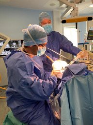 Corentin Dauleac合作论文数Hospices Civils de Lyon, Hôpital neurologique Pierre Wertheimer5
 Ayache Samar S合作论文数excitabilité nerveuse et thérapeutique, université Paris-Est-Créteil5
 Cedric Barrey合作论文数Hospices Civils de Lyon, France Hospices Civils de Lyon5
 François Cotton合作论文数Hospices Civils de Lyon5Catherine Fischer合作论文数5
 Perrine Ruby合作论文数5
 Emmanuel Poulet合作论文数4
 Veronique Mutschler合作论文数4
 Baptiste Balanca合作论文数4Jean-Christophe Antoine合作论文数4
 Laurent Vercueil合作论文数Grenoble Inst Neurosci GIN, Univ Grenoble Alpes4
 Jérôme Honnorat合作论文数University Claude Bernard Lyon 14Fabien Perrin合作论文数Unité Mixte de Recherche 5020, Université Claude Bernard Lyon 1 - CNRS4
 Roland Peyron合作论文数4Laurence Tell合作论文数4
 Herve Devanne合作论文数Clinical Neurophysiology, Hospital R. Salengro4j fournetfayard合作论文数3
 Lionel Naccache合作论文数Paris Brain Institute;Pitié-Salpêtrière Hospital;Sorbonne Université3Gianluca Deiana合作论文数3
 P. Garassus合作论文数3Marine Loizon合作论文数3Martina Corazzol合作论文数3Gérard Bollini合作论文数3
 Ines Merida合作论文数CERMEP, Bron, France.3Nathalie Kubis合作论文数3Birgit Frauscher合作论文数Department of Neurology & Neurosurgery, Faculty of Medicine and Health Sciences, McGill University3Farcy J P合作论文数3




