"Inflammaging", the chronic increase in inflammatory signaling with age, remains poorly understood in hematopoietic aging. Here, we identify the innate immune RNA sensor melanoma differentiation-associated protein 5 (MDA5) as an important factor of hematopoietic stem cell (HSC) aging. Aged Mda5-/- mice exhibit reduced HSC accumulation and myeloid bias. Importantly, aged Mda5-/- HSCs retain greater quiescence and superior repopulation capacity in noncompetitive transplants compared to wild-type counterparts. Multiomic analyses- including chromatin accessibility, transcriptomics, and metabolomics-reveal decreased inflammatory signaling, a youthful metabolic profile, and improved proteostasis in Mda5-/- HSCs, through regulation of HSF1 and phospho-EIF2A, key proteostasis regulators. Activation of HSF1 in aged wild-type HSCs partially restores youthful features, supporting a causal role for proteostasis maintenance. Collectively, our findings demonstrate that attenuating MDA5-dependent inflammation preserves HSC function during aging by maintaining metabolic fitness and proteostasis and provide insight into potential therapeutic strategies for mitigating hematopoietic aging.
Abstract The precise regulation of protein synthesis is essential for cellular function and survival. In particular, in neurons, dysregulated mRNA translation is linked to impaired memory formation and is a hallmark of neurodegenerative diseases. Neurons are characterized by tissue-specific, long 3′ untranslated regions (3′UTRs); in this study, we demonstrate that mRNA isoforms with these neuronal 3′UTRs are less efficiently translated than their short counterparts in Drosophila and mammalian brains. 3′UTR-dependent translation is based on a negative feedback mechanism centered around the two neural-enriched proteins ELAV and Pumilio. The long elav 3′UTR inhibits production of the neuronal ELAV protein, which in turn mediates 3′UTR extension of hundreds of neuronal genes. Those long 3′UTR isoforms are preferentially bound and translationally inhibited by Pumilio. The regulatory loop maintains optimal neuronal 3′UTR and protein levels in conditions of genetic and environmental perturbations; its disruption reduces animal viability and lowers stress resilience, and causes severe developmental phenotypes in flies and in human brain organoids. We propose 3′UTR-mediated translational control as an evolutionarily conserved mechanism for the maintenance of cell-type-specific proteostasis.
The BCL-2 inhibitor venetoclax has transformed the treatment of acute myeloid leukemia (AML), but relapse due to resistance of leukemic stem cells (LSCs) remains a major challenge. By molecular and functional profiling of LSCs from >150 patients, we identify four LSC subtypes. These mirror distinct hematopoietic lineage stages, which determine the expression ratio between the venetoclax target BCL-2 and resistance-inducing proteins MCL-1 and BCL-xL (MAC-score). Longitudinal analyses reveal that venetoclax resistance mostly arises in LSCs through plasticity toward a megakaryocytic/erythroid-progenitor (MEP)-LSC state that switches survival dependency from BCL-2 to BCL-xL. In rare cases, mature monocytic/dendritic (MoDe)-LSCs, found within LAMP5+ monocytic AMLs, drive venetoclax resistance. LSC subtyping improves genetic risk stratification and provides subtype-specific therapies: venetoclax-resistant MEP-LSCs respond to BCL-xL inhibitors, whereas MoDe-LSCs are sensitive to MEK1/2 inhibition. Our findings reveal four distinct LSC types with unique vulnerabilities and propose biomarker-guided treatment strategies that complement genetic profiling to overcome venetoclax resistance.
Myocardial infarction (MI) triggers emergency hematopoiesis, driving an acute inflammatory response critical for tissue repair. However, excessive myelopoiesis impairs cardiac remodeling and function. Despite their pivotal role in hematopoiesis, the contribution of bone marrow (BM) hematopoietic stem cells (HSCs) to post-MI inflammation remains poorly understood. Here, by analyzing BM samples from patients with a history of MI undergoing cardiac surgery, we showed that MI induces detrimental transcriptional and functional changes in human BM HSCs, priming them toward inflammatory myeloid differentiation. Using lineage-tracing in mice, we demonstrated that activated HSCs contribute directly to the infiltration of proinflammatory myeloid cells into the injured myocardium. Therapeutically, we showed that enforcing HSC quiescence with the vitamin A metabolite 4-oxo-retinoic acid (4-oxo-RA) dampens emergency hematopoiesis, reducing inflammatory leukocyte infiltration and preserving long-term cardiac function after MI. In contrast, treatment with the vitamin A metabolite all-trans RA, an agent already clinically used in hematologic and dermatologic diseases, was associated with exacerbated myocardial inflammation, underscoring the need for targeted approaches. These findings establish HSCs as key orchestrators of inflammatory responses after MI and identify 4-oxo-RA as a promising strategy to modulate emergency hematopoiesis and improve cardiac repair. Our study introduces a novel concept for the selective regulation of HSC activity as a new therapeutic avenue.(Rettkowski et al., Nature Cell Biology, 2025)
Acute graft-versus-host disease (aGVHD) can affect the central nervous system (CNS) through microglial activation and T cell infiltration, but the role of gut microbiota in CNS-aGVHD remains unclear. Here, we investigated the role of microbiota in microglial activation during aGVHD using antibiotic-treated specific pathogen-free (SPF), germ-free (GF), and wildling mice. Antibiotic-mediated microbiota depletion led to infiltration of IFN-γ-producing T cells in the brain, activation of microglia via the TLR4/p38 MAPK pathway, and neurocognitive deficits in SPF aGVHD mice. Microglial depletion reversed the neurocognitive deficits. GF and wildling mice treated with antibiotics exhibited similar microglial activation after allogeneic hematopoietic cell transplantation (allo-HCT). Mechanistically, the bacteria-derived metabolite N,N,N-trimethyl-5-aminovaleric acid (TMAVA) was decreased in microglia following antibiotic treatment. TMAVA administration suppressed TLR4/p38 MAPK pathway activity in microglia and alleviated gut microbiota depletion-mediated neurocognitive deficits. Additionally, TMAVA abundance decreased in patient blood after allo-HCT and after GVHD onset. In summary, we identify TMAVA loss as a central causative factor for CNS-aGVHD, opening new perspectives for a metabolite-based therapy.
Metabolic cues are crucial for regulating haematopoietic stem and progenitor cells (HSPCs). However, the metabolic profile of human HSPCs remains poorly understood due to the limited number of cells and the scarcity of bone marrow samples. Here we present the integrated metabolome, lipidome and transcriptome of human adult HSPCs (lineage-, CD34+, CD38-) upon differentiation, ageing and acute myeloid leukaemia. The combination of low-input targeted metabolomics with our newly optimized low-input untargeted lipidomics workflow allows us to detect up to 193 metabolites and lipids from a starting material of 3,000 and 5,000 HSPCs, respectively. Among other findings, we observe elevated levels of the essential nutrient choline in HSPCs compared with downstream progenitors, which decline upon ageing and further decrease in acute myeloid leukaemia. Functionally, we show that choline supplementation fuels lipid production in HSPCs and enhances stemness. Overall, our study provides a comprehensive resource identifying metabolic changes that can be utilized to promote and enhance human stem cell function.
Satellite cells (SCs), the skeletal muscle resident stem cells, maintain a state of quiescence yet exhibit robust circadian oscillations at the transcriptional level. How SC circadian rhythms are controlled is not well understood. Here, we use SC-specific reconstitution of the essential clock gene Bmal1 in mice to elucidate the role of the local SC clock and its interplay with the central clock in the brain. We find that 24-h rhythmicity of metabolic genes in SCs depends on central clock inputs, independent of the SC clock, and identify rhythmic feeding-fasting cycles as the key brain clock-dependent output controlling their oscillation. Functionally, central signals regulate SC metabolic state and SC-mediated muscle repair, and we identify intact autophagic function as a prerequisite for correct oscillation of metabolic transcripts. Overall, we show that the central clock acts dominantly via feeding-fasting cycles to control rhythmic gene expression and metabolic state in quiescent SCs.
The establishment of germ layers during early development is crucial for body formation. The Drosophila zygote serves as a model for investigating these transitions in relation to the chromatin landscape. However, the cellular heterogeneity of the blastoderm embryo poses a challenge for gaining mechanistic insights. Using 10× Multiome, we simultaneously analyzed the in vivo epigenomic and transcriptomic states of wild-type, E(z)-, and CBP-depleted embryos during zygotic genome activation at single-cell resolution. We found that pre-zygotic H3K27me3 safeguards tissue-specific gene expression by modulating cis-regulatory elements. Furthermore, we demonstrate that CBP is essential for cell fate specification functioning as a transcriptional activator by stabilizing transcriptional factors binding at key developmental genes. Surprisingly, while CBP depletion leads to transcriptional arrest, chromatin accessibility continues to progress independently through the retention of stalled RNA Polymerase II. Our study reveals fundamental principles of chromatin-mediated gene regulation essential for establishing and maintaining cellular identities during early embryogenesis.
Stem cells are undifferentiated cells capable of self-renewal and differentiation into specialized cell types, forming the foundation of tissue maintenance and repair. In the blood system, this process is known as hematopoiesis. Hematopoietic stem cells (HSCs), positioned at the apex of the hematopoietic hierarchy, have the unique ability to reconstitute the hematopoietic system long-term. HSC stemness is defined by multipotency, allowing differentiation into all blood lineages, and self-renewal, maintaining the stem cell pool. A fundamental property of HSCs is quiescence, which refers to a reversible inactive cell cycle state that preserves their self-renewal potential. Dormant HSCs represent a subset of quiescent stem cells with minimal division rates and the most potent stemness. Dysregulation of dormancy and quiescence is linked to HSC dysfunction. Here, we explore mechanisms regulating HSC dormancy and quiescence under homeostatic and stress conditions. Finally, we describe how factors such as aging, inflammation, and malignancies disrupt these states.
Understanding the interplay between oncogenic mutations and the tumor microenvironment could help improve therapy for hematological malignancies. We found that the STAT5-activating oncogenes JAK2 p.V617F, FLT3-ITD, and BCR::ABL1 induce oncostatin M (OSM), which triggers disease progression and immunosuppression. The OSM receptor was predominantly expressed on nonhematopoietic bone marrow (BM) stromal cells. OSM reprogrammed these cells via STAT3 and induced the secretion of cytokines connected to T-cell exhaustion, including IL-6 and MCP-1. Compared with control mice, OSM-overexpressing mice presented reduced T-cell numbers, increased levels of inhibitory receptors on T cells, and elevated lactic acid production by BM stromal cells. OSM induced the expansion of myeloid cells which suppressed T cells. Conversely, genetic deletion of Osm in a JAK2 p.V617F-driven polycythemia vera mouse model reduced polycythemia, BM fibrosis, inflammatory cytokine levels and the expression of inhibitory markers on T cells. Transcriptomic analyses of T cells from OSM-overexpressing mice revealed enrichment of IL6–JAK–STAT3 and inflammatory signaling pathways. Additionally, pharmacological inhibition of OSM reduced disease activity and cytokine production. These findings establish OSM as a key mediator linking oncogenic STAT5 activation to remodeling of the microenvironment and immune suppression. Targeting OSM signaling therefore represents a promising therapeutic strategy to alleviate disease progression in myeloproliferative neoplasms and related malignancies.
Metabolic signals play a critical role in the regulation of hematopoietic stem and progenitor cells (HSPCs). Despite their importance, the metabolic landscape of human HSPCs remains largely unexplored, primarily due to the scarcity of bone marrow samples and the limited availability of these cells. In this study (Lalioti*, Romero-Mulero* et al., Nature Cell Biology 2025; *first-shared authorship), we provide a comprehensive profile of the metabolome, lipidome, and transcriptome of human adult HSPCs (lineage−, CD34+, and CD38−) as they undergo differentiation, aging, and transformation in acute myeloid leukemia (AML). By integrating low-input targeted metabolomics with a newly refined, low-input untargeted lipidomics approach, we successfully quantified up to 193 distinct metabolites and lipids from as few as 3,000 and 5,000 HSPCs, respectively. Notably, our analysis revealed higher levels of the essential nutrient choline in HSPCs relative to their downstream progeny, with levels diminishing during aging and dropping further in AML. Functionally, we demonstrated that supplementing HSPCs with choline promotes lipid biosynthesis and enhances stemness, highlighting its potential in stem cell regulation. Overall, this work established a valuable resource for understanding the metabolic shifts that influence human stem cell function.
Myocardial infarction (MI) is a major global health concern. Although myeloid cells are crucial for tissue repair in emergency haematopoiesis after MI, excessive myelopoiesis can exacerbate scarring and impair cardiac function. Bone marrow (BM) haematopoietic stem cells (HSCs) have the unique capability to replenish the haematopoietic system, but their role in emergency haematopoiesis after MI has not yet been established. Here we collected human sternal BM samples from over 150 cardiac surgery patients, selecting 49 with preserved cardiac function. We show that MI causes detrimental transcriptional and functional changes in human BM HSCs. Lineage tracing experiments suggest that HSCs are contributors of pro-inflammatory myeloid cells infiltrating cardiac tissue after MI. Therapeutically, enforcing HSC quiescence with the vitamin A metabolite 4-oxo-retinoic acid dampens inflammatory myelopoiesis, thereby modulating tissue remodelling and preserving long-term cardiac function after MI.
Hallmarks of hematopoietic aging include dysfunction of hematopoietic stem cells (HSCs), characterized by reduced regenerative capacity, decreased differentiation potential, and a skewed bias toward the myeloid lineage. These aging-induced changes in HSCs result from the accumulation of dysregulated inflammatory signaling and DNA damage throughout their lifetime. In this study, we found the innate immune RNA sensor, melanoma differentiation-associated protein 5 (MDA5), as a critical regulator of HSC aging. Several hallmarks of aging were alleviated in the hematopoietic system of Mda5−/−. Mda5−/− HSCs displayed reduced inflammation, both intrinsically and within their bone marrow (BM) microenvironment, alongside diminished accumulation of myeloid-biased and aged HSCs. Moreover, aged Mda5−/− HSCs maintained enhanced quiescence and superior repopulation capacity compared with wild-type (WT) HSCs during transplantation assays. Mechanistically, integrated genome-wide and single-cell transcriptomic analyses revealed that heat shock factor 1 (HSF1), a master regulator of protein homeostasis (or proteostasis ), acts as a key upstream modulator of aging-associated gene dysregulation in Mda5−/− HSCs. Consistent with this, aged Mda5−/− HSCs exhibited improved proteostasis, and inhibition of HSF1 reversed their youthful phenotypic traits in in vitro. Conversely, activating HSF1 in aged WT HSCs ameliorated age-related dysfunction, confirming HSF1’s central role in mediating these effects.Collectively, our findings demonstrate that MDA5 deletion mitigates HSC aging by tempering inflammatory signaling and sustaining proteostatic resilience through HSF1 activation. This highlights MDA5 as a potential therapeutic target to counteract the decline of hematopoietic function during aging.
Hematopoietic stem cell (HSC) ex vivo culture is a fundamental step for HSC-based gene therapy (GT). However, a major clinical challenge for HSC GT arises from the loss of HSC function during ex vivo culture, posing a high risk of delayed or failed blood recovery in patients. Recent work from our laboratory has demonstrated that more than 66% of functional HSCs are lost during the early adaptation to ex vivo culture (approximately 24 hours) with significant transcriptional remodeling in genes linked to metabolism (Johnson et al., 2024). Metabolic switches are known key regulators of HSC function in vivo, but if and how they globally contribute to early loss of human HSC function during ex vivo culture is less well understood. Here we have combined scRNAseq and low-input metabolomics to generate a kinetically resolved predictive model of metabolomic switches occurring during the first division of human HSC ex vivo in GT culture conditions and upon induction of differentiation. We identified that metabolic shifts occur dynamically over 72 hours of culture but are most pronounced in the first 24 hours and are independent of cell cycle progression. Flux through one-carbon metabolism was dynamically changed in the first 24 hours of culture. Pharmacologic inhibition of key one-carbon metabolism enzymes changed HSC and progenitor survival, cell cycle, and regenerative capacity ex vivo.Collectively, our data contribute to a better understanding of the interplay between stem cell stress, metabolism, and regeneration during clinically relevant human HSC culture and will aid in the identification of new preclinical strategies to improve HSC function ex vivo.
All-trans retinoic acid, the active form of retinoid, is a potent antiaging and anti-inflammatory agent with pleiotropic effects on various skin conditions. In the interfollicular epidermis, tissue turnover is maintained by heterogeneous epidermal stem cell populations located at the basal layer. Mouse tail skin contains slow- and fast-cycling epidermal stem cell populations. The reduction of fast-cycling epidermal regions after all-trans retinoic acid application was first documented in 1987; however, stem cell-level changes have remained largely unexplored. This study demonstrates that all-trans retinoic acid treatment leads to reversible changes that decrease the fast-cycling epidermal compartment while expanding the slow-cycling one. All-trans retinoic acid biases both slow- and fast-cycling epidermal stem cell populations toward differentiation, with the remaining Slc1a3-CreER+ fast-cycling clones in the basal layers biasing to the slow-cycling lineage. Similar changes in slow- and fast-cycling epidermal stem cell populations are also evident in human primary cultures in vitro. These findings shed light on the role of retinoic acid signaling in regulating the balance of epidermal stem cell heterogeneity and lineages.
Nature Cell Biology turns 25 years old, we asked cell biologists across the globe to share their thoughts on what a productive mentor–mentee relationship looks like and their views on training the next generation of cell biologists.
Cellular function critically depends on metabolism, and the function of the underlying metabolic networks can be studied by measuring small molecule intermediates. However, obtaining accurate and reliable measurements of cellular metabolism, particularly in rare cell types like hematopoietic stem cells, has traditionally required pooling cells from multiple animals. A protocol now enables researchers to measure metabolites in rare cell types using only one mouse per sample while generating multiple replicates for more abundant cell types. This reduces the number of animals that are required for a given project. The protocol presented here involves several key differences over traditional metabolomics protocols, such as using 5 g/L NaCl as a sheath fluid, sorting directly into acetonitrile, and utilizing targeted quantification with rigorous use of internal standards, allowing for more accurate and comprehensive measurements of cellular metabolism. Despite the time required for the isolation of single cells, fluorescent staining, and sorting, the protocol can preserve differences among cell types and drug treatments to a large extent.
 Andreas Trumpp合作论文数Division of Stem Cells and Cancer, The Heidelberg Institute for Stem Cell Technology and Experimental Medicine;German Cancer Research Center32
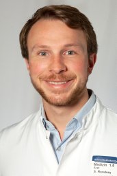 Simon Renders合作论文数German Canc Res Ctr, Div Stem Cells & Canc, Heidelberg, Germany18
 Katharina Schönberger合作论文数15
 Daniel Klimmeck合作论文数14Pia Sommerkamp合作论文数German Canc Res Ctr, Div Stem Cells & Canc, D-69120 Heidelberg, Germany13Zeisberger Petra合作论文数13Jasmin Rettkowski合作论文数12Luisa Ladel合作论文数12Robert Zeiser合作论文数12Katharina Schoenberger合作论文数11Julian Mess合作论文数9Geoffroy Andrieux合作论文数9
 Weiyu Zhang (张为宇)合作论文数College of Animal Science and Technology, Guangxi University9Melanie Boerries合作论文数Center for Biological Systems Analysis, University of Freiburg9
 Anna Lena Illert合作论文数8Mari Carmen Romero-Mulero合作论文数7
 Nadine Obier合作论文数7Buescher Joerg Martin合作论文数7
 Justus Duyster合作论文数Department of Hematology and Oncology, University Medical Center Freiburg, Freiburg, Germany7
 Sandro Altamura合作论文数University of Heidelberg7Karin Jacklein合作论文数6Adriana Przybylla合作论文数German Canc Res Ctr, Div Stem Cells & Canc, D-69120 Heidelberg, Germany6Michael Mitterer合作论文数6
 Khalid Shoumariyeh合作论文数Univ Freiburg, Med Ctr, Dept Hematol Oncol & Stem Cell Transplantat, Freiburg, Germany6
 Trompouki Eirini合作论文数Department of Cellular and Molecular Immunology, Max Planck Institute of Immunobiology and Epigenetics6Rosario Heck合作论文数5Carolin Wadle合作论文数5
 Maria-Eleni Lalioti合作论文数Medical School, University of Patras5
 Aikaterini Polyzou合作论文数Department of Cellular and Molecular Immunology, Max Planck Institute of Immunobiology and Epigenetics, Freiburg, Germany5
 Laurenti Elisa合作论文数Wellcome - MRC Cambridge Stem Cell Institute, University of Cambridge/Department of Haematology, University of Cambridge5
 Rodríguez-Fraticelli Alejo E合作论文数Ctr Biol Mol Severo Ochoa, CSIC5
 Teresa Poggio合作论文数5
 Jenny Hansson合作论文数European Molecular Biology Laboratory (EMBL), Genome Biology Unit, 69117 Heidelberg, Germany.5
 Wolfgang Huber合作论文数Molecular Medicine Partnership Unit, European Molecular Biology Laboratory5
 Dominic Grun合作论文数Würzburg Institute of Systems Immunology, The Julius Maximilians University of Würzburg;Max-Planck Institute of Immunobiology and Epigenetics5Marie Follo合作论文数Department of Medicine I, Medical Center – University of Freiburg and Faculty of Medicine, University Hospital Freiburg, Freiburg, Germany5
 Jeroen Krijgsveld合作论文数Medical Faculty, Heidelberg University;German Cancer Research Center5Ernesto Bockamp合作论文数5
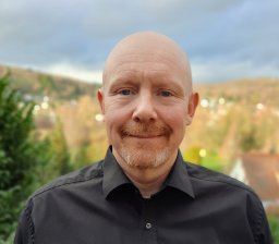 Michael Milsom合作论文数Harvard Medical School, Children’s Hospital Boston5
 Alejandro Reyes合作论文数Molecular Ecology and Evolution Laboratory and Centre for Coral Reef Biodiversity, James Cook University5
 Leonid Eshkind合作论文数5Wrobel Jan合作论文数4
 Carys Johnson合作论文数4Melissa Zwick合作论文数4Andreas Narr合作论文数Division of Stem Cells and Cancer, German Cancer Research Center (DKFZ) and DKFZ-ZMBH Alliance, 69120 Heidelberg, Germany4
 Jasper Panten合作论文数Division of Stem Cells and Cancer, Deutsches Krebsforschungszentrum (DKFZ), Heidelberg, Germany4
 Petya Apostolova合作论文数Univ Freiburg, Fac Med, Med Ctr, Dept Med 1, Freiburg, Germany4
 Erika Pearce合作论文数Department of Oncology, School of Medicine, Johns Hopkins University;Department of Biochemistry and Molecular Biology, Bloomberg School of Public Health, Johns Hopkins University4
 Heidt Timo合作论文数Department of Cardiology and Angiology, University of Freiburg4




