Although not a major determinant of receptivity, underexpression of the vitamin D receptor (VDR) is associated with endometrial dysfunction. This study aimed to determine VDR expression in the endometrium of patients with recurrent implantation failure (RIF) and unexplained infertility (UEI) using polymerase chain reaction (PCR) and immunohistochemistry. A total of 75 patients (25 RIF, 25 UEI, and 25 fertile controls) were enrolled in the study. In the control group, only patients with early luteal phase histology according to Noyes’ criteria were included to match the endometrial sampling timing of the RIF and UEI groups; patients with follicular and mid-luteal phase histology were excluded, and enrollment continued until 25 eligible patients were identified. Serum vitamin D (VD) levels were measured in all participants. Endometrial tissue was collected using a Pipelle cannula at the time of oocyte retrieval. VDR transcript variant 2 (VDR2) and 4 (VDR4) expression was analyzed using PCR. Endometrial tissue was subjected to VDR immunostaining. VDR2 expression in the RIF group (0.37 ± 0.02 vs. 1.01 ± 0.03, p < 0.001) and UEI group (0.28 ± 0.01 vs. 1.01 ± 0.03, p < 0.001) was significantly lower compared to the control group. VDR2 expression was similar in the RIF and UEI groups. VDR4 expression in the RIF group (0.46 ± 0.02) was significantly lower than that in the UEI (1.16 ± 0.07) and control (1.02 ± 0.05) groups (p < 0.001 for each comparison). VDR4 expression in the UEI and control groups was similar. The histo-score values of the RIF (0.49 ± 0.03) and UEI (0.35 ± 0.03) groups were significantly lower than those of the control group (1.24 ± 0.13) (p < 0.001 for both). In the RIF group, VDR2 was positively correlated with VD (r = 0.593, p = 0.002) and endometrial thickness (r = 0.627, p < 0.001). Low serum vitamin D levels in patients with RIF and UEI were found concomitantly with low VDR expression in the endometrium. Further research is needed to determine whether VD supplementation and adjustment of serum VD concentrations modulate VDR expression and its effects on embryo implantation.
To analyze the effects of hyperandrogenemia (HA) in polycystic ovary syndrome (PCOS) on negative and positive regulators of endometrial receptivity. Fifty-four women with PCOS undergoing total embryo freezing were classified into four phenotypes: classical type A, classical type B, ovulatory type C, and normoandrogenic type D. Hyperandrogenemia was present in phenotypes A-C and absent in phenotype D. Twenty-five age-matched infertile women without clinical or biochemical features of PCOS served as controls. Endometrial sampling was performed on day five after oocyte retrieval. Relative PCX mRNA and protein levels, together with HOXA10 and HOXA11 mRNA expression, were evaluated. Immunohistochemical analysis was performed to assess the spatial distribution of PCX, and H-score values were compared between the PCOS and control groups. PCX mRNA expression and protein concentration were significantly higher in the PCOS group compared with controls (all p < 0.001), consistent with the significantly higher H-score values observed in the PCOS group (p < 0.001). In contrast, HOXA10 and HOXA11 mRNA expression levels were significantly reduced in PCOS patients (all p < 0.001). Among PCOS phenotypes, those associated with hyperandrogenemia exhibited significantly lower HOXA10 and HOXA11 expression than the normoandrogenic phenotype (all p < 0.005). PCX mRNA and protein levels were significantly higher in classical and ovulatory hyperandrogenic phenotypes compared with the normoandrogenic group (all p < 0.005). PCX was identified as a negative predictor of HOXA10 and HOXA11 expression, while testosterone negatively predicted HOXA10 and HOXA11. Insulin resistance and testosterone were positive predictors of PCX expression, whereas progesterone levels on the day of oocyte retrieval and on day five after retrieval were negative predictors of PCX expression. Hyperandrogenemia disrupts the physiological mid-luteal downregulation of PCX and the upregulation of HOXA10 and HOXA11, leading to impaired endometrial receptivity and displacement of the implantation window in PCOS.
PROBLEM:Immunomodulatory agents (IVIG, intralipid, corticosteroids, and G-CSF) and platelet-rich plasma (PRP) are increasingly used in infertility despite inconsistent clinical outcomes. A critical but overlooked question is whether these agents exert their effects through systemic immune modulation or require direct access to target cells within the ovary and endometrium. HYPOTHESIS:We propose that insufficient tissue accessibility, rather than intrinsic biological inefficacy, may contribute to inconsistent clinical outcomes. This framework examines the physical constraints governing therapeutic effectiveness if local action were necessary, while acknowledging that benefits may also be mediated systemically. FINDINGS:Anatomical and biophysical barriers differ by administration route. For systemic agents, the vascular endothelium (continuous capillaries) restricts macromolecular extravasation via capacity-limited transcytosis. For local agents, epithelial tight junctions (0.5-2 nm pores), follicular basal lamina (40-100 nm), granulosa cell layers (∼20 nm), and the zona pellucida (5-10 nm) create sequential barriers. Molecular size analysis suggests that large immunomodulators like IVIG (∼150 kDa, 10-15 nm) and intralipid particles (200-500 nm) face substantial limitations in reaching target compartments. PRP growth factors face identical barriers. By contrast, smaller molecules (<1 kDa) and FSH (∼30 kDa, 5 nm) penetrate more readily. Ovarian tissue stiffness (1.5-5 kPa in normal ovaries; 0.9-11.3 kPa in premature ovarian insufficiency) further impedes macromolecular diffusion. The proposed Mathematical Access Index integrates molecular size, barrier pore dimensions, and tissue stiffness to predict accessibility. CONCLUSION:Accessibility of immunomodulators and PRP growth factors to reproductive tissues is more limited than commonly assumed. If these therapies act predominantly through systemic immune modulation (e.g., Fc receptor saturation, Th1/Th2 modulation), direct tissue penetration may not be necessary. For PRP, observed effects likely result from indirect mechanisms (paracrine signaling, angiogenesis). Distinguishing between systemic and local mechanisms is essential. Until pharmacokinetic studies quantify drug concentrations in reproductive tissues, clinical use should remain cautious.
Reproductive aging in the ovary, endometrium, and testis is characterized by progressive decline in regenerative capacity, yet the upstream mechanisms coordinating stem cell dysfunction across these tissues remain incompletely understood. Emerging evidence suggests that alterations in extracellular matrix (ECM) mechanics, particularly age and disease associated increases in tissue stiffness, may contribute to impaired stem cell function through mechanotransductive signaling pathways. In the testis, increased matrix stiffness has been associated with activation of mechanosensitive pathways such as Piezo1, leading to calcium influx, mitochondrial stress, and downstream effects on steroidogenic and stem cell populations. In the ovary, age-related fibrosis and cortical stiffening correlate with disrupted follicular development and altered signaling through Hippo-YAP/TAZ pathways. Similarly, the endometrium exhibits stiffness-dependent functional changes, where excessive ECM remodeling is associated with impaired decidualization and reduced regenerative potential. Across these reproductive tissues, mechanotransduction pathways involving calcium signaling, reactive oxygen species generation, and YAP/TAZ activity appear to integrate biomechanical cues with cellular stress responses, potentially contributing to stem cell exhaustion. However, current evidence remains largely correlative, and direct causal links between tissue stiffness and stem cell failure in human reproductive tissues are still limited. We propose the “Hardened Ground” framework as a unifying working model suggesting that increased ECM stiffness may represent a contributory biomechanical factor influencing stem cell function across the reproductive axis. Importantly, this model does not replace established molecular and hormonal mechanisms of aging but instead integrates mechanical properties of the tissue microenvironment as an additional regulatory layer. Notably, clinical observations such as preserved follicular reserve in polycystic ovary syndrome despite increased ovarian stiffness, and dynamic changes in endometrial stiffness across the menstrual cycle, indicate that mechanotransduction outcomes are context-dependent and influenced by hormonal and inflammatory states. Future studies integrating quantitative elastography with stem cell and molecular markers in human tissues are needed to clarify the causal role of stiffness in reproductive aging. This framework generates testable predictions and highlights potential therapeutic avenues targeting extracellular matrix remodeling and mechanosensitive signaling pathways to preserve reproductive tissue function.
Anti-Müllerian hormone (AMH) is widely used as a clinical biomarker of ovarian reserve and is traditionallyinterpreted as a surrogate measure of remaining oocyte quantity. However, accumulating biological and clinicalevidence challenges this quantitative paradigm. AMH is exclusively produced by granulosa cells of growing folliclesrather than by primordial follicles themselves, suggesting that circulating AMH primarily refl ects somatic follicularactivity instead of dormant oocyte pool size. Here, we propose a conceptual framework redefi ning ovarian aging as aprocess that may be strongly infl uenced by progressive somatic ovarian dysfunction. In this model, granulosa cells, stromal integrity, vascular support, immune regulation, and metabolicenvironment collectively form a somatic support network that determines follicular survival and developmentalcompetence. Disruption of this somatic ecosystem, through aging, surgery, chemotherapy, autoimmunity,environmental toxicants, smoking, or metabolic stress, results in reduced granulosa cell functionality, declining AMHsecretion, impaired follicle maturation, and secondary oocyte loss. Evidence from granulosa cell biology, controlledovarian stimulation, ovarian surgery, autoimmune ovarian disease, chemotherapy exposure, and fertility outcomestudies consistently demonstrates that AMH responds dynamically to changes in somatic ovarian health and doesnot reliably predict natural fecundability or absolute follicle number. Primordial follicle depletion progresses continuously throughout life, yet circulating AMH levels often showabrupt declines in response to somatic ovarian injury such as surgery, chemotherapy, or metabolic stress.Continuous primordial follicle attrition therefore does not translate into continuous AMH decline, supporting the viewthat AMH represents the functional cohort of biologically supported follicles rather than the total ovarian reserve. It isimportant to recognize, however, that ovarian reserve markers including AMH have limited predictive value fornatural fecundability with area under the curve values ranging from 0.60 to 0.65. We introduce the concept of somatic ovarian function as an integrated framework for AMHinterpretation, proposing AMH as a biomarker of ovarian functional capacity. Reframing AMH from a purelyquantitative reserve marker to a functional systems biomarker that refl ects granulosa cell integrity, metabolichealth, and environmental infl uences may help reconcile longstanding clinical paradoxes and open new translationalavenues for fertility preservation, ovarian aging research, and therapeutic intervention.
BACKGROUND:Homeobox genes (HOX) are the basic molecules that regulate endometrial receptivity, decidualization, and progesterone response. This study was designed to investigate the effects of the classical hysterosalpingography (HSG) procedure on endometrial HOXA-10 and 11 mRNA expression. METHODS:Thirty-five primary infertile patients who applied for investigation of infertility etiology and were approved for conventional HSG were included in the study. Ten fertile patients who required biopsy due to an endometrial pathology were accepted as a control group. Women in HSG group were eligible to participate in the study if they were between 22 and 34 years of age, had spontaneous menstrual cycles, and had been trying to conceive for at least 1.5 year and if there was an indication for evaluation of tubal patency with hysterosalpingography. First endometrial sampling was performed with a pipelle before the contrast-medium infusion during the HSG. Endometrial sampling was collected for the second time with a pipelle cannula in the midluteal phase of the next cycle in all cases undergoing HSG. HOXA-10 and 11 mRNA expressions in the endometrial tissues obtained before HSG and respective endometrial tissues after HSG were measured with RT-PCR. RESULTS:Pre-HSG average ΔCt values of HOXA-10 and HOXA-11 mRNA were found to be significantly lower than fertile controls (4.30 vs 6.74, P < .001; 3.93 vs 6.74, P < .002). Post-HSG HOXA-10 mRNA levels increased significantly compared to pre-HSG levels (5.66 vs 4.30, P < .01). Post-HSG HOXA-10 mRNA levels increased approximately 4.2-fold compared to pre-HSG levels. Post-HSG HOXA-11 mRNA levels increased significantly compared to pre-HSG levels (4.94 vs 3.93, P < .03). Post-HSG HOXA-11 mRNA levels increased approximately 3.5-fold compared to pre-HSG levels. When the pre-HSG and post-HSG HOXA-10 and HOXA-11 mRNA levels were compared among themselves, the increase in HOXA-10 average ΔCt was significantly higher than the increase in HOXA-11 average ΔCt (5.66 vs 4.94, P < .02.). Similarly, the fold increase in post-HSG mRNA levels was significantly higher in the HOXA-10 group (4.2-fold) than the HOXA-11 group (3.5-fold) (P < .001). CONCLUSIONS:Conventional HSG procedure improves fertility by increasing endometrial HOXA-10 and HOXA-11 mRNA levels.
Research question: Does endometrial thinning affect the physiological transition of the endometrium from a non-receptive to a receptive state? Design: Fifty-eight women who underwent total embryo freezing were divided into three groups according to their endometrial thickness (EMT): group 1, EMT <= 7 mm; group 2, 7 mm < EMT <= 10 mm; and group 3, EMT > 10 mm. Group 1 was considered as having a thin EMT and groups 2 and 3 as a normal EMT. Endometrial sampling was performed at the time of oocyte retrieval. Anti-adhesive podocalyxin (PDX), adhesive homeobox A10 and A11 (HOXA10, HOXA11) and leukaemia inhibitory factor (LIF) mRNA, proinflammatory cytokines, oxidative stress markers and collagen deposition were determined. Results: Compared with groups 2 and 3, the relative expression of HOXA10, HOXA11 and LIF mRNA was down-regulated in group 1 (all P < 0.001), while PDX levels significantly increased (P < 0.001). The nuclear factor-kappa B, long pentraxin 3 (PTX3), tumour necrosis factor-alpha and total oxidant status of the thin endometrium group were significantly higher than those of participants with normal endometrium (all P < 0.001), while total antioxidant status was significantly lower (P < 0.001). The histoscore values for PTX3, PDX and Masson's trichrome were significantly higher in the thin endometrium group (P < 0.001 for each). Each millimetre increase in EMT decreased the risk of down-regulation of adhesive receptivity genes. The adjusted odds ratio for PDX was 1.69, representing a 69% increase in PDX expression. Conclusion: Endometrial thinning causes defective expression of anti-adhesive and adhesive receptivity modulators, restricting the transition of the endometrium from a non-receptive to a receptive state.
(Abstracted from Reprod Biomed Online 2025;51(2):104697, doi:10.1016/j.rbmo.2024.104697) Receptivity modulators control and facilitate cell differentiation and embryo development as well as inflammatory pathways and suppression of paternal alloantigens. Molecular changes driven by the microenvironment of the endometrium drive endometrial thickening that prepares the uterus for implantation.
Background A fine-tuned pro-inflammatory and anti-inflammatory balance in the follicular unit is essential for cumulus expansion and successful ovulation. While the long pentraxin 3 (PTX3) gene is required for the expansion of cumulus cells (CCs), ovulation, resumption of meiosis and fertilization, the vitamin D receptor gene (VDR-X2) is required for intra-follicle redox balance. This study was planned to determine the expression pattern of VDR-X2 and PTX3 mRNA in CCs isolated from germinal vesicle (GV), metaphase I (MI), and metaphase II (MII) oocytes of PCOS patients with ovulatory dysfunction. Methods The relative expression of CC-PTX3 and CC-VDR-X2 mRNA were evaluated using qRT-PCR in a total of 79 CC samples collected from individual cumulus-oocyte complex of 40 infertile patients (20 PCOS and 20 non-PCOS normal responders) who underwent ovarian stimulation with the GnRH antagonist protocol. Results Relative PTX3 mRNA expressions of CCMI-control and CCMII-control showed 3- and 9-fold significant upregulation compared to CCGV-control, respectively. The relative PTX3 mRNA expression of CCMII-control increased approximately three fold compared to CCMI-control. Compared to CCGV-pcos, a 3-fold increase was noted in the relative PTX3 mRNA expression of CCMI-pcos and an approximately 4-fold increase in the PTX3 mRNA expression of CCMII-pcos. Relative PTX3 mRNA expression values of CCMII-pcos and CCMI-pcos were similar. A 6-fold upregulation of relative PTX3 mRNA and a 4-fold upregulation of VDR-X2 mRNA were detected in CCMII-control compared to CCMII-pcos. CC-VDR-X2 expression patterns of the PCOS and control groups overlapped with the CC-PTX3 pattern. Fertilization rates of the PCOS group exhibiting failed transcript expression were similar to normal responders. Conclusion The fact that relative CC-PTX3 and CC-VDR mRNA expression does not increase during the transition from MI to MII stage in PCOS as in normal responders suggests that PTX3 and VDR expression may be defective in cumulus cells of PCOS patients with ovulatory dysfunction.
Objective: To determine whether the urinary excretion of podocyte degradation products varies according to PCOS phenotype and metabolic syndrome (MetS). Methods: The concentrations of podocalyxin (PDX) and nephrin, chronic markers of podocyte damage, and neutrophil gelatinase-associated lipocalin (NGAL), a marker of acute glomerular damage, were analyzed in the morning urine samples of 50 PCOS patients and 50 healthy controls matched by age and BMI. Albuminuria was assessed by calculating the urine albumin–creatinine ratio (uACR). Results: The PDX, nephrin and NGAL concentrations of PCOS participants were significantly higher than those of the control group. While PDX, nephrin and NGAL levels of classic phenotypes were similar, they were higher than ovulatory and non-hyperandrogenic phenotypes. Significant increases in urinary levels of each podocyte protein were detected in PCOS patients with MetS compared to patients without MetS. A positive significant correlation between podocyte proteins and BMI, systolic blood pressure, testosterone, glucose, HOMA-IR and uACR. After adjusting for age and BMI, podocyte proteins were an independent risk factor for microalbuminuria. The incidence of microalbuminuria in PCOS increased 6-fold compared to controls. The frequency of microalbuminuria was higher in classical phenotypes than in ovulatory phenotype. The frequency of microalbuminuria in PCOS patients with MetS was 6.5 times higher than in PCOS patients without MetS. Conclusions: In PCOS accompanied by hyperandrogenemia or metabolic syndrome, leakage of acute and chronic podocyte breakdown products into the urine becomes more pronounced.
OBJECTIVE:The aim of the study was to determine the expression pattern of long pentraxin 3 (PTX3) mRNA in cumulus cells (CCs) isolated from metaphase II oocytes of women with unilateral endometrioma undergoing controlled ovarian stimulation using a gonadotropin-releasing hormone antagonist (GnRHa) protocol. PATIENTS AND METHODS:A total of 60 CC samples, 30 from the affected ovary and 30 from the contralateral ovary, were collected from 12 patients with unilateral endometrioma who underwent flexible GnRHa protocol with recombinant human chorionic gonadotropin (rhCG) trigger. Thirty CC samples collected from the left ovary of 12 women with male factor infertility were used as external controls. Long PTX3 mRNA expression in each group was analyzed by real-time polymerase chain reaction (RT-PCR). Relative gene expression (fold-change) was calculated according to the 2-ΔΔCt equation. Fertilization rates after intracytoplasmic sperm injection (ICSI) were recorded for each group. RESULTS:CC-PTX3 mRNA expression in the unilateral endometrioma group was significantly lower than the mRNA expression of the disease-free ovary (0.90±0.01 vs. 0.25±0.02, p<0.01). CC-PTX3 mRNA expression of MII oocytes of the disease-free ovary was found to be similar to the control group (1.02±0.03 vs. 0.90±0.01, p=0.107). A significant 3.6-fold downregulation was observed in the CC-PTX3 mRNA expression of the endometrioma group compared to the CC-PTX3 mRNA expression in the contralateral ovary. CC-PTX3 mRNA expression in the endometrioma group was downregulated 4.08-fold compared to the CC-PTX3 mRNA expression of the control group (1.02±0.03 vs. 0.25±0.02, p<0.001). The cumulus morphologies of the endometrioma group with low CC-PTX3 expression and the groups with normal CC-PTX3 levels were similar. Fertilization rates of the endometrioma group were similar to the contralateral ovary and control groups. CONCLUSIONS:Unilateral endometrioma reduces CC-PTX3 expression but does not affect disease-free ovaries. The GnRHa protocol improved the fertilization rates, suggesting that failed CC-PTX3 expression is an in vivo pathology.
Objectives To determine the effects of hyperandrogenemia and other phenotypic parameters on endometrial vitamin D receptor (VDR-X2 and VDR-X4) expression in women with polycystic ovary syndrome (PCOS) undergoing ovarian stimulation and total embryo freezing.Methods Forty-four PCOS patients were divided into four phenotypes according to the criteria for hyperandrogenemia (HA), ovulatory dysfunction (OD), and polycystic ovary morphology (PCOM): phenotype A (HA+OD+PCOM), phenotype B (HA+OD), phenotype C (HA+PCOM), and phenotype D (OD+PCOM). Endometrial VDR expression was determined by real-time PCR and immunohistochemistry. Twenty age- and body mass index (BMI)-matched couples with male infertility were included as controls.Results VDR-X2 and VDR-X4 expression levels were significantly lower in the PCOS group than in the control group. A significant downregulation was detected in the relative VDR-X2 and X4 expression in phenotypes A, B, and C compared to the control group. VDR-X2 and X4 expression in phenotype D was significantly higher than in phenotypes A and B. A significant negative correlation was detected among VDR-X2, VDR-X4, serum testosterone (T), androstenedione (A), DHEAS, and insulin resistance (IR). Multivariate analysis revealed that serum T, A, DHEAS, and IR levels were independently associated with both VDR-X2 and VDR X4 relative gene expression after adjusting for age and BMI. The VDR mRNA and immunoreactivity of each phenotype overlapped. The clinical pregnancy rates for each phenotype were similar.Conclusion VDR expression in the endometria of patients with PCOS was defective. Hyperandrogenemia and insulin resistance are the key drivers of defective VDR expression in the endometrium of patients with PCOS.
BACKGROUND:Homeobox genes A10 (HOXA10) and A11 (HOXA11), members of the abdominal B gene family, are responsible for embryonic survival and implantation. This study was planned to investigate whether endometrial injury alters the expression of both transcripts in women with implantation failure.METHODS:A total of 54 women with implantation failure were divided into two equal groups as experimental (scratching) and sham (no scratching). Participants in the scratching group were exposed to endometrial injury in the mid-luteal phase, and those in the sham group were exposed to endometrial flushing. The scratching group, but not the sham group, underwent prior endometrial sampling. A second endometrial sampling was performed on the scratching group in the mid-luteal phase of the following cycle. The mRNA and protein levels of the HOXA10 and 11 transcripts were determined in endometrial samples collected before and after injury/flushing. Participants in each group underwent IVF/ET in the cycle after the second endometrial sampling.RESULTS:Endometrial injury caused a 60.1-fold (p < 0.01) increase in HOXA10 mRNA and a 9.0-fold increase in HOXA11 mRNA (p < 0.02). Injury resulted in a significant increase in both HOXA10 (p < 0.001) and HOXA11 protein expression (p < 0.003). There was no significant change in HOXA10 and 11 mRNA expressions after flushing. Clinical pregnancy, live birth, and miscarriage rates of the both groups were similar.CONCLUSIONS:Endometrial injury increases homeobox transcript expression at both mRNA and protein levels.
Background : Analyzing expression patterns of receptivity genes is a minimally invasive diagnostic method to identify the underlying cause of subfertility in women with uterine fibroid with a history of implantation failure or recurrent pregnancy loss. This updated systematic review was designed to determine the molecular and genetic changes in the endometrium of women with fibroid and how myomctomy affect the outcome of spontaneous or assisted conception treatment. We also discussed the extent to which we should consider the effects of fibroids on endometrial receptivity when deciding whether or not to perform myomectomy. Methods : A total of 184 articles reached as a result of PubMed research and meeting the selection criteria, were evaluated. Of these, 28 full text articles on uterine leiomyoma and endometrium, leiomyoma and receptivity, fibroid and implantation, myomectomy and implantation, fibroid and genes, fibroid surgery and receptivity, fibroid and uterine peristalsis, fibroid and immune cell were evaluated. Results : The endometrium of subfertile women with fibroid appears to have a disease specific pattern according to the type of the fibroid. The response of the endometrium to a fibroid may vary depending on whether the fibroid is close or far from it. Leiomyomas that contribute to subfertility must be near to or in contact with the endometrium, as is the case for Types 0, 1 and 2 leiomyomas. The proximity to the endometrial cavity makes the effect of fibroid on the endometrium more pronounced. While Type 3 fibroid causes subfertility similar to submucosal fibroids, the subfertility-producing effects of Type 4 fibroids have not been clearly clarified. However, the fact that the fibroid is far from the cavity should not mean that it has no effect on the endometrium. The mechanical stress created by a Type 4 fibroid that is not connected to the endometrium may be converted into biological signal and disrupt receptivity. Data on whether myomectomy restores impaired receptivity are mostly based on clinical observations, and studies evaluating endometrial receptivity before and after myomectomy are very few. Conclusions : Analysis of receptivity genes in subfertile women with fibroid may assist the clinician in deciding whether or not to perform myomectomy. If it is determined whether fibroids affect receptivity other than their mechanical effects, the indications for myomectomy may expand or narrow.
The objective of our study was to investigate the possible relationship between poor perinatal outcome and foetal cardiac functions in pregnant women with reduced foetal movements (RFM). This cross-sectional study included 126 pregnant women with normal foetal movements (Group 1, Controls) and 42 pregnant women over 32 weeks gestation with RFM (Group 2). Group 2 was further divided into two subgroups according to their perinatal outcome: normal perinatal outcome (Group 2a) and poor perinatal outcome (Group 2b). Cardiotocography, the E/A ratio in both atrioventricular valves, myocardial performance index (MPI) and foetal tricuspid annular plane systolic excursion (f-TAPSE) were evaluated. Foetuses with poor perinatal outcome had a higher MPI (p = .003), higher tricuspid and mitral E/A (p < .001), and lower f-TAPSE values (p < .001). In regression analysis, f-TAPSE was the only parameter (p = .04) independently associated with poor perinatal outcome. In conclusion, examining f-TAPSE may predict adverse perinatal outcome in pregnancies with RFM.IMPACT STATEMENT What is already known on this subject? Reduced foetal movement (RFM) is associated with adverse pregnancy outcome. Cardiotocography, amniotic fluid assessment, estimated birthweight, foetal Doppler and formal foetal movement count (kick chart) are generally used in the clinical assessment of pregnancies with reduced foetal movements. These tests, we currently use to assess foetal wellbeing in women with reduced foetal movements, have limited sensitivity in predicting foetal compromise. What do the results of this study add? Foetal cardiac Doppler may potentially be used as an important adjunct to the conventional management of women with a perception of reduced foetal movements. What are the implications of these findings for clinical practice and/or further research? Foetal echocardiographic evaluation, such as f-TAPSE, may influence clinical practice by enabling improved risk stratification for poor perinatal outcome, thus allowing more timely definitive intervention. This could help to decrease the rate of stillbirth related to reduced foetal movements. The few established echocardiographically derived parameters, which can asses global right ventricle function, are not always easy to obtain, however, f-TAPSE is easily obtainable using ultrasound and it appears to be a clinically useful echocardiographic measurement of right ventricular function.
OBJECTIVE: The aim of the study was to investigate the existence of neuroendocrine cells and to compare the density of those in normal ovarian tissue, endometriotic and non-endometriotic benign ovarian cysts. PATIENTS AND METHODS: Twenty patients with the diagnosis of endometrioma and 30 control subjects consisting of ovarian serous cystadenoma (n=10), ovarian mucinous cystadenoma (n=10) and normal ovarian tissue (n=10) were included. The tissues were prepared and assessed according to staining density by using the H-score method. RESULTS: Tissues with mucinous cystadenoma were significantly more stained with PAS and VanGieson, when compared to women with endometrioma. Macrophage deposition was higher in cyst samples with endometrioma and in normal ovarian tissue when compared to serous cystadenoma and mucinous cystadenoma. Normal ovarian tissue was significantly more stained with PGP9.5, NSE and SYN when com-pared to endometrioma and non-endometriotic benign ovarian cyst. PGP9.5 staining was higher in normal ovarian tissue when compared with endometriotic lesions (p <.001). Endometrioma samples were significantly more stained with p53 when compared to non-endometriotic cysts and normal ovarian tissue. c-Kit staining was mild and not statistically significant among all groups. CONCLUSIONS: During endometrioma trans-formation, expression intensity of neuroendocrine markers decreases compared to normal ovarian tissue and other benign ovarian cysts.
OBJECTIVE: DNA repair genes may be related with the onset of primary ovarian failure (POF). The study was planned to investigate whether the polymorphisms in the DNA repair genes modulate the risk of POF. PATIENTS AND METHODS: This prospective study included 25 women diagnosed with POF and 25 healthy controls. The genotyping and allele of XRCC1 and XPD genes were determined by using Polymerase Chain Reaction and fluorescence melting curve analysis. RESULTS: The genotype and allele distribution of the Arg194Trp and Arg399Lys polymorphism of the XRCC1 gene did not differ statistically between those of the POF and control groups. The frequency of the C (Gln) allele was significantly lower in patients diagnosed with POF when compared to that in healthy controls [48% vs. 76%, p=0.040, OR: 3.43 (CI: 1.03-11.48)]. The Lys/Lys genotype for XPD-Lys751Gln polymorphism in patients diagnosed with POF was more common than in healthy controls (p=0.028, 52% vs. 24%). CONCLUSIONS: The genotype distribution and allele frequency of XPD-Lys751Gln, XRCC1-Arg194Trp and XRCC1-Arg399 Gln did not regulate the risk of developing POF. Gln/Gln+Lys/Gln and XPD-Lys751Gln polymorphism may have a possible protective role against the development of POF.
Myometrium cells are an important reproductive niche in which cyclic mechanical forces of a pico-newton range are produced continuously at millisecond and second intervals. Overproduction and/or underproduction of micro-forces, due to point or epigenetic mutation, aberrant methylation, and abnormal response to hypoxia, may lead to the transformation of fibroid stem cells into fibroid-initiating stem cells. Fibroids are tumors with a high modulus of stiffness disturbing the critical homeostasis of the myometrium and they may cause unfavorable and strong mechanical forces. Micro-mechanical forces and soluble-chemical signals play a critical role in transcriptional and translational processes’ maintenance, by regulating communication between the cell nucleus and its organelles. Signals coming from the external environment can stimulate cells in the format of both soluble biochemical signals and mechanical ones. The shape of the cell and the plasma membrane have a significant character in sensing electro-chemical signals, through specialized receptors and generating responses, accordingly. In order for mechanical signals to be perceived by the cell, they must be converted into biological stimuli, through a process called mechanotransduction. Transmission of fibroid-derived mechanical signals to the endometrium and their effects on receptivity modulators are mediated through a pathway known as solid-state signaling. It is not sufficiently clear which type of receptors and mechanical signals impair endometrial receptivity. However, it is known that biomechanical signals reaching the endometrium affect epithelial sodium channels, lysophosphatidic acid receptors or Rho GTPases, leading to conformational changes in endometrial proteins. Translational changes in receptivity modulators may disrupt the selectivity and receptivity functions of the endometrium, resulting in failed implantation or early pregnancy loss. By hypermethylation of the receptivity genes, micro-forces can also negatively affect decidualization and implantation. The purpose of this narrative review is to summarize the state of the art of the biomechanical forces which can determine fibroid stem cell transformation and, thus, affect the receptivity status of the endometrium with regard to fertilization and pregnancy.
Although many pregnant women have been infected by coronavirus, the presence of intrauterine vertical transmission has not been conclusively reported yet. What prevents this highly contagious virus from reaching the fetus? Is it only the presence of a strong placental barrier, or is it the natural absence of the some receptor that the viruses use for transmission? We, therefore, need to comprehensively understand the mechanism of action of the mammalian epithelial barriers located in two different organs with functional similarity. The barriers selected as potential targets by SARS-CoV-2 are the alveolo-capillary barrier (ACB), and the syncytio-capillary barrier (SCB). Caveolae are omega-shaped structures located on the cell membrane. They consist of caveolin-1 protein (Cav-1) and are involved in the internalisation of some viruses. By activating leukocytes and nuclear factor-κB, Cav-1 initiates inflammatory reactions. The presence of more than one Cav-1 binding sites on coronavirus is an important finding supporting the possible relationship between SARS-CoV-2-mediated lung injury. While the ACB cells express Cav-1 there is no caveolin expression in syncytiotrophoblasts. In this short review, we will try to explain our hypothesis that the lack of caveolin expression in the SCB is one of the most important physiological mechanisms that prevents vertical transmission of SARS-CoV-2. Since the physiological Cav-1 deficiency appears to prevent acute cell damage treatment algorithms could potentially be developed to block this pathway in the non-pregnant population affected by SARS-CoV-2.
 Seyma Hascalik合作论文数80
 Nilufer Celik合作论文数56
 Yavuz Simsek合作论文数Faculty of Medicine, Inonu University36Ebru Celik合作论文数Sch Med, Koc Univ32Abdullah Karaer合作论文数Department of Obstetrics and Gynecology, Dr. Zekai Tahir Burak Woman Health Education and Research Hospital28Suleyman Aydin合作论文数27Bulent Mizrak合作论文数School of Medicine, Inonu University26
 Yilmaz Ercan合作论文数Department of Obstetrics and Gynecology, Inonu University School of Medicine25
 Ilgın Turkcuoglu合作论文数Faculty of Medicine, Inonu University23
 Aydin Nasuhi Engin合作论文数Tibbi Patoloji Anabilim Dali, Inönü Universitesi18
 Aynur Ersahin合作论文数16
 Hakki Muammer Karakas合作论文数Faculty of Medicine, İnonu University16Elif Ozerol合作论文数Department of Biochemistry, Inonu University Medical School16Sudenaz Celik合作论文数15
 Yusuf Turkoz合作论文数15Saim Yologlu合作论文数14Mehmet Hascalik合作论文数14
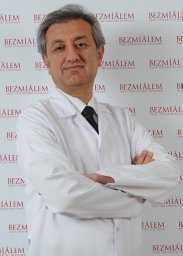 Alpay Alkan合作论文数Turgut Ozal Medical Center, Inonu University School of Medicine13
 Erdal Yilmaz合作论文数Medical School, Gazi University;Department of Surgery;Medical School, Gazi University12
 Ibrahim Sahin合作论文数Ankara Postgraduate Training Hospital12Gulnur Erdem合作论文数Department of Radiology, Inonu University School of Medicine11
 Meltem Yardim合作论文数10
 Nur DOKUZEYLÜL GÜNGÖR合作论文数Medical Park Hospital, Bahcesehir University Göztepe10
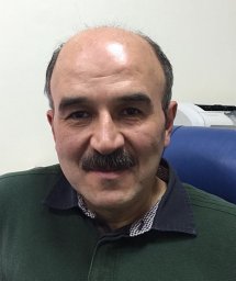 Kaya Sarac合作论文数Department of Radiology, Inonu University School of Medicine10
 Semih Dalkilic合作论文数10Yilmaz Cigremis合作论文数Art and Science Faculty, Inonu University10Cihat Unlu合作论文数10
 Ibrahim Halil Ozerol合作论文数Department of Microbiology & Clinical Microbiology, Inonu University School of Medicine9Yagmur Minareci合作论文数8
 Bulent Duran合作论文数Faculty of Medicine, Abant Izzet Baysal University8Mehmet Emin Tagluk合作论文数Electrical Electronics Engineering Department, Inonu University7
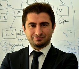 Mustafa Ulas合作论文数7Tanbek Kevser合作论文数7Mustafa Tamser合作论文数7
 Safak Hatirnaz合作论文数ART and Genetics Center, Istanbul Memorial Hospital7
 Pinar Kadioglu合作论文数University of Istanbul Division of Endocrinology and Metabolism, Cerrahpasa Medical School Ic Hastalıkları Anabilim Dali7
 Mustafa Acet合作论文数6
 Kader Ugur合作论文数Dept Internal Med Endocrinol & Metab Dis, Firat Univ6
 Kagan Gungor合作论文数Goztepe Prof Dr Suleyman Yalcin City Hosp, Istanbul Medeniyet Univ6
 Tuncay Kuloglu合作论文数Dept Histol & Embryol, Firat Univ6
 Banu Kumbak合作论文数Department of Obstetric and Gynecology, Firat Universitesi Hastanesi6Ahmet Kemal Firat合作论文数6
 Baris Otlu合作论文数Tıp Fakültesi, Temel Tıp Bilimleri, Inonu University6
 Muhittin Yurekli合作论文数Faculty of Arts and Sciences, Inonu University6
 Ulun Ulug合作论文数Bahçeci Women Health Care Centre and German Hospital at Istanbul6Ugur Turhan合作论文数5Mehmet Gul合作论文数5
 Arzu Yurci合作论文数Andrology and Genetics Center, Memorial Bahcelievler Hospital5
 Ahmet Tektemur合作论文数Faculty of Medicine, Firat University5




