The major spliceosome contains five small nuclear RNAs (snRNAs; U1, U2, U4, U5 and U6) essential for splicing. Variants in RNU4-2, encoding U4, cause a neurodevelopmental disorder called ReNU syndrome. We investigated de novo variants in 50 snRNA-encoding genes in a French cohort of 23,649 individuals with rare disorders and gathered additional cases through international collaborations. Altogether, we identified 145 previously unreported probands with (likely) pathogenic variants in RNU4-2 and 21 individuals with de novo and/or recurrent variants in RNU5B-1 and RNU5A-1, encoding U5. Pathogenic variants typically arose de novo on the maternal allele and cluster in regions critical for splicing. RNU4-2 variants mainly localize to two structures, the stem III and T-loop/quasi-pseudoknot, which position the U6 ACAGAGA box for 5' splice site recognition and associate with different phenotypic severity. RNU4-2 variants result in specific defects in alternative 5' splice site usage and methylation patterns (episignatures) that correlate with variant location and clinical severity. This study establishes RNU5B-1 as a neurodevelopmental disorder gene, suggests RNU5A-1 as a strong candidate and highlights the role of de novo variants in snRNAs.
BACKGROUND:Decisions to split two or more phenotypic manifestations related to genetic variations within the same gene can be challenging, especially during the early stages of syndrome discovery. Genotype-based diagnostics with artificial intelligence (AI)-driven approaches using next-generation phenotyping (NGP) and DNA methylation (DNAm) can be utilized to expedite syndrome delineation within a single gene. METHODS:We utilized an expanded cohort of 56 patients (22 previously unpublished individuals) with truncating variants in the MN1 gene and attempted different methods to assess plausible strategies to objectively delineate phenotypic differences between the C-Terminal Truncation (CTT) and N-Terminal Truncation (NTT) groups. This involved transcriptomics analysis on available patient fibroblast samples and AI-assisted approaches, including a new statistical method of GestaltMatcher on facial photos and blood DNAm analysis using a support vector machine (SVM) model. FINDINGS:RNA-seq analysis was unable to show a significant difference in transcript expression despite our previous hypothesis that NTT variants would induce nonsense mediated decay. DNAm analysis on nine blood DNA samples revealed an episignature for the CTT group. In parallel, the new statistical method of GestaltMatcher objectively distinguished the CTT and NTT groups with a low requirement for cohort number. Validation of this approach was performed on syndromes with known DNAm signatures of SRCAP, SMARCA2 and ADNP to demonstrate the effectiveness of this approach. INTERPRETATION:We demonstrate the potential of using AI-based technologies to leverage genotype, phenotype and epigenetics data in facilitating splitting decisions in diagnosis of syndromes with minimal sample requirement. FUNDING:The specific funding of this article is provided in the acknowledgements section.
The Mediator complex regulates protein-coding gene transcription by coordinating the interaction of upstream enhancers with the basal transcription machinery at the promoter. Pathogenic variants in Mediator subunits typically lead to neurodevelopmental or neurodegenerative disorders with variable clinical presentations, designated as MEDopathies. Here, we report the identification of 25 individuals from 18 families with bi-allelic MED16 variants who have a multiple congenital anomalies (MCAs)-intellectual disability syndrome. Intellectual disability, speech delay, and/or motor delay of variable severity were constant and associated with variable combinations of craniofacial defects (micro/retrognathia, cleft palate, and preauricular tags), anomalies of the extremities, and heart defects (predominantly tetralogy of Fallot). Visual impairment, deafness, and magnetic resonance imaging (MRI) abnormalities were also frequent. The 26 variants identified were comprised of eight predicted protein-truncating (three intragenic deletions, two frameshifts, and one nonsense and two essential splice site variants) and 18 missense or in-frame duplication variants affecting conserved residues, without clear correlation between phenotypic severity and variant type combination. Three-dimensional modeling indicated that the missense and duplication variants likely have a destabilizing effect on the structural elements of the protein. Immunofluorescence assays demonstrated protein mislocalization from the nucleus to the cytoplasm for 16 of the 17 variants studied. Homozygous mutant med16 zebrafish presented growth delay and increased mortality compared with wild-type fish, and Med16 knockout mice are preweaning lethal, highlighting the conserved requirement of MED16 for development. Overall, we describe an autosomal recessive MCAs-intellectual disability MEDopathy, emphasizing the importance of Mediator during neurodevelopment and suggesting that some tissues are particularly sensitive to the loss of certain subunits.
OBJECTIVE:To report the incidental detection of maternal somatic mosaicism during the development of exclusion-based non-invasive prenatal diagnosis for monogenic disorders (NIPD-MD) initially indicated for apparently de novo pathogenic or likely pathogenic variants. METHOD:A droplet digital PCR (ddPCR)-based exclusion NIPD_MD assay was developed for four couples, each with a prior pregnancy affected by a rare autosomal dominant or X-linked condition due to a de novo pathogenic or likely pathogenic variant. Assays were designed to detect fetal-specific variants in maternal plasma, with validation performed on parental and proband samples. RESULTS:In four cases, maternal somatic mosaicism (3%-9%) among 70 personalized NIPD_MD (5.7%) was identified during assay validation, rendering NIPD_MD infeasible due to interference from maternal alleles. Each couple was informed of the elevated recurrence risk. Depending on their preferences, invasive prenatal testing or intensive ultrasound follow-up was undertaken. Retrospective analysis of maternal sequencing data confirmed low-level mosaicism that had been filtered out during routine analysis. CONCLUSION:These cases underscore a key limitation of exclusion NIPD_MD when maternal mosaicism is present. Its identification is essential for accurate recurrence risk estimation and genetic counseling. Sensitive detection methods, careful pre-test evaluation, and transparent communication are critical to ensure informed reproductive decision-making.
In 30-40% of fetuses with structural defects, the causal variant remains undiagnosed after karyotype, chromosomal microarray, and exome sequencing. This study presents the results of a reanalysis of unsolved prenatal ES (pES) cases and investigates how postnatal/postmortem phenotyping contributes to identifying relevant variants. pES data was prospectively reanalyzed for unsolved cases enrolled in the AnDDI-Prénatome cohort study. Postnatal/postmortem data were included with prenatal features using Human Phenotype Ontology terms up to 3 years after pES. The reanalysis involved updating bioinformatic processing and querying raw data using a GREP query. We reanalyzed 58/94 (62%) unsolved pES cases, including 8 variants of unknown significance. Data for clinical examination at birth was available for all live newborns, and postmortem examination was available in 12 terminated fetuses. Additional features were identified at birth in 27/58 cases (44%): 9 terminated fetuses, 2 stillbirths, and 16 live newborns. One diagnosis (SNAPC4) was obtained through a periodic query following recent associations with human disease, and without additional clinical data. Three additional VUS were identified through reanalysis with the addition of new clinical features, illustrating the limited contribution of updated postnatal/postmortem phenotyping in identifying relevant variants after negative pES. In conclusion, the benefit of prospective reanalysis of unsolved pES is limited, even over time. Postnatal genome sequencing may be a more appropriate option than reanalysis with postnatal/postmortem phenotyping to establish a causal diagnosis.
Neurodevelopmental disorders (NDD) with brain malformations have recently been associated with de novo variants in the DPYSL5 gene, which encodes a member of the dihydropyrimidinase-like proteins family. Here, we aimed to understand its role in NDD by characterizing novel or recurrent de novo variants at the molecular and cellular levels. We collected clinical data on individuals in whom DPYSL5 missense variants were identified through clinical genetic assessment of NDD or following the identification of brain malformations in fetuses. Functional analyses of wild-type and variant DPYSL5 proteins were performed to evaluate their impact on in vitro neuronal development and maturation, using primary neuronal cultures from mouse embryonic brains or hiPSC-derived human neural stem cells. We describe six different missense variants in DPYSL5 in nine individuals (including three fetuses), including the previously identified p.(Glu41Lys) recurrent mutation (in 2 individuals), a novel recurrent missense p.(Glu25Lys) de novo variant (in 3 individuals including 2 fetuses), and 3 novel candidates. Common features were developmental delay, intellectual disability, as well as brain malformations including agenesis of the corpus callosum for the N-terminal variants. Functional assays in differentiating mouse or human neuronal cultures revealed impairments in dendritic arborization, axonal elongation, and synaptic density. We thus expanded the functional characterization of DPYSL5 variants in NDD with brain malformations, including at the fetal stage, highlighting a fundamental role of the DPYSL5 gene in brain formation and functioning.
OBJECTIVE:Prenatal exome sequencing (pES) is now commonly used in clinical practice. It can be used to identifiy an additional diagnosis in around 30% of fetuses with structural defects and normal chromosomal microarray analysis (CMA). However, interpretation remains challenging due to the limited prenatal data for genetic disorders. METHOD:We conducted an ancillary study including fetuses with pathogenic/likely pathogenic variants identified by trio-pES from the "AnDDI-Prenatome" study. The prenatal phenotype of each patient was categorized as typical, uncommon, or unreported based on the comparison of the prenatal findings with documented findings in the literature and public phenotype-genotype databases (ClinVar, HGMD, OMIM, and Decipher). RESULTS:Prenatal phenotypes were typical for 38/56 fetuses (67.9%). For the others, genotype-phenotype associations were challenging due to uncommon prenatal features (absence of recurrent hallmark, rare, or unreported). We report the first prenatal features associated with LINS1 and PGM1 variants. In addition, a double diagnosis was identified in three fetuses. CONCLUSION:Standardizing the description of prenatal features, implementing longitudinal prenatal follow-up, and large-scale collection of prenatal features are essential steps to improving pES data interpretation.
Telangiectasia-ectodermal dysplasia-brachydactyly-cardiac anomaly (TEBC) syndrome is a rare autosomal dominant condition, recently linked to the protein kinase D1 (PRKD1) gene. The phenotype of TEBC remains incomplete at this point. Our aim is to improve the characterization of the clinical and molecular aspects of the TEBC syndrome.We report on the 8th patient carrying a heterozygous de novo variation of PRKD1 c.2134G>A, p.(Val712Met) identified by trio exome sequencing. The proband presents with partial atrioventricular septal defect, brachydactyly, ectodermal dysplasia, telangiectasia that developed in childhood, intellectual disability with microcephaly, multicystic renal dysplasia and moderate hormonal resistance. In view of this 8th description and review of the literature, it appears that neurodevelopmental disorders and microcephaly are frequently associated with PRKD1 missense variants, adding to the four main clinical signs described initially in the TEBC syndrome. Further descriptions are required to confirm the observed endocrine and kidney abnormalities. This should contribute to a more comprehensive understanding of the phenotypic spectrum and may help establish genotype-phenotype correlations.In the context of genotype-first strategy, accurate patient descriptions are fundamental. Characterization of specific syndromic associations is essential for variant interpretation support and patient follow-up, even in very rare diseases, such as the TEBC syndrome.
PURPOSE:MYRF-related cardiac-urogenital syndrome (MYRF-CUGS) is a rare condition associated with heterozygous MYRF variants. The description of MYRF-CUGS phenotype is mostly based on postnatal cases and 36 affected individuals have been published so far. We aim now to delineate the prenatal phenotype of MYRF-CUGS by reporting clinical data from fetuses and neonates with a pathogenic MYRF variant. METHODS:Detailed radiographic, pathological, clinical, and molecular data from 12 prenatal cases were collected through an international collaborative study. Adding the five fetuses previously published, we were able to study a cohort of 17 cases. RESULTS:Main ultrasound-accessible manifestations of MYRF-CUGS include congenital heart defects (13/17, 76%), congenital diaphragmatic hernia (10/17, 59%) and disorders of sexual differentiation in 46, XY fetuses (7/14; 50%). Postnatal examination and/or autopsy data highlighted additional birth defects and neurological findings with a large spectrum of severity. Molecular results revealed ten previously unpublished variants, one missense and nine predicted truncating variants (three frameshift, three nonsense and three splice site variants). CONCLUSION:We report the first prenatal cohort of MYRF-CUGS, allowing us to further characterize the variable expressivity of this rare disorder in fetuses. Severe congenital anomalies with a poor prognosis are more frequent than previously described in postnatal cases. Our data suggest that MYRF-CUGS is characterized by a recurrent recognizable malformative association, accessible to prenatal diagnosis, with a significant intrafamilial phenotypic variability making genetic counseling challenging.
Xq28 int22h-1/int22h-2 duplication is the result of non-allelic homologous recombination between int22h-1/int22h-2 repeats separated by 0.5 Mb. It is responsible for a syndromic form of intellectual disability (ID), with recurrent infections and atopic diseases. Minor defects, nonspecific facial dysmorphic features, and overweight have also been described. Half of female carriers have been reported with ID, whereas all reported evaluated born males present mild to moderate ID, suggesting complete penetrance. We collected data on 15 families from eight university hospitals. Among them, 40 patients, 21 females (one fetus), and 19 males (two fetuses), were carriers of typical or atypical Xq28 int22h-1/int22h-2 duplication. Twenty-one individuals were considered asymptomatic (16 females and 5 males), without significantly higher rate of recurrent infections, atopia, overweight, or facial dysmorphism. Approximately 67% live-born males and 23% live-born female carriers of the typical duplication did not have obvious signs of intellectual disability, suggesting previously undescribed incomplete penetrance or low expression in certain carriers. The possibility of a second-hit or modifying factors to this possible susceptibility locus is yet to be studied but a possible observational bias should be considered in assessing such challenging X-chromosome copy number gains. Additional segregation studies should help to quantify this newly described incomplete penetrance.
Purpose: BCL11B-related disorder (BCL11B-RD) arises from rare genetic variants within the BCL11B gene, resulting in a distinctive clinical spectrum encompassing syndromic neurodevelopmental disorder, with or without intellectual disability, associated with facial features and impaired immune function. This study presents an in-depth clinico-biological analysis of 20 newly reported individuals with BCL11B-RD, coupled with a characterization of genome-wide DNA methylation patterns of this genetic condition.Methods: Through an international collaboration, clinical and molecular data from 20 individuals were systematically gathered, and a comparative analysis was conducted between thisseries and existing literature. We further scrutinized peripheral blood DNA methylation profile of individuals with BCL11B-RD, contrasting them with healthy controls and other neurodevelopmental disorders marked by established episignature.Results: Our findings unveil rarely documented clinical manifestations, notably including RubinsteinTaybi-like facial features, craniosynostosis, and autoimmune disorders, all manifesting within the realm of BCL11B-RD. We refine the intricacies of T cell compartment alterations of BCL11B-RD, revealing decreased levels naive CD4+ T cells and recent thymic emigrants while concurrently observing an elevated proportion of effector-memory expressing CD45RA CD8+ T cells (TEMRA). Finally, a distinct DNA methylation episignature exclusive to BCL11B-RD is unveiled. Conclusion: This study serves to enrich our comprehension of the clinico-biological landscape of BCL11B-RD, potentially furnishing a more precise framework for diagnosis and follow-up of individuals carrying pathogenic BCL11B variant. Moreover, the identification of a unique DNA methylation episignature offers a valuable diagnosis tool for BCL11B-RD, thereby facilitating routine clinical practice by empowering physicians to reevaluate variants of uncertain significance within the BCL11B gene.(c) 2023 by American College of Medical Genetics and Genomics. Published by Elsevier Inc.
Introduction: Prenatal ultrasound (US) anomalies are detected in around 5%-10% of pregnancies. In prenatal diagnosis, exome sequencing (ES) diagnostic yield ranges from 6% to 80% depending on the inclusion criteria. We describe the first French national multicenter pilot study aiming to implement ES in prenatal diagnosis following the detection of anomalies on US. Patients and methods: We prospectively performed prenatal trio-ES in 150 fetuses with at least two US anomalies or one US anomaly known to be frequently linked to a genetic disorder. Trio-ES was only performed if the results could influence pregnancy management. Chromosomal microarray (CMA) was performed before or in parallel. Results: A causal diagnosis was identified in 52/150 fetuses (34%) with a median time to diagnosis of 28 days, which rose to 56/150 fetuses (37%) after additional investigation. Sporadic occurrences were identified in 34/56 (60%) fetuses and unfavorable vital and/or neurodevelopmental prognosis was made in 13/56 (24%) fetuses. The overall diagnostic yield was 41% (37/89) with first-line trio-ES versus 31% (19/61) after normal CMA. Trio-ES and CMA were systematically concordant for identification of pathogenic CNV. Conclusion: Trio-ES provided a substantial prenatal diagnostic yield, similar to postnatal diagnosis with a median turnaround of approximately 1 month, supporting its routine implementation during the detection of prenatal US anomalies.
The PIK3CA-related overgrowth spectrum (PROS) encompasses various conditions caused by mosaic activating PIK3CA variants. PIK3CA somatic variants are also involved in various cancer types. Some generalized overgrowth syndromes are associated with an increased risk of Wilms tumor (WT). In PROS, abdominal ultrasound surveillance has been advocated to detect WT. We aimed to determine the risk of embryonic and other types of tumors in patients with PROS in order to evaluate surveillance relevance. We searched the clinical charts from 267 PROS patients for the diagnosis of cancer, and reviewed the medical literature for the risk of cancer. In our cohort, six patients developed a cancer (2.2%), and Kaplan Meier analyses estimated cumulative probabilities of cancer occurrence at 45 years of age was 5.6% (95% CI = 1.35%-21.8%). The presence of the PIK3CA variant was only confirmed in two out of four tumor samples. In the literature and our cohort, six cases of Wilms tumor/nephrogenic rests (0.12%) and four cases of other cancers have been reported out of 483 proven PIK3CA patients, in particular the p.(His1047Leu/Arg) variant. The risk of WT in PROS being lower than 5%, this is insufficient evidence to recommend routine abdominal imaging. Long-term follow-up studies are needed to evaluate the risk of other cancer types, as well as the relationship with the extent of tissue mosaicism and the presence or not of the variant in the tumor samples.
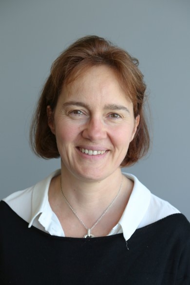 Faivre Laurence合作论文数Faculty of Medicine, University of Bourgogne;Medical Genetics Department, Hopital d'Enfants;Genetics Centre, Children's Hospital, Dijon University Hospital23
 Sylvie Odent合作论文数19Bertrand Isidor合作论文数Unité de Génétique clinique, Service de génétique médicale, Centre Hospitalier Universitaire de Nantes19
 Florence Petit合作论文数16Alice Goldenberg合作论文数15
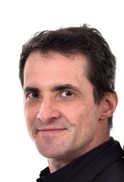 David Geneviève合作论文数Département de génétique médicale, Centre Hospitalier Universitaire de Montpellier;Faculté de Médecine, Université de Montpellier15Valérie Cormier-Daire合作论文数13
 Jeanne Amiel合作论文数Université Paris Cité;Institut Imagine13
 Didier Lacombe合作论文数Service de Génétique Médicale, CHU Bordeaux;INSERM U1211;Centre de Référence Anomalies du Développement et Syndromes Malformatifs Sud-Ouest Occitanie-Réunion;Centre Régional de Dépistage Néonatal12Marlène Rio合作论文数12Catherine Vincent-Delorme合作论文数12Schaefer Elise合作论文数12Busa Tiffany合作论文数12Joris Andrieux合作论文数12
 Sylvie Hanu Manouvrier合作论文数and Unité INSERM 16, Centre Hospitalier Régional12
 Massimiliano Rossi合作论文数11
 Stanislas Lyonnet合作论文数Institut Imagine;Paris Descartes University;Hôpital Necker-Enfants Malades11
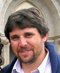 Damien Sanlaville合作论文数From Assistance Publique-Hôpitaux de Paris, Hôpital Européen Georges Pompidou;Assistance Publique des Hôpitaux de Paris, Hôpital Necker Enfants Malades;CHRU, Hôpital de Hautepierre;CHRU, Hôpital de Hautepierre11Tania Attié-Bitach合作论文数10Delphine Héron合作论文数10Chloe Quelin合作论文数CHU Rennes10Alain Verloes合作论文数10Guerrot Anne-Marie合作论文数10Annick Toutain合作论文数Université de Tours10Sabine Sigaudy合作论文数10Geneviève Baujat合作论文数9Frederic Tran Mau-Them合作论文数9Sandra Whalen合作论文数9Nicole Philip合作论文数9Marie Vincent合作论文数9
 Yannis Duffourd合作论文数Genet Anomalies Dev, Univ Bourgogne9Willems Marjolaine合作论文数9
 Patrick Edery合作论文数Université Claude Bernard9Christine Francannet合作论文数9Boris Keren合作论文数9Mélanie Fradin合作论文数9
 Christel Thauvin-Robinet合作论文数Centre de génétique, Hôpital Mitterrand;Déficiences intellectuelles de causes rares du CHU de Dijon9
 Christophe Philippe合作论文数9Caroline Rooryck合作论文数9Marion Gérard合作论文数8Thomas Smol合作论文数8Piard Juliette合作论文数Centre de Référence «Déficiences Intellectuelles de Causes Rares»,, Groupe Hospitalier CRicm8Nizon Mathilde合作论文数8
 Ange-Line Bruel合作论文数Génétique des Anomalies du Développement, Centre de Recherche Inserm 1231;CHU Dijon Bourgogne;Université de Bourgogne8
 Arthur Sorlin合作论文数8
 Paul Kuentz合作论文数Institut de Génétique et de Biologie Moléculaire et Cellulaire, Université de Strasbourg8Laurent Pasquier合作论文数8Christèle Dubourg合作论文数8Godelieve Morel合作论文数7




