Diffuse hemispheric gliomas, H3G34R/V-mutant (DHG-H3G34), are lethal brain tumors lacking targeted therapies. They originate from interneuronal precursors; however, leveraging this origin for therapeutic insights remains unexplored. Here, we delineate a cellular hierarchy along the interneuron lineage development continuum, revealing that DHG-H3G34 mirror spatial patterns of progenitor streams surrounding interneuron nests, as seen during human brain development. Integrating these findings with genome-wide CRISPR-Cas9 screens identifies genes upregulated in interneuron lineage progenitors as major dependencies. Among these, CDK6 emerges as a targetable vulnerability: DHG-H3G34 tumor cells show enhanced sensitivity to CDK4/6 inhibitors and a CDK6-specific degrader, promoting a shift toward more mature interneuron-like states, reducing tumor growth, and prolonging xenograft survival. Notably, a patient with progressive DHG-H3G34 treated with a CDK4/6 inhibitor achieved 17 months of stable disease. This study underscores interneuronal progenitor-like states, organized in characteristic niches, as a distinct vulnerability in DHG-H3G34, highlighting CDK6 as a promising clinically actionable target.
Abstract Supratentorial ependymomas (ST-EPN) are aggressive pediatric brain tumors that are categorized into distinct molecular subgroups. However, the developmental origins, tumor microenvironment, and phenotypic characteristics of tumor subpopulations across these subgroups are still poorly understood. In this study, we explored the human developmental signatures, global spatial organization, and the morphological and migratory tumor cell state behaviors in ST-EPN tumors at unprecedented resolution. We profiled 42 ST-EPN patients encompassing ZFTA-RELA (n = 20), ZFTA-Clusters 1 to 4 (n=20), and ST-YAP1 (n=4) subgroups by single-cell RNA sequencing (scRNA-seq), spatial transcriptomics, and live-cell imaging. We identified recurrent tumor cell states across tumors, that cover both generic cell processes (including cycling and mesenchymal/hypoxia) and human embryonic and fetal brain developmental signatures. ST-EPN subgroups displayed distinct developmental signatures, mapping to two separate temporally restricted progenitors-neuroepithelial-like and embryonic-like cells. Additionally, tumors showed diverging patterns of neuronal or ependymal differentiation, with ZFTA-Cluster 3 tumors as especially distinct from other subgroups in both developmental origin and differentiation. Furthermore, by utilizing 10X Xenium and novel algorithms we discovered that mesenchymal/hypoxia is critical for driving global tissue architecture to become more organized. Notably, recurrent tumors exhibited higher proportion of mesenchymal/hypoxia cells and greater structure than primary tumors. Lastly, by superimposing molecular and functional assays in both in vivo and in vitro models, we found that cell states exhibit distinct morphological and migratory behaviors, with neuronal-like cells being the most invasive. Neuronal-like cells especially exhibited distinct behavioral patterns reminiscent of neuronal migration during development. Moreover, we demonstrated the crucial role of neuronal microenvironment in promoting plasticity of cells toward this neuronal lineage. Taken together, we present a multidimensional framework for investigating cellular states within ST-EPN tumors, offering novel insights into their developmental origins, patterns of spatial organization, and cellular behavioral characteristics.
Supratentorial ependymomas are aggressive childhood brain cancers that retain features of neurodevelopmental cell types and segregate into molecularly and clinically distinct subgroups, suggesting different developmental roots. The developmental signatures as well as microenvironmental factors underlying aberrant cellular transformation and behavior across each supratentorial ependymoma subgroup are unknown. Here we integrated single cell- and spatial transcriptomics, as well as in vitro and in vivo live-cell imaging to define supratentorial ependymoma cell states, spatial organization, and dynamic behavior within the neural microenvironment. We find that individual tumor subgroups harbor two distinct progenitor-like cell states reminiscent of early human brain development and diverge in the extent of neuronal or ependymal differentiation. We further uncover several modes of spatial organization of these tumors, including a high order architecture influenced by mesenchymal and hypoxia signatures. Finally, we identify an unappreciated role for brain-resident cells in shifting supratentorial ependymoma cellular heterogeneity towards neuronal-like cells that co-opt immature neuronal morphology and invasion mechanisms. Collectively, these findings provide a multidimensional framework to integrate transcriptional and phenotypic characterization of tumor heterogeneity in supratentorial ependymoma and its potential clinical implications.
Abstract Despite the growing insights into the cellular heterogeneity of pediatric brain tumors, the transcriptional changes and spatial reorganization during disease progression remain largely unexplored. Here, we present an in-depth single-cell and 10X Xenium spatial transcriptomic characterization of 24 matched pediatric high-grade gliomas (pHGGs) in children, adolescents and young adults (AYA) profiled at both diagnosis and recurrence - constituting a total of 55 tissue samples. By stratifying the transcriptional programs activated at recurrence, we identify two distinct global response sets. Hemispheric AYA tumors predominantly exhibit upregulation of extracellular matrix pathways at recurrence, whereas midline tumors preferentially activate generic stress response programs. On a cellular level, we find that heterogeneity within the malignant tumor compartment remains largely unchanged at recurrence. However, a subset of AYA patients with hemispheric pHGG shows enrichment of the neural-progenitor-like (NPC-like) tumor cell state, and compositional changes in the immune and normal cell landscape, characterized by a global decrease in myeloid cells and an increase in oligodendrocyte fractions. Those changes lead, in turn, to enhanced NPC-like tumor cell-to-oligodendrocyte interactions. Spatial analysis of our extensive cohort at unprecedented subcellular resolution further reveals distinct cellular and spatial features associated with tumor progression, particularly in the remodeling of cell-cell interactions and tumor cell neighborhoods. Overall, this study provides a comprehensive longitudinal single-cell and spatial atlas of pHGG, uncovering the extensive cellular heterogeneity associated with tumor progression. Our findings highlight the role of intrinsic and extrinsic tumor adaptations in shaping disease progression in pHGG, offering potential novel therapeutic targets. Citation Format: Sara G. Danielli, Sina Neyazi, Olivia A. Hack, Li Jiang, Costanza Lo Cascio, Andrezza Nascimento, Cuong Nguyen, Jacob Rozowsky, Ilon Liu, Owen Hoare, Karin Shamardani, Kennedy Cunliffe-Koehler, Johannes Gojo, Keith L. Ligon, Lissa Baird, Sanda Alexandrescu, Jennifer Cotter, Michael Prados, Adam Resnick, Lin Wang, Michelle Monje, Aaron Diaz, Mariella G. Filbin. Project HOPE: A spatiotemporal single-cell landscape of high-grade gliomas in children, adolescents and young adults [abstract]. In: Proceedings of the AACR Special Conference in Cancer Research: Advances in Pediatric Cancer Research; 2024 Sep 5-8; Toronto, Ontario, Canada. Philadelphia (PA): AACR; Cancer Res 2024;84(17 Suppl):Abstract nr A052.
Abstract Epigenetic dysregulation resulting in stalled development plays a crucial role in pediatric cancer tumorigenesis. Diffuse midline gliomas (DMG) are universally fatal pediatric brain cancers refractory to standard of care treatment modalities. These malignancies are driven by heterozygous mutations in genes encoding histone 3 (H3K27M) which create an aberrant epigenetic landscape that keeps glioma cells in an undifferentiated stem-like state. Consequently, targeting epigenetic regulators to restore the epigenome and force glioma cells to exit this stem-like cell state represents a promising new therapeutic strategy for H3K27M-DMG. To interrogate for epigenetic dependencies, we performed a CRISPR/Cas9 inactivation screen in patient-derived H3K27M-DMG neurospheres using an epigenetically focused sgRNA library and identified several core components of the mammalian BAF (SWI/SNF) chromatin remodeling complex as genetic vulnerabilities. Validation assays revealed that knockout of the BAF catalytic subunit BRG1 results in decreased glioma cell proliferation and tumor growth in orthotopic mouse models. Mechanistically, genome wide localization and DNA accessibility studies combined with regulatory network analysis demonstrated that BRG1 controls the transcription factor and enhancer landscapes that maintain H3K27M-DMG cells in a cycling, oligodendrocyte precursor cell-like state. Single cell transcriptome analysis in vitro and immunofluorescence studies in vivo confirmed that genetic perturbation of this chromatin remodeler promotes progression of differentiation along the astrocytic lineage. Similarly, pharmacological suppression of BRG1 activity, using both catalytic inhibitors as well as recently developed degraders, opposes tumor cell proliferation, stimulates cell state transition, and improves overall survival of patient-derived xenograft models. Interestingly, these effects seem to be restricted to H3K27M mutant glioma, as H3 wildtype glioma cells were less sensitive to BRG1 inhibition both in vitro and in vivo. In summary, we demonstrate that the BAF complex contributes to the maintenance of glioma cells in a proliferative stem-like state and that its therapeutic inhibition has translational potential for children bearing H3K27M-DMG.
Abstract Gliomatosis cerebri (GC) is a rare, lethal glioma that is radiologically diagnosed and characterised by its diffuse infiltration throughout the cerebral lobes of the brain. It is no longer recognised as a separate entity by the WHO classification, but its growth pattern and invasive phenotype differ from other types of glioma. In order to understand what underlies this unique presentation, we sought to unravel these complex tumours in children through single cell and spatial approaches. 26 paediatric cases (1.3-19 years, median=11.3) were collected for DNA methylation, whole exome sequencing and single cell RNA-sequencing. Of these, 16 cases were used for Imaging Mass Cytometry (IMC). 5 patient-derived cell lines were used to model migration and invasion in vitro, whereby we have also been able to explore gene expression differences associated with the invading cells by RNAseq. We analyzed the transcriptional landscape of all 26 GC patients at single-cell resolution, and identified shared tumor cellular programs of glial and neuronal lineage. We observed notable heterogeneity of neuronal lineage tumor cells that were composed of different states, such as metabolic active and synaptogenic states. We described tumor cell states associated with higher invasivity, as corroborated from integrated analysis with our in vitro invasion-associated gene expression profiles. Next, we stratified all tumors by their DNA methylation profiles, and defined 16 archetypal GC cases, for which we show specific transcriptional features. Furthermore, we analyzed multiple regions of interest within FFPE patient tissue sections by IMC and demonstrate spatial patterns in the interaction between the tumour and normal brain tissue interface, and inter-regional heterogeneity for stem, invasive and proliferative markers. Together, we dissect the characteristic invasive phenotype of GC at multiple molecular, spatial and functional layers, thereby providing the basis for modelling and therapeutic avenues much needed for this lethal disease.
Abstract Embryonal tumors with multilayered rosettes (ETMR) are malignant brain tumors that occur predominantly in infants and young children. Most patients die within two years of diagnosis, and more effective, targeted therapies are urgently needed. To better characterize the oncogenic mechanisms of key driver alterations and to identify novel therapeutic targets, we set out to study the cellular heterogeneity of ETMR using single-cell RNA sequencing. Analyses conducted on >4,000 high-quality cells collected from eleven primary and relapse specimens revealed a common cellular hierarchy across all tumors: A highly proliferative neural stem cell-like population (SOX2+) that gives rise to intermediate progenitors (NEUROD1/NEUROG1+) and more differentiated neuron-like cells (STMN2/4+). These malignant cell populations closely overlap with histological patterns of ETMRs, as confirmed by multiplexed immunofluorescence microscopy on patients’ tumors. Comparison to single-cell datasets from human fetuses indicated high resemblance to normal cortical neurogenesis but also revealed key tumor-specific differences. These include expression of the chromosome 19 miRNA cluster (C19MC, the presumed driver in ~90% of ETMRs), which was restricted to the malignant stem cell-like population. Investigating oncogenic mechanisms of C19MC (comprising 46 miRNA genes) through transcriptome-wide RNA immunoprecipitation analysis, we identified extensive target gene regulation for most C19MC members, including distinct regulators of cell cycle, pluripotency, and neuronal differentiation. Silencing of C19MC families with antisense oligonucleotides resulted in pronounced reduction of ETMR cell line growth, indicating potential avenues for therapeutic targeting in the future. To identify more immediately actionable targets, we investigated inter-cellular signaling between malignant cell populations of ETMRs. Interestingly, we identified marked FGFR and NOTCH receptor-ligand interactions common to all tumors. An in vitro screen of experimental and approved small molecule inhibitors designed to target these interactions nominated several promising candidates for clinical evaluation. Our unpublished results provide much needed insight into targeting ETMR cellular states using multiple modes of action.
Abstract Diffuse hemispheric gliomas, H3G34R/V-mutant (DHG-H3G34), are uniformly lethal malignancies with currently no targeted therapies available. They exclusively occur in the cerebral hemispheres of adolescents and young adults, and have been linked to a distinct interneuronal lineage of origin. The developmental spectrum and functional role of this interneuronal lineage in DHG-H3G34 remain incompletely understood. Here, through integrating bulk and single-cell multi-omics with genome-wide CRISPR-Cas9 screens, we resolve a putative cellular hierarchy that follows a continuum of interneuronal lineage development, ranging from a self-renewing progenitor-like cell to a more differentiated cell resembling early immature GABAergic interneurons, along with quiescent astrocyte-like and mesenchymal-like cells. We validate these single-cell states in patient DHG-H3G34 tissue sections by multiplexed immunofluorescence, and describe spatial structures that resemble nests of early migratory interneurons surrounded by progenitor cells, characteristic of human embryonal interneuron development. Intriguingly, we reveal the majority of CRISPR-Cas9 screen-derived gene dependencies are upregulated in interneuronal lineage tumor cells, specifically in less differentiated progenitor-like cells, highlighting these as a driver of DHG-H3G34. We validate the essentiality of these interneuronal lineage associated targets in patient-derived in vitro and in vivo models, and highlight CDK6 as a druggable target selectively upregulated in DHG-H3G34. Inhibition of CDK6 leads to a decrease of undifferentiated progenitor-like signatures, reduced tumor growth, and prolonged survival of patient-derived xenograft models. Encouraged by these findings, we treated a patient upon a second relapse of a DHG-H3G34 with ribociclib on a compassionate use basis, who, as of the time of submission, has shown stable disease within four cycles of ribociclib treatment after progression on PCV chemotherapy. In sum, we reveal CDK6 inhibition as a rationally informed and clinically actionable therapeutic avenue that selectively perturbs the unique interneuronal lineage in DHG-H3G34, paving the way for rapid clinical translation.
PDGFRA has been shown to be commonly altered in high-grade gliomas (HGGs), including histone 3 lysine 27-mutated diffuse midline gliomas (H3K27M DMG), a disease with almost no long-term survivors. Here, we performed comprehensive genomic and transcriptomic analysis of 260 high-grade glioma cases, which revealed PDGFRA genomic alterations (mutations and/or amplifications) in 13% of patients. H3K27M DMGs had significantly higher PDGFRA expression compared to H3 wild-type tumors, and PDGFRA gene amplification resulted in even higher expression levels in H3K27M DMGs as well as H3 wild-type HGGs. We tested a panel of patient- derived pHGG/H3K27M DMG models against a range of PDGFRA inhibitors, including avapritinib, a potent small molecule inhibitor with relatively selective activity against both wild-type and mutant PDGFRA. Avapritinib showed supra-micromolar blood-brain barrier penetration in our pre-clinical models and demonstrated significant survival impact in an aggressive patient-derived H3K27M DMG mouse xenograft model. Finally, building on this preclinical activity, we report here the first clinical experience using avapritinib in eight pediatric and young adult patients with high-grade glioma (H3K27M DMG and/or PDGFRA altered). Avapritinib has thus far been well tolerated with no significant acute toxicities. Most importantly, our preliminary data reveal radiographic response evaluated by RAPNO criteria in 50% of patients, a striking outcome rarely seen in this patient population. In summary, we report that avapritinib is a selective, CNS-penetrant small molecule inhibitor of PDGFRA that shows potent activity in preclinical models and produces promising clinical responses with good tolerability in patients with high-grade glioma. This suggests a promising role for avapritinib therapy in this population with previously dismal outcomes. Citation Format: Lisa Mayr, Maria Trissal, Kallen Schwark, Jenna Labelle, Andrew Groves, Julia Furtner-Srajer, Jeffrey Supko, Liesa Weiler-Wichtl, Olivia Hack, Jacob Rozowsky, Joana G. Marques, Eshini Pandatharatna, Ulrike Leiss, Verena Rosenmayr, Frank Dubois, Noah F. Greenwald, Sibylle Madlener, Armin S. Guntner, Hana Pálová, Natalia Stepien, Daniela Lötsch-Gojo, Christian Dorfer, Karin Dieckmann, Andreas Peyrl, Amedeo A. Azizi, Alicia Baumgartner, Ondřej Slabý, Petra Pokorná, Pratiti Bandopadhayay, Rameen Beroukhim, Keith Ligon, Christof Kramm, Annika Bronsema, Simon Bailey, Ana Guerreiro Stücklin, Sabine Mueller, David T. Jones, Natalie Jäger, Jaroslav Štěrba, Leonhard Müllauer, Christine Haberler, Chandan Kumar-Sinha, Arul Chinnaiyan, Rajen Mody, Mary Skrypek, Nina Martinez, Daniel C. Bowers, Carl Koschmann, Johannes Gojo, Mariella Filbin. Clinical response to the PDGFRα inhibitor avapritinib in high-grade glioma patients. [abstract]. In: Proceedings of the American Association for Cancer Research Annual Meeting 2023; Part 1 (Regular and Invited Abstracts); 2023 Apr 14-19; Orlando, FL. Philadelphia (PA): AACR; Cancer Res 2023;83(7_Suppl):Abstract nr 5719.
Abstract Embryonal tumor with multilayered rosettes (ETMR) is a highly aggressive brain tumor that predominantly occurs in children under the age of four years. Most children succumb within 10 months of diagnosis, typically due to invasive tumor growth and therapy-refractory behavior. 95% of ETMR harbor activating genetic alterations of the chromosome 19 microRNA cluster (C19MC). Despite the identification of this presumed oncogenic driving event, its biological consequences remain understudied and targeted therapy options are lacking. Here, we set out to further explore the intracellular heterogeneity of ETMR and shed light onto its oncogenic mechanisms with the goal to identify novel therapeutic targets for this dismal disease. We have compiled and analyzed a cohort of 11 ETMR tumors using single-cell RNA sequencing and multiplexed spatial imaging. We reveal a common, spatially distinct cellular hierarchy, closely resembling physiological brain development. This hierarchy spans a highly proliferative neural stem cell-like population that gives rise to more differentiated neuron-like cells. C19MC, predominantly expressed in the malignant stem-like population, controls a transcriptional network governing stemness and lineage commitment, as resolved by genome-wide analysis of microRNA-mRNA interactions. Targeting specific C19MC members by antisense oligonucleotides results in reduced cell proliferation, suggesting potent new avenues for future therapeutic approaches. Seeking more immediate therapeutic targets, we performed comprehensive cell-cell interaction analyses and reveal that FGF-FGFR and Delta-Notch interactions are important to maintain oncogenic signaling across malignant cell populations. These interactions were successfully targeted using small molecule inhibitors in preclinical models and in one ETMR patient. Taken together, we identify different malignant ETMR cell populations that follow a common developmental hierarchy, explore the regulatory reach of C19MC activation, and identify an oncogenic receptor-ligand interaction network. Targeting this interaction network opens a powerful new rationale for more effective ETMR therapies.
Abstract Histone 3 lysine27-to-methionine mutations (H3-K27M) frequently occur in childhood diffuse midline gliomas (DMGs) of the pons, thalamus, and spinal cord, presumed to be driven by the specific spatiotemporal context of these midline locations during postnatal development. While most common in the pons and at mid-childhood ages, the same oncohistone mutation is recurrently detected in adult DMGs and throughout different midline regions. The potential heterogeneity of tumors at different ages and in different anatomical locations of the midline are vastly understudied. Through dissecting the transcriptomic, epigenomic and spatial architectures of a comprehensive cohort of patient H3-K27M DMGs - spanning the age range from 2-68 years and locations from spinal cord to thalamus - at single cell resolution, we delineate how age- and location-dependent contexts shape glioma cell-intrinsic and -extrinsic features in light of the shared driver mutation. We identify that oligodendrocyte precursor (OPC)-like cells constitute the stem-like compartment in H3-K27M DMGs across all clinico-anatomical groups, however, depending on location, display varying levels of maturity resembling less differentiated pre-OPCs or more mature OPCs further differentiated along the oligodendroglial lineage. We further demonstrate increased mesenchymal cell states in adult tumors, which we link to age-related differences in glioma-associated immune cell compartments, in particular an increase of macrophages in adult compared to pediatric tumors. Furthermore, we resolve the spatial organization of H3-K27M DMG cell types and states in intact patient tissues, identifying a local niche of the oligodendroglial lineage. Our study provides a powerful resource for rational modeling and therapeutic frameworks taking into account determinants of age and location in this lethal glioma group.
Abstract Ependymomas (EPN) are among the most common and fatal pediatric brain tumors with currently no targeted therapies available. Molecular studies including genome-wide DNA methylation profiling have shown that supratentorial ependymoma is subdivided into subgroups characterized by distinct fusion proteins: ZFTA-RELA fusion-positive (ZFTA-RELA), YAP1 fusion-positive (ST-YAP1), and four alternative ZFTA fusion-positive (ZFTA-Cluster 1, ZFTA-Cluster 2, ZFTA-Cluster 3, and ZFTA-Cluster 4). ZFTA-RELA tumors have been identified as the most common and aggressive molecular group with high intra-tumoral heterogeneity. However, limited sample size and interpatient variability in previous studies highlight a critical need for comprehensive profiling of supratentorial ependymomas across a larger patient cohort. In particular, how ZFTA-RELA compares to other subgroups of supratentorial ependymoma is unknown. Here, we collected and profiled 44 supratentorial patient tumors encompassing ZFTA-RELA (n=21), ZFTA-Cluster 1 (n=6), ZFTA-Cluster 2 (n=4), ZFTA-Cluster 3 (n=7), ZFTA-Cluster 4 (n=2), and ST-YAP1 (n=4). We profiled all tumors by single cell/single nucleus RNA-sequencing, and further characterized ZFTA-RELA by single cell spatial transcriptomics. Within each patient’s tumor we find multiple normal and aberrant differentiation trajectories from neural progenitor-like cells towards glial-like, mesenchymal-like and ependymal-like cells. Using sequencing-based lineage tracing, we experimentally validate these computationally inferred cellular hierarchies in vitro. Notably, compared to ZFTA-RELA ependymomas we observed predominantly higher proportion of early neural progenitor-like cells in ZFTA-Cluster 2 and ZFTA-Cluster 3, while in ST-YAP1 greater representation of mature ependymal-like cells. These findings highlight differences in developmental and aberrant differentiation trajectories driven by combination of oncogenic fusions and cell of origin in supratentorial ependymoma subgroups. In addition, neural progenitor-like cells most highly express ZFTA-fusions and are spatially distinct from other cell states, highlighting oncogenic fusions driving tumorigenesis organized in spatial niches. Together, this study reveals previously unknown intra- and intertumoral heterogeneity across subgroups of supratentorial ependymoma, as well as distinct cellular and spatial architecture.
Diffuse hemispheric gliomas, H3G34R/V-mutant (DHG-H3G34), are uniformly lethal malignancies with currently no targeted therapies available. They exclusively occur in the cerebral hemispheres of adolescents and young adults, and have been linked to a distinct interneuronal lineage of origin. The developmental spectrum and functional role of this interneuronal lineage in DHG-H3G34 remain incompletely understood. Here, through integrating bulk and single-cell multi-omics with genome-wide CRISPR-Cas9 screens, we resolve a putative cellular hierarchy that follows a continuum of interneuronal lineage development, ranging from a self-renewing progenitor-like cell to a more differentiated cell resembling early immature GABAergic interneurons, along with quiescent astrocyte-like and mesenchymal-like cells. We validate these single-cell states in patient DHG-H3G34 tissue sections by multiplexed immunofluorescence, and describe spatial structures that resemble nests of early migratory interneurons surrounded by progenitor cells, characteristic of human embryonal interneuron development. Intriguingly, we reveal the majority of CRISPR-Cas9 screen-derived gene dependencies are upregulated in interneuronal lineage tumor cells, specifically in less differentiated progenitor-like cells, highlighting these as a driver of DHG-H3G34. We validate the essentiality of these interneuronal lineage associated targets in patient-derived in vitro and in vivo models, and highlight CDK6 as a druggable target selectively upregulated in DHG-H3G34. Inhibition of CDK6 leads to a decrease of undifferentiated progenitor-like signatures, reduced tumor growth, and prolonged survival of patient-derived xenograft models. Encouraged by these findings, we treated a patient upon a second relapse of a DHG-H3G34 with ribociclib on a compassionate use basis, who, as of the time of submission, has shown stable disease within four cycles of ribociclib treatment after progression on PCV chemotherapy. In sum, we reveal CDK6 inhibition as a rationally informed and clinically actionable therapeutic avenue that selectively perturbs the unique interneuronal lineage in DHG-H3G34, paving the way for rapid clinical translation.
Supplementary Table from BAF Complex Maintains Glioma Stem Cells in Pediatric H3K27M Glioma
Abstract PDGFRA is commonly altered in pediatric and young adult high-grade gliomas (pHGGs) including histone 3 lysine 27-mutated diffuse midline gliomas (H3K27M DMG), a fatal disease with no current options for cure. We performed comprehensive genomic and transcriptomic analyses of n=259 pediatric high-grade glioma cases which revealed PDGFRA genomic alterations in ~15% of patients. H3K27M DMGs had significantly higher PDGFRA expression compared to H3 wild-type tumors regardless of genomic alteration. Tumors with PDGFRA gene amplification demonstrated significantly elevated PDGFRA expression in both H3K27M DMGs and H3 wild-type pHGGs relative to tumors with wild-type or point mutated PDGFRA. We tested a range of PDGFRA inhibitors against a panel of patient derived pediatric H3K27M DMG, pHGG, and adult HGG. Amongst the inhibitors tested, avapritinib, a potent small molecule inhibitor with relatively selective activity against both wild-type and mutant PDGFRA showed potent toxicity against a wide array of pediatric and adult cell lines. This molecule also demonstrated supra-micromolar blood brain barrier penetration in pre-clinical in vivo models, and demonstrated significant decrease in tumor growth and improved survival in orthotopic mouse xenograft models. Finally, building on this preclinical activity, we report the first clinical experience using avapritinib in eight pediatric and young adult patients with high-grade glioma (H3K27M DMG and/or PDGFRA altered). Avapritinib usage showed no significant acute toxicities within this patient cohort. Most importantly, our preliminary data reveal radiographic response evaluated by RAPNO criteria in 50% of patients, a striking outcome rarely seen in this patient population. In summary, we report that avapritinib, a selective, CNS penetrant small molecule inhibitor of PDGFRA has potent activity in preclinical models and produced promising clinical responses with good tolerability in pediatric and young adult patients with high-grade glioma, suggesting a promising role for avapritinib therapy in pediatric high grade glioma.
Abstract Diffuse midline gliomas are uniformly fatal pediatric central nervous system cancers that are refractory to standard-of-care therapeutic modalities. The primary genetic drivers are a set of recurrent amino acid substitutions in genes encoding histone H3 (H3K27M), which are currently undruggable. These H3K27M oncohistones perturb normal chromatin architecture, resulting in an aberrant epigenetic landscape. To interrogate for epigenetic dependencies, we performed a CRISPR screen and show that patient-derived H3K27M-glioma neurospheres are dependent on core components of the mammalian BAF (SWI/SNF) chromatin remodeling complex. The BAF complex maintains glioma stem cells in a cycling, oligodendrocyte precursor cell–like state, in which genetic perturbation of the BAF catalytic subunit SMARCA4 (BRG1), as well as pharmacologic suppression, opposes proliferation, promotes progression of differentiation along the astrocytic lineage, and improves overall survival of patient-derived xenograft models. In summary, we demonstrate that therapeutic inhibition of the BAF complex has translational potential for children with H3K27M gliomas. Significance: Epigenetic dysregulation is at the core of H3K27M-glioma tumorigenesis. Here, we identify the BRG1–BAF complex as a critical regulator of enhancer and transcription factor landscapes, which maintain H3K27M glioma in their progenitor state, precluding glial differentiation, and establish pharmacologic targeting of the BAF complex as a novel treatment strategy for pediatric H3K27M glioma. See related commentary by Beytagh and Weiss, p. 2730. See related article by Mo et al., p. 2906. This article is highlighted in the In This Issue feature, p. 2711
Abstract Background Histone 3 lysine27-to-methionine mutations (H3-K27M) frequently occur in childhood diffuse midline gliomas (DMGs) of the pons, thalamus and spinal cord, presumed to be driven by the specific spatiotemporal context of these midline locations during postnatal development. While most common in the pons and at mid-childhood ages, the same oncohistone mutation is recurrently detected in adult DMGs and throughout different midline regions. The potential heterogeneity of tumors at different ages and in different anatomical locations of the midline are vastly understudied. Material and Methods Through dissecting the transcriptomic, epigenomic and spatial architectures of a comprehensive cohort of patient H3-K27M DMGs - spanning the age range from 2-68 years and locations from spinal cord to thalamus - at single cell resolution, we delineate how age- and location-dependent contexts shape glioma cell-intrinsic and -extrinsic features in light of the shared driver mutation. Results We identify that oligodendrocyte precursor (OPC)-like cells constitute the stem-like compartment in H3-K27M DMGs across all clinico-anatomical groups, however, depending on location, display varying levels of maturity resembling less differentiated pre-OPCs or more mature OPCs further differentiated along the oligodendroglial lineage. We further demonstrate increased mesenchymal cell states in adult tumors, which we link to age-related differences in glioma-associated immune cell compartments. We for the first time resolve the spatial organization of H3-K27M DMG cell types in intact patient tissues, identifying a local niche of the oligodendroglial lineage. Conclusion Our study provides a powerful resource for rational modeling and therapeutic frameworks taking into account determinants of age and location in this lethal glioma group.
Pediatric high-grade glioma (pHGG) is an incurable disease with a median survival of less than 6 months post-progression and no effective targeted therapy. PDGFRA is commonly altered in pHGG, but targeting PDGFRA in this disease has been unsuccessful, likely due to poor central nervous system (CNS) penetrance. Avapritinib is a novel and CNS-penetrant PDGFRA/KIT inhibitor that is FDA-approved for adults with unresectable or metastatic PDGFRA exon 18-mutant gastrointestinal stromal tumor (GIST) and is being studied in CNS tumors. We performed a pre-clinical and clinical assessment to determine the potential suitability of avapritinib therapy in PDGFRA-driven glioma. A multi-institutional cohort genetic analysis revealed PDGFRA amplification and mutation in 10.2% and 6.1% of pHGG, respectively. Additionally, PDGFRA expression in the absence of genetic events was significantly increased in H3K27-altered diffuse midline glioma (DMG) compared to H3-wildtype pHGG. Avapritinib performed well in: (i) mutant PDGFRA enzyme inhibition and wildtype inhibition at high dose, (ii) minimal off-target kinase inhibition, (iii) brain penetration (peak 10 µM), and (iv) proliferation/pPDGFRA reduction in PDGFRA-amplified and mutant pHGG cell lines. Avapritinib treatment in an aggressive PDX model of pHGG resulted in significant survival benefit. We pursued treatment of eight pediatric and young adult HGG patients with avapritinib across seven institutions. Patients were a mixture of local (N = 4) and metastatic disease (N = 4); all patients were post-initial radiation, with 7/8 having progressed on prior treatment. 7/8 patients had PDGFRA amplifications or mutations, and 7/8 had H3K27M mutations. Therapy was generally well-tolerated. 4/8 patients showed radiographic response to avapritinib, with one patient demonstrating complete response of target lesion and remains on therapy. Avapritinib levels in patients’ CSF and brain tumor tissue reached micromolar levels. These results demonstrate that avapritinib is a potent, selective, and CNS-penetrant PDGFRA/KIT inhibitor that is promising for further study in pHGG with relevant alterations.
 Mariella G Filbin合作论文数Department of Pediatric Oncology, Dana-Farber Boston Children's Cancer and Blood Disorders Center, Boston, MA 02215, USA; Broad Institute of Harvard and MIT, Cambridge, MA 02142, USA.35
 McKenzie Shaw合作论文数Broad Institute of Harvard and MIT , Cambridge , USA28
 Johannes Gojo合作论文数24
 Ilon Liu合作论文数22Bernhard Englinger合作论文数Institute of Cancer Research and Comprehensive Cancer Center of the Medical University, Medical University of Vienna20
 Sanda Alexandrescu合作论文数19
 Keith L. Ligon合作论文数Center for Patient Derived Models, Dana-Farber Cancer Institute;Division of Neuropathology, Brigham and Women's Hospital;Medical School, Harvard University;Travera;Broad Institute, Massachusetts Institute of Technology;Boston Children's Hospital18
 Michelle Monje合作论文数Department of Psychiatry and Behavioral Sciences, School of Medicine, Stanford University17Sibylle Madlener合作论文数Institute of Clinical Pathology, Medical University of Vienna16
 Christine Haberler合作论文数16
 Lisa Mayr合作论文数15Irene Slavc合作论文数Dept Pediat & Adolescent Med, Med Univ Vienna14Beck Alexander合作论文数14Li Jiang合作论文数13Eshini Panditharatna合作论文数13Trissal Maria合作论文数13Michael A Quezada合作论文数Stanford University12
 Christian Dorfer合作论文数Department of Neurosurgery, Medical University of Vienna12Hafsa Mire合作论文数11
 Rene Geyeregger合作论文数Department of Clinical Cell Biology and FACS Core Unit, Children's Cancer Research Institute (CCRI),11Jiang Li合作论文数11
 Sameer Agnihotri合作论文数Hospital for Sick Childrens Research Institute, University Health Network11
 Mario Suva合作论文数Harvard Stem Cell Institute, Harvard University;Massachusetts General Hospital10
 Stefan Pfister合作论文数Hopp Children's Cancer Center Heidelberg10
 Marcel Kool合作论文数Division of Pediatric Neurooncology, German Cancer Research Center10Jenna Labelle合作论文数9
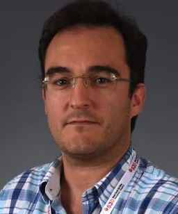 Ángel Montero Carcaboso合作论文数Pediatric Cancer Center Barcelona, Hospital Sant Joan de Déu Barcelona9Da Eun Jeong合作论文数8
 Carl Koschmann合作论文数Department of Pediatrics, Medical School, University of Michigan8
 Mathewson Nathan合作论文数8
 Thomas Czech合作论文数8Kristian W. Pajtler合作论文数7
 Samantha E Hoffman合作论文数Dept Neurosurg, Brigham & Womens Hosp7
 Joana Marques合作论文数Porto Pharmacovigilance Ctr, Univ Porto7
 Nguyen Quang-Dé合作论文数Univ London Imperial, Hammersmith Hosp7Groves Andrew合作论文数7
 Hovestadt Volker合作论文数Division of Molecular Genetics, German Cancer Research Center (DKFZ), Im Neuenheimer Feld 280, Heidelberg 69120, Germany.7
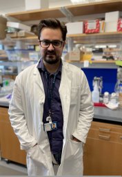 Gustavo Alencastro Veiga Cruzeiro合作论文数7
 Adam C. Resnick合作论文数Department of Neurosurgery, Perelman School of Medicine, University of Pennsylvania;Center for Data Driven Discovery in Biomedicine, The Children's Hospital of Philadelphia7Valeria Molinari合作论文数6Sara Temelso合作论文数6Jacob Rozowsky合作论文数6Hana Palova合作论文数6Alan Mackay合作论文数6
 Joshua M Dempster合作论文数Broad Inst MIT & Harvard, Cambridge, MA 02142 USA6
 Yura Grabovska合作论文数Newcastle Univ, Northern Inst Canc Res, Wolfson Childhood Canc Res Ctr, Newcastle Upon Tyne, Tyne & Wear, England6
 Maria Vinci合作论文数6
 Aaron Diaz合作论文数University of California, University of California6
 Daniela Lotsch合作论文数Department of Neurosurgery, Wagner-Jauregg Hospital;Institute of Pathology and Neuropathology, Wagner-Jauregg Hospital;Department of Internal Medicine, Wagner-Jauregg Hospital;Institute of Cancer Research and Comprehensive Cancer Center-Central Nervous System Tumors Unit, Medical University of Vienna;Department of Surgery, Medical University of Vienna;Laboratory of Molecular Biology and Tumorcytogenetics, Hospital Barmherzige Schwestern;Medical University of Vienna, Medical University of Vienna6




