DNA methylation is a fundamental epigenetic mark that governs gene expression and chromatin organization, thus providing a window into cellular identity and developmental processes 1 . Current datasets typically include only a fraction of methylation sites and are often based either on cell lines that underwent massive changes in culture or on tissues containing unspecified mixtures of cells 2 – 5 . Here we describe a human methylome atlas, based on deep whole-genome bisulfite sequencing, allowing fragment-level analysis across thousands of unique markers for 39 cell types sorted from 205 healthy tissue samples. Replicates of the same cell type are more than 99.5% identical, demonstrating the robustness of cell identity programmes to environmental perturbation. Unsupervised clustering of the atlas recapitulates key elements of tissue ontogeny and identifies methylation patterns retained since embryonic development. Loci uniquely unmethylated in an individual cell type often reside in transcriptional enhancers and contain DNA binding sites for tissue-specific transcriptional regulators. Uniquely hypermethylated loci are rare and are enriched for CpG islands, Polycomb targets and CTCF binding sites, suggesting a new role in shaping cell-type-specific chromatin looping. The atlas provides an essential resource for study of gene regulation and disease-associated genetic variants, and a wealth of potential tissue-specific biomarkers for use in liquid biopsies.
This study aimed to examine the incidence rate of early reoperations following hip fracture surgery and determine the safety of resuming direct oral anticoagulants. Many orthopedic surgeons are reluctant to resume chronic anticoagulation therapy for patients after surgical intervention for hip fractures. One of the main reasons is the potential for reoperation in the case of surgical complications. We conducted a retrospective cohort study at an Academic Level I trauma center, reviewing the records of 425 geriatric patients (age > 60) who underwent hip fracture surgery between 2018 and 2020, including a subgroup treated with direct oral anticoagulants prior to hospitalization. The study assessed the incidence rate of complications requiring early reoperation. Out of the 425 patients, only nine (2%) required reoperation within a month after discharge, with two (0.5%) on chronic anticoagulation therapy. None of the reoperations were urgent, and all were performed at least 24 h after re-admission. The findings revealed a very low incidence rate of reoperations in patients who underwent hip fracture surgery, with no reoperations performed within 24 h of re-admission. Consequently, we believe that resuming chronic direct oral anticoagulants is a safe and effective approach when discharging patients after hip fracture surgery.
The increase in hip fractures (HF) due to aging of the population and the rise in attractiveness of services provided at home following the COVID-19 pandemic, emphasize the need to compare outcomes of home versus hospital HF rehabilitation. To date, studies comparing the 2 services have focused primarily on clinical outcomes rather than patient-reported outcomes (PROs). This longitudinal observational study evaluated PROs of older adults with HF in the 2 settings. The SF36 questionnaire was used to measure PROs 3 times after surgery. The first PRO was retrospective and reflected pre-fracture health status. Descriptive statistics and mixed-effect logistic regression were used. Of 86 patients participating in the study, 41 had home rehabilitation and 45 had hospital rehabilitation. In both groups, the mental and physical scores plummeted 2 weeks after the HF, compared to pre-fracture status. The difference in improvement from pre-fracture status to recovery in both groups, were not significantly ( P < .05) different, except for the pain domain. PROs of home versus hospital rehabilitation were similar, suggesting that rehabilitation at home can be as effective as hospital rehabilitation for suitable patients. This knowledge can improve quality of care in an aging global population.
Sarcoma classification is challenging and can lead to treatment delays. Previous studies used DNA aberrations and machine-learning classifiers based on methylation profiles for diagnosis. We aimed to classify sarcomas by analyzing methylation signatures obtained from low-coverage whole-genome sequencing, which also identifies copy-number alterations. DNA was extracted from 23 suspected sarcoma samples and sequenced on an Oxford Nanopore sequencer. The methylation-based classifier, applied in the nanoDx pipeline, was customized using a reference set based on processed Illumina-based methylation data. Classification analysis utilized the Random Forest algorithm and t-distributed stochastic neighbor embedding, while copy-number alterations were detected using a designated R package. Out of the 23 samples encompassing a restricted range of sarcoma types, 20 were successfully sequenced, but two did not contain tumor tissue, according to the pathologist. Among the 18 tumor samples, 14 were classified as reported in the pathology results. Four classifications were discordant with the pathological report, with one compatible and three showing discrepancies. Improving tissue handling, DNA extraction methods, and detecting point mutations and translocations could enhance accuracy. We envision that rapid, accurate, point-of-care sarcoma classification using nanopore sequencing could be achieved through additional validation in a diverse tumor cohort and the integration of methylation-based classification and other DNA aberrations.
Background Goal-oriented patientcare is a key element in qualityhealthcare. Medical-caregiver’s (MC) are expected to generate a shared decision-making process with patients regarding goals and expected health-outcomes. Hip-fracture patients (HFP) are usually older-adults with multiple health-conditions, necessitating that agreed-upon goals regarding the rehabilitation process, take these conditions into consideration. This topic has yet to be investigated by pairing and comparing the perception of expected outcomes and therapeutic goals of multidisciplinary MCs and their HF patient’s. Our aim was to assess in a quantitative method whether HFPs and their multidisciplinary MCs agree upon target health-outcomes and their most important goals as they are reflected in the SF12 questionnaire. Methods This was a cross-sectional, multi-center, study of HFPs and their MCs. Patients and MCs were asked to rate their top three most important goals for rehabilitation from the SF12 eight subscales: physical functioning, physical role limitation, bodily pain, general health, vitality, social functioning, emotional role limitation and mental health, and indicate their expected outcome. Descriptive statistics and mixed effect logistic-regression were used to compare concordance of the ratings. Agreement between patients and MCs was assessed using interclass coefficients (ICCs). Results A total of 378 ratings were collected from 52 patients, 12 nurses, 12 physicians and 6 paramedical personnel. Each patient had between 3 and 9 raters. Patients considered physical functioning and physical role limitation more important than did MCs. Physicians and nurses emphasized the importance of bodily pain while patients referred to it as relatively less significant. The total ICC was low (2%) indicating poor agreement between MCs and patients. With the exception of physical-functioning, MCs predicted a less optimistic outcome in all of the SF12's subscales in comparison to HFPs. Conclusion Effective intervention in HFPs requires constructive communication between MCs and patients. The study suggests that caregivers have an insufficient understanding of the expectations of HFPs. More effective communication channels are required in order to better understand HFPs’ needs and expectations.
Background Hip fracture patients (HFPs) frequently have multiple underlying conditions, necessitating that agreed-upon goals take these complications into consideration. Communication regarding goals between medical-personnel and patients is not always effective. Patient-reported outcomes (PROs) can outline personal goals and help promote quality health care in HFPs. Few studies have been published on this topic. The study’s aim was to outline the process of using PROs for goal-directed therapy among HFPs.Methods This sequential controlled trial was conducted among HFPs from two medical centres. The control and the intervention group received integrative rehabilitation. PROs were measured in both groups using the SF36 questionnaire three times postsurgery: 24–48 hours, 2 weeks and 3 months. During the first round of questioning, only the intervention group was asked ‘what matters most to you?’ during the rehabilitative process. Accordingly, agreed-upon goals that were determined by the SF36’s eight topics and were incorporated into the HFP’s rehabilitative process. A Likert scale of 1–5, ‘1’ indicating no-achievement and ‘5’ full-achievement, was used to assess the goal achievement 4–6 months post-fracture.Results 84 HFPs participated in the study: 40 and 44 in the intervention and control group, respectively. In both groups, PROs declined after the HF, then improved somewhat 3 months later, but did not return to prefracture scores. Among the intervention group, 39% reached their specific goals (Likert level 5). Patients who achieved their goals had better PROs in comparison to others. The intervention group indicated PROs helped them articulate their desires and introduced them to new areas of care.Conclusions Shifting from asking ‘what’s the matter?’ to ‘what matters most to you?’ can improve the understanding of HFPs’ own priorities, promote quality outcomes and enhance patient-centred care. Using PROs as a guide for goal-directed therapy can create a more inclusive process that includes the patients’ most important health determinants and needs.
DNA methylation is a fundamental epigenetic mark that governs chromatin organization, cell identity, and gene expression. Here we describe a human methylome atlas, based on deep whole-genome bisulfite sequencing allowing fragment-level analysis across thousands of unique markers for 39 cell types sorted from 207 healthy tissue samples. Replicates of the same cell-type are >99.5% identical, demonstrating robustness of cell identity programs to genetic variation and environmental perturbation. Unsupervised clustering of the atlas recapitulates key elements of tissue ontogeny, and identifies methylation patterns retained since gastrulation. Loci uniquely unmethylated in an individual cell type often reside in transcriptional enhancers and contain DNA binding sites for tissue-specific transcriptional regulators. Uniquely hyper-methylated loci are rare and are enriched for CpG islands, polycomb targets, and CTCF binding sites, suggesting a novel role in shaping cell type-specific chromatin looping. The atlas provides an essential resource for interpretation of disease-associated genetic variants, and a wealth of potential tissue-specific biomarkers for use in liquid biopsies. Summary paragraph DNA methylation, a fundamental epigenetic mark, governs chromatin organization and gene expression 1 , thus defining the molecular identity of cells and providing a window into developmental processes with wide-ranging physiologic and clinical ramifications. Current DNA methylation datasets have limitations, typically including only a fraction of methylation sites, many from cell lines that underwent massive changes in culture or from tissues containing unspecified mixtures of cells 2–6 . We present a human methylome atlas based on deep whole-genome bisulfite sequencing of 39 sorted, primary cell types and use this dataset to address fundamental questions in developmental biology, physiology and pathology. Biological replicates are >99.5% identical, demonstrating unappreciated robustness to genetic variation and environmental perturbations. Clustering recapitulates key elements of tissue ontogeny, identifying methylation patterns retained since gastrulation. Loci uniquely unmethylated in individual cell types identify novel transcriptional enhancers and are enriched for tissue-specific transcription factors binding motifs. In contrast, loci uniquely hyper-methylated in specific cell types are rare, enriched for CpG islands and polycomb targets, and overlap CTCF binding sites, suggesting a novel role in shaping cell-type-specific chromatin looping. Finally, the atlas facilitates fragment-level deconvolution of tissue and plasma methylomes across thousands of cell-type specific regions to quantify their individual components at unprecedented resolution. The human cell-type-specific methylation atlas provides an essential resource for studying gene regulation by defining cell-type-specific distal enhancers and regulators of 3D organization, for identifying pathological changes in DNA methylation, and for the interpretation of methylation-based liquid biopsies. A deep methylation atlas of 39 human cell types, sorted from healthy samples Methylomes record developmental history of cells Thousands of novel cell type-specific methylation markers Hypo-methylation uncovers cell type-specific regulatory map of distal enhancers Hyper-methylation across CTCF sites Cell type-specific biomarkers facilitate fragment-level deconvolution of tissues and cfDNA
A woman in her 70s presented with a small subcutaneous retrosacrococcygeal mass and a history of elevated erythrocyte sedimentation rate present for several years. It was misdiagnosed as an inflammatory process of unclear origin. She underwent further investigation with the appearance of weight loss and weakness. A sacrococcygeal mass was noted on CT scan. A core needle biopsy was inconclusive for chordoma versus myoepithelioma. Wide surgical resection of the tumour including the coccygeal bone was performed. Following surgery, all the systemic symptoms resolved with normalisation of inflammatory markers. The pathological examination showed a relatively circumscribed multinodular myxoid tumour with lymphatic tissue cuff. Pan-sarcoma fusion analysis detected an EWSR1 (Exon7)-CREB1 (Exon7) fusion gene. The lesion was diagnosed as angiomatoid fibrous histiocytoma with paraneoplastic syndrome presentation of several years’ duration.
CASE:A 7-year-old boy presented with osteosarcoma of the ulna that required a transhumeral amputation. The patient completed neoadjuvant chemotherapy before surgery. To prevent bone overgrowth and improved prosthetic fitting, a modified amputation was performed with acute shortening and distal epiphysis preservation. CONCLUSION:In this modified amputation, bone overgrowth was not seen and repeated surgeries for stump shaping were prevented. The modified technique enabled length for the prosthetic elbow and a wide stump for better fitting. In cases of transhumeral amputation in children, the modified technique should be considered.
Abstract. Osteoporosis is a common disease of the elderly. Many patients at high risk are neither identified nor treated. A Fracture Liaison Service is a coordinated model of care for secondary fracture prevention. Several national quality indicators have been published in each country to improve surgical treatment and osteoporosis medical treatment. Fracture Liaison Services in both countries have been created by local clinicians with different models depending on the medical geographic locations of patients and the local setup. The objective of this review is to describe the national guidelines and the current clinical treatment models for fragility fractures in South Africa and Israel. Successes and barriers to successful implementation have been identified and are summarized.
BACKGROUND Osteoporosis is a common medical condition in older ages. A devastating result of osteoporosis may be a hip fracture with up to 30% mortality rate in one year. The compliance rate of osteoporotic medication following a hip fracture is 20% in the western world. OBJECTIVES To evaluate the impact of the fracture liaison service (FLS) model in the orthopedic department on patient compliance following hip fracture. METHODS We performed a retrospective review of all patients with hip fracture who were involved with FLS. We collected data regarding kidney function, calcium levels, parathyroid hormone levels, and vitamin D levels at admission. We educated the patient and family, started vitamin D and calcium supplementation and recommended osteoporotic medical treatment. We phoned the patient 6-12 weeks following the fracture to ensure treatment initiation. RESULTS From June 2018 to June 2019 we identified 166 patients with hip fracture who completed at least one year of follow-up. Over 75% of the patients had low vitamin D levels and 22% had low calcium levels at admission. Nine patients (5%) died at median of 109 days. Following our intervention, 161 patients (96%) were discharged with a specific osteoporotic treatment recommendation; 121 (73%) received medication for osteoporosis on average of < 3 months after surgery. We recommended on injectable medications; however, 51 (42%) were treated with oral biphsophonate. CONCLUSIONS FLS improved the compliance rate of osteoporotic medical treatment and should be a clinical routine in every medical center.
Background Multiple myeloma (MM) affects the long bones in 25% of patients. The advent of positron-emission tomography/computed tomography (PET/CT) scanners offers the possibility of both metabolic and radiographic information and may help determine fracture risk. To the best of our knowledge, no published study correlates these two factors with long bone fractures. Objectives To evaluate the impact of PET/CT on fracture risk assessment in multiple myeloma patients. Methods We identified all bone marrow biopsy proven multiple myeloma patients from 1 January 2010 to 31 January 2015 at a single institution. We prospectively followed patients with long bone lesions using PET/CT scan images. Results We identified 119 patients (59 males/60 females) with 256 long bone lesions. Mean age at diagnosis was 58 years. The majority of lesions were in the femur (n=150, 59%) and humerus (n=84, 33%); 13 lesions in 10 patients (8%) required surgery for impending (n=4) or actual fracture (n=9). Higher median SUVmax was measured for those with cortical involvement (8.05, range 0-50.8) vs. no involvement (5.0, range 2.1-18.1). SUVmax was found to be a predictor of cortical involvement (odds ratio = 1.17, P = 0.026). No significant correlation was found between SUVmax and pain or fracture (P = 0.43). Conclusions Improved medical treatment resulted improvement in 8% of patients with an actual or impending fracture. The orthopedic surgeons commonly use the Mirels classification for long bone fracture prediction. Adding PET/CT imaging to study in myeloma long bone lesions did not predict fracture risk directly but suggested it indirectly by cortical erosion.
OBJECTIVE:Postoperative compartment syndrome is a reported complication with known patient- and treatment-specific risk factors. Cancer patients carry unique risk factors associated with their underlying disease and long, complex procedures. While elevated serum lactate in traumatic and intensive care settings portends higher risk, no laboratory parameter has demonstrated utility in postoperative risk stratification. Postoperative extremity compartment syndrome in the study institution's cancer population was examined and whether intraoperative serum lactate correlates with postoperative compartment syndrome risk was investigated. METHODS:A 1:2 case-control study was performed, which compared cancer patients with postoperative compartment syndrome to those who underwent similar surgical procedures without this complication. Twelve patients were matched to 24 controls by sex, age, surgical procedures, and duration of surgery. Patient and operative variables were analyzed for prognostic significance. RESULTS:The compartment syndrome rate was 0.09% of all cases (n = 13,491); 0.12% of cases ≥ 3 h' duration (n = 9,979), and 0.25% of cases ≥ 5 h (n = 4,811). Compared with controls, the case group had higher median BMI (31.7 kg/m2 vs. 25.4 kg/m2, P = 0.001), and median intraoperative lactate level (4.05 mmol/L vs. 1.5 mmol/L, P = 0.047). CONCLUSION:Our institutional incidence of postoperative compartment syndrome was similar to that of non-oncologic institutions. While many traditional risk factors did not prove to be influential in our patients, elevated median body mass index and intraoperative serum lactate were identified as risk factors for postoperative compartment syndrome in a cancer population.
Aneurysmal bone cyst (ABC) is a benign locally destructive lesion that usually developed in the bone cavity of bones, less commonly on the surface of cortical bones and very rarely develop outside the bone. There are only 35 reports of extraskeletal aneurysmal bone cyst (ESABC) in the English literature. We report a case of a 12-year-old female with no history of trauma who presented with knee pain. Imaging studies revealed an infrapatellar mass that was fast to calcify during a period of 3 months. MRI showed high T2 center, low T1 signal, and heterogenic enhancement with a rim of low intensity consistent with calcified boarders surrounded by severe soft tissue edema. The lesion was surgically excised, and a histological examination revealed an aneurysmal bone cyst possibly arising within myositis ossificans or heterotopic ossification. In her last follow-up 1.5 years after the surgical excision, the patient was symptom free and without signs of recurrence. To the best of our knowledge, this is the first reported case of an intra-articular ESABC located in the knee.
CASE:A 16-year-old girl presented with pain and swelling of the medial edge of the clavicle. She denied previous trauma and was evaluated by multiple physicians for a possible infection or neoplastic lesion. The patient underwent multiple studies and procedures, including blood tests, imaging studies, and biopsy. After 6 months, a diagnosis of osteochondrosis of the medial clavicle was made. After 1 year of observation, the symptoms resolved.CONCLUSIONS:The rarity of Friedrich disease and the scarcity of literature may lead to delayed diagnosis of this pathology. Awareness of medical personnel of this condition may facilitate accurate diagnosis.
Background & Objectives Primary chest wall sarcomas are rare and therapeutically challenging tumors. Herein we report the outcomes of a surgery-based multimodality therapy for these pathologies over an 11-year period. In addition, we present a case that illustrates the surgical challenges that extensive chest wall resection may pose. Methods Using the Society of Thoracic Surgeons general thoracic surgery database, we have prospectively collected data in our institute on all patients undergoing chest wall resection and reconstruction for primary chest wall sarcomas between June 2008–October 2019. Results We performed 28 surgical procedures on 25 patients aged 5 to 91 years (median age 33). Eleven tumors were bone- and cartilage-derived and 14 tumors originated from soft tissue elements. Seven patients (7/25, 28%) received neo-adjuvant therapy and 14 patients (14/25, 56%) received adjuvant therapy. The median number of ribs that were resected was 2.5 (range 0 to 6). In 18/28 (64%) of surgeries, additional skeletal or visceral organs were removed, including: diaphragm [1], scapula [2], sternum [2], lung [2], vertebra [1], clavicle [1] and colon [1]. Chest wall reconstruction was deemed necessary in 16/28 (57%) of cases, polytetrafluoroethylene (PTFE) Gore-Tex patches was used in 13/28 (46%) of cases and biological flaps where used in 4/28 (14%) of cases. R0, R1 and R2 resection margins were achieved in 19/28 (68%), 9/28 (32%) and 0/28 (0%) of cases, respectively. The median follow up time was 33 months (range 2 to 138). During the study period, disease recurred in 8/25 (32%) of patients. Of these, 3 were re-operated on and are free of disease. At date of last follow up, 5/25 (20%) of patients have died due to their disease and in contrast, 20/25 (80%) were alive with no evidence of disease. Conclusions Surgery-based multimodality therapy is an effective treatment approach for primary chest wall sarcomas. Resection of additional skeletal or visceral organs and reconstruction with synthetic and/or biological flaps is often required in order to obtain R0 resection margins. Ultimately, long-term survival in this clinical scenario is an achievable goal.
Osteosarcoma (OS) is an aggressive malignancy affecting mostly children and adolescents. MicroRNAs (miRNAs) play important roles in OS development and progression. Here we found that miR‐16‐1‐3p and miR‐16‐2‐3p “passenger” strands, as well as the “lead” miR‐16‐5p strand, are frequently downregulated and possess strong tumor suppressive functions in human OS. Furthermore, we report different although strongly overlapping functions for miR‐16‐1‐3p and miR‐16‐2‐3p in OS cells. Ectopic expression of these miRNAs affected primary tumor growth, metastasis seeding and chemoresistance and invasiveness of human OS cells. Loss‐of‐function experiments verified tumor suppressive functions of these miRNAs at endogenous levels of expression. Using RNA immunoprecipitation (RIP) assays, we identify direct targets of miR‐16‐1‐3p and miR‐16‐2‐3p in OS cells. Moreover, validation experiments identified FGFR2 as a direct target for miR‐16‐1‐3p and miR‐16‐2‐3p. Overall, our findings underscore the importance of passenger strand miRNAs, at least some, in osteosarcomagenesis.
Sarcopenia is defined as an age associated decline in skeletal muscle mass. The pathophysiology of sarcopenia is multifactorial, with decreased caloric intake, muscle fiber denervation, intracellular oxidative stress, hormonal decline, and enhanced myostatin signaling all thought to contribute. Prevalence rates are as high as 29% and 33% in elderly community dwelling and long-term care populations, respectively, with advanced age, low body mass index, and low physical activity as significant risk factors. Sarcopenia shares many characteristics with other disease states typically associated with risk of fall and fracture, including osteoporosis, frailty, and obesity. There is no current universally accepted definition of sarcopenia. Diagnosing sarcopenia with contemporary operational definitions requires assessments of muscle mass, muscle strength, and physical performance. Screening is recommended for both elderly patients and those with conditions that noticeably reduce physical function. Sarcopenia is highly prevalent in orthopedic patient populations and correlates with higher hospital costs and rates of falling, fracture, and mortality. As no muscle building agents are currently approved in the United States, resistance training and nutritional supplementation are the primary methods for treating sarcopenia. Trials with various agents, including selective androgen receptor modulators and myostatin inhibitors, show promise as future treatment options. Increased awareness of sarcopenia is of great importance to begin reaching consensus on diagnosis and to contribute to finding a cure for this condition.
 Ora Paltiel合作论文数6Eric Marty合作论文数4Olshinka Noam合作论文数4
 Joseph M. Lane合作论文数Metabolic Bone Disease Service, Hospital for Special Surgery4
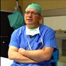 Meir Liebergall合作论文数Hadassah University Medical Center4Yusef Azraq合作论文数3Eldad Erez合作论文数3Yair Barzilay合作论文数3
 Joshua Moss合作论文数Hebrew Univ Jerusalem, Hadassah Med Sch, Inst Med Res Israel Canada, Dept Dev Biol & Canc Res, IL-91120 Jerusalem, Israel3
 Bella Savitsky合作论文数Dept Nursing, Ashkelon Acad Coll3
 Zimlichman Eyal合作论文数Dept Med B, Chaim Sheba Med Ctr3
 Shussman Noam合作论文数Department of Surgery, Hadassah Hebrew University Medical Center3
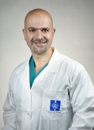 Weil Yoram A合作论文数Department of Orthopaedics, Hadassah Hebrew University Hospital;fax: +972 2 6423074;mobile: +972 50 7874314.;Department of Orthopaedics, Hadassah Hebrew University Hospital3Goldschmidt Neta合作论文数3Avi Israeli合作论文数3
 Uzi Izhar合作论文数3Martine Klein合作论文数3Amit Korach合作论文数3Dan Justo合作论文数Department of Internal Medicine D, Tel-Aviv Sourasky Medical Center3
 Ori Wald合作论文数Department of Cardiothoracic Surgery, Hadassah University Hospital3Tsuria Pelet合作论文数2Zeina Drawshy合作论文数2Machmud Abu Gazala合作论文数2Patriss Moradi合作论文数2
 Dvora Bauman合作论文数2Sudharani Nagaraju合作论文数2Ayelet Peretz合作论文数2Einat Carmon合作论文数2Alon J Pikarsky合作论文数2Judith Diment合作论文数2Wiessam Abu Ahmed PhD合作论文数2Devorah Rotnemer Golinkin合作论文数2Sapir Shabi-Porat合作论文数2
 Hanna Schroeder合作论文数2Gordon Cann合作论文数2Rehan Saiyed合作论文数2Yaacov Samet合作论文数2Arash Jamshidi合作论文数GRAIL, Inc., Menlo Park, CA;2Netanel Loyfer合作论文数School of Computer Science and Engineering, The Hebrew University of Jerusalem, Israel.2Ilana Fox-Fisher合作论文数Dept Dev Biol & Canc Res, Hebrew Univ Jerusalem2
 Jose E Cohen合作论文数2
 Itshayek Eyal合作论文数Department of Neurosurgery, Hadassah-Hebrew University Medical Center2Khalaileh Abed合作论文数2
 Peter Arner合作论文数Mikael Rydén & Niklas Mejhert lab, Endocrinology Unit, Department of Medicine, Huddinge, Karolinska Institutet2
 Andrew Mark James合作论文数Department of Surgery, Faculty of Medicine & Dentistry, University of Alberta;Division of General Surgery, Faculty of Medicine & Dentistry, UniversityofAlberta2
 Agnes Klochendler合作论文数2Fraifeld Shifra合作论文数Department of Neurosurgery, Hadassah-Hebrew University Medical Center;Department of Radiology, Hadassah-Hebrew University Medical Center;Hadassah-Hebrew University Medical Center, Hadassah-Hebrew University Medical Center2Hecht Merav合作论文数2Oliver Venn合作论文数Wellcome Trust Centre for Human Genetics2




