Paroxysmal nocturnal hemoglobinuria (PNH) is a rare hematologic disease characterized by a deregulated complement system, chronic Coombs-negative, intravascular hemolysis, and a variable clinical course with substantial risk to develop thromboembolic events. We analyzed diagnostic and prognostic parameters as well as clinical endpoints in 59 adult patients suffering from PNH in 5 hematology centers in Austria (observation period: 1978–2015). Median follow-up time was 5.6 years. The median clone size at diagnosis amounted to 55% and was higher in patients with classical PNH (81%) compared to patients with PNH associated with aplastic anemia (AA) or myelodysplastic syndromes (MDS) (50%). The clone size also correlated with lactate dehydrogenase (LDH) levels. In one patient, anemia improved spontaneously and disappeared with complete normalization of LDH after 16 years. Seventeen patients received therapy with eculizumab. The rate of thromboembolic events was higher in the pre-eculizumab era compared with eculizumab-treated patients but did not correlate with the presence of age-related clonal hematopoiesis or any other clinical or laboratory parameters. Peripheral blood colony-forming progenitor cell counts were lower in PNH patients compared with healthy controls. Only two patients with classical PNH developed MDS. Overall, 7/59 patients died after 0.5–32 years. Causes of death were acute pulmonary hypertension, Budd-Chiari syndrome, and septicemia. Overall survival (OS) was mainly influenced by age and was similar to OS measured in an age-matched healthy Austrian control cohort. Together, compared with previous times, the clinical course and OS in PNH are favorable, which may be due to better diagnosis, early recognition, and eculizumab therapy.
Background: The interstitial deletion of the long arm of chromosome 5—del(5q)—is the most common cytogenetic abnormality in MDS occurring either as a sole aberration or as a part of complex karyotypes (CK). MDS with isolated del(5q) are associated with a favorable outcome while MDS with CK relate to poor prognosis. Extent of deleted segment is highly variable. However, it remains unclear whether its size matters for the different MDS phenotypes. Aims: The aim was to compare the extent of deletion in 264 MDS patients [116 with isolated del(5q) and 148 with del(5q) involved in CK] and to assess the relationship of del(5q) extent and TP53 gene mutation. Methods: Extent of del(5q) was analyzed using I-FISH (Abbott, MetaSystems), mBAND (MetaSystems) and array CGH/SNP (CytoChip Cancer SNP 180K, Illumina or SurePrint G3 Cancer CGH+SNP 4x180K, Agilent). CK were analyzed with mFISH (MetaSystems). Sequence analysis of TP53 gene was performed in 84 cases using amplicon deep sequencing (exons 4–11) on a 454 GS Junior system (Roche) or with TruSight Myeloid Panel Kit on MiSeq sequencing instruments (Illumina). Results: In the group with isolated del(5q), the most frequently deleted segment ranged between the bands 5q14 and 5q33.3, with the smallest deletion encompassing the 5q31.1–5q31.3 region (18.527 Mb). The TP53 mutation was proved in 19.4% cases. The size of the deletion did not significantly differ in patients with and without TP53 mutation. In the group with CK, the deletion often involved entire long arm including the telomeric region. In some cases, a part of short arm was also deleted, but we never observed the loss of the entire chromosome 5. The commonly conserved region (CCR) was localized in bands 5p12 to 5p14.1. In this group, the mutation of TP53 and/or LOH17p was detected in 49% of patients. The commonly deleted regions (CDR) were in the band 5q31.1 (5.522 Mb) in cases with isolated del(5q) and between the bands 5q31.1 and 5q31.3 (18.527 Mb) in patients with CK. Summary/Conclusion: Patients with isolated del(5q) had a smaller size of the deleted segment. More extensive 5q deletion was associated with higher karyotype complexity, increased frequency of TP53 aberrations and worse prognosis. Accurate analysis of breakpoints and range of del(5q) points out to the correlation of deletion size with increasing genomic instability in MDS and contributes to a better understanding of the MDS pathogenesis. Supported by RVO-VFN64165, GACR P302/12/G157, ProgresQ28/LF1, MHCR 00023736.
Basophils form a distinct cell lineage within the hematopoietic cell family. In various myeloid neoplasms, including chronic myeloid leukemia, basophilia is frequently seen. Acute and chronic basophilic leukemias, albeit rare, have also been described. However, no generally accepted criteria and classification of basophilic leukemias have been presented to date. To address this unmet need, a series of Working Conferences and other meetings were organized between March 2015 and March 2016. The current article provides a summary of consensus statements from these meetings, together with proposed criteria to delineate acute basophilic leukemia (ABL) from chronic basophilic leukemia (CBL) and primary forms of the disease where no preceding myeloid malignancy is detected, from the more common ‘secondary’ variants. Moreover, the term hyperbasophilia (HB) is proposed for cases with a persistent peripheral basophil count ⩾1000 per μl of blood. This condition, HB, is highly indicative of the presence of an underlying myeloid neoplasm. Therefore, HB is an important checkpoint in the diagnostic algorithm and requires a detailed hematologic investigation. In these patients, an underlying myeloid malignancy is often found and is then labeled with the appendix -baso, whereas primary cases of ABL or CBL are very rare. The criteria and classification proposed in this article should facilitate the diagnosis and management of patients with unexplained basophilia and basophil neoplasms in routine practice, and in clinical studies.
Definite progress has been made in the exploration of myelodysplastic syndromes (MDS) by flow cytometry (FCM) since the publication of the World Health Organization 2008 classification of myeloid neoplasms. An international working party initiated within the European LeukemiaNet and extended to include members from Australia, Canada, Japan, Taiwan and the United States has, through several workshops, developed and subsequently published consensus recommendations. The latter deal with preanalytical precautions, and propose small and large panels, which allow evaluating immunophenotypic anomalies and calculating myelodysplasia scores. The current paper provides guidelines that strongly recommend the integration of FCM data with other diagnostic tools in the diagnostic work-up of MDS.
Special attention has recently been drawn to the molecular network of different genes that are responsible for the development of erythroid cells. The aim of the present study was to establish in detail the immunophenotype of early erythroid cells and to compare the gene expression profile of freshly isolated early erythroid precursors with that of the CD34-positive (CD34(+)) compartment. Multiparameter flow cytometric analyses of human bone marrow mononuclear cell fractions (n=20) defined three distinct early erythroid stages. The gene expression profile of sorted early erythroid cells was analyzed by Affymetrix array technology. For 4524 genes, a differential regulation was found in CD105-positive erythroid cells as compared with the CD34(+) progenitor compartment (2362 upregulated genes). A highly significant difference was observed in the expression level of genes involved in transcription, heme synthesis, iron and mitochondrial metabolism and transforming growth factor-β signaling. A comparison with recently published data showed over 1000 genes that as yet have not been reported to be upregulated in the early erythroid lineage. The gene expression level within distinct pathways could be illustrated directly by applying the Ingenuity software program. The results of gene expression analyses can be seen at the Gene Expression Omnibus repository.
Flow cytometry (FC) is increasingly recognized as an important tool in the diagnosis and prognosis of myelodysplastic syndromes (MDS). However, validation of current assays and agreement upon the techniques are prerequisites for its widespread acceptance and application in clinical practice. Therefore, a working group was initiated (Amsterdam, 2008) to discuss and propose standards for FC in MDS. In 2009 and 2010, representatives from 23, mainly European, institutes participated in the second and third European LeukemiaNet (ELN) MDS workshops. In the present report, minimal requirements to analyze dysplasia are refined. The proposed core markers should enable a categorization of FC results in cytopenic patients as ‘normal’, ‘suggestive of’, or ‘diagnostic of’ MDS. An FC report should include a description of validated FC abnormalities such as aberrant marker expression on myeloid progenitors and, furthermore, dysgranulopoiesis and/or dysmonocytopoiesis, if at least two abnormalities are evidenced. The working group is dedicated to initiate further studies to establish robust diagnostic and prognostic FC panels in MDS. An ultimate goal is to refine and improve diagnosis and prognostic scoring systems. Finally, the working group stresses that FC should be part of an integrated diagnosis rather than a separate technique.
 K. Lechner合作论文数119Hinterberger W合作论文数88
 Peter Valent合作论文数Department of Medicine, Medizinische Universitat Wien;Ludwig Boltzmrnn Institute70
 Klaus Geissler合作论文数Hosp Hietzing, Med Dept 5, Vienna, Austria61Walter Knapp合作论文数49Otto Majdic合作论文数49I. Schwarzinger合作论文数40
 Herwig Niessner合作论文数35
 Oskar Haas合作论文数St. Anna Children's Hospital;Labdia Labordiagnostik GmbH33Simon Panzer合作论文数28Erich Neumann合作论文数28Christian Korninger合作论文数27Ulrich Jäger合作论文数25
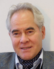 Eberhard Neumann合作论文数Fakultat fur Chemie, Universitat Bielefeld25
 Ursula Koller合作论文数23D Lutz合作论文数20
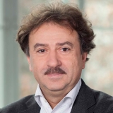 Hannes Stockinger合作论文数Department of Molecular Immunology, Institute for Hygiene and Applied Immunology, Center for Pathophysiology, Infectiology and Immunology, Medical University of Vienna20Paul Hocker合作论文数19christian sillaber合作论文数18
 Ingrid Pabinger合作论文数Clinical Division of Haematology and Haemostaseology, Department of Medicine I, Medical University of Vienna18Thaddaus Radaszkiewicz合作论文数17K LISZKA合作论文数16
 Sigrid Machherndl-Spandl合作论文数15
 Helmut Gadner合作论文数15W Schmidmeier合作论文数13Ansgar Weltermann合作论文数13
 Christian Gabriel合作论文数12
 Wolfgang R Sperr合作论文数Division of Hematology and Hemostaseology and Ludwig Boltzmann Institute for Hematology and Oncology, Medical University of Vienna12C STAIN合作论文数12H GOSSINGER合作论文数12E Liehl合作论文数11O Krieger合作论文数10P Kalhs合作论文数10M KOS合作论文数10Elisabeth Paietta合作论文数10C. Pollak合作论文数10J. Proell合作论文数9
 Werner Linkesch合作论文数9P Mayer合作论文数9
 Dieter Maurer合作论文数9Jurgen Besemer合作论文数9
 Susanne Suessner合作论文数8W Speiser合作论文数8R Scherrer合作论文数8Otto Zach合作论文数8J. Lauf合作论文数7
 Martin Danzer合作论文数7P Kyrle合作论文数7Jd Schwarzmeier合作论文数7




