Phosphorylation is one of the most common of the reversible post-translational modifications, but also one of the most challenging. During the last few years, phosphopeptide analysis has benefited from the rapid improvement in biological mass spectrometry, but also from innovations such as immobilized metal affinity chromatography, the development of antibodies specific for phosphorylated amino acids, and most recently, chromatography using TiO2. During our analysis of the saliva phosphoproteome, we were concerned that there is a bias against monophosphorylated peptides when using TiO2 columns. We therefore decided to explore alternatives that rely on fractionation rather than affinity purification. Using the approach of Roepstorff and colleagues of packing columns in pipette tips to make disposable microcolumns, and combining strong cationic exchange (SCX) together with hydrophilic interaction liquid chromatography (HILIC), we have been able to fractionate peptides from digests of total saliva, and isolate phosphopeptides in a few fractions with a minimum of non-phosphorylated peptides. In preliminary experiments using about 6 μg of saliva, we have been able to map out phosphorylation sites from proteins such as the salivary acid proline-rich phosphoprotein 1/2, the salivary α-amylase, and statherin. A comparison of isolation of phosphopeptides using TiO2 vs. a combination of SCX and HILIC will be presented.
Introduction. The Bronchopulmonary Dysplasia (BPD) or Mikity Wilson Syndrome (MWS) are chronic lung diseases with several pathogenic mechanisms. They can be associated with atypical infections by U. urealyticum, C. trachomatis, and Mycoplasma. Objective. We researched pulmonary infections in 10 VLBWN with BPD or MWS and had negative cell cultures by PCR in tracheobronchial samples. Methods. We studied VLBWN in our institution with BPD (n 6) or WMS (n 4) (with oxygen dependence and images lung radiographic of lung damage with fibrosis and cystic areas) as diagnosis and negative cell culture to atypical pathogens. We took new tracheobronchial samples at 28 days of life and send them Virology Department to PCR and infection determination. First. Tracheobronchial aspirate was took with saline solution 0.9% and stored in aliquots at 20°C until needed. Second. DNA isolation by proteinase K method and we used gel electrophoresis and densitometer to determine DNA concentration. Third. Determinate polymerase chain reaction (PCR). DNA concentration and extraction process were included as samples to identify background bacterial sequences present in reagents and supplies. Results. We found 7/10 positive infections, 3/6 C. trachomatis infections in neonates with BPD and 4/4 with MWS: U. urealyticum (1), Mycoplasma genitalium (1), C. trachomatis (1) and M. pneumoniae (1). Conclusions. We should researched atypical pulmonary infections by intracellular pathogens by another methods with more sensitivity as PCR to explain inflammatory response and lung damage. 1125 Comparative Proteomic Analyses of Normal and Normal Pressure Hydrocephalus in Human Cerebrospinal Fluid
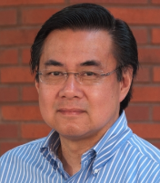 Joseph A. Loo合作论文数Department of Chemistry and Biochemistry, David Geffen School of Medicine, University of California, Los Angeles;Molecular Instrumentation Center, University of California, Los Angeles7
 David T.W Wong合作论文数Center for Oral/Head & Neck Oncology Research, School of Dentistry, University of California Los Angeles5
 Shen Hu (胡深)合作论文数4M Arellano-Garcia合作论文数3
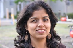 Prasanna Ramachandran合作论文数Molecular Biology Institute, University of California—Los Angeles3
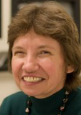 Rachel Ogorzalek Loo合作论文数Molecular Biology Institute, University of California, Los Angeles2S. Hu合作论文数1M Sondej合作论文数1Y. Xie合作论文数1R. Gunsalus合作论文数1Jr Strahler合作论文数1R. Loo合作论文数1
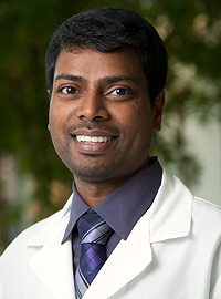 Saravana Mohan M. Dhanasekaran合作论文数Department of Pathology, Medical School, University of Michigan1
 Alexey Nesvizhskii合作论文数Department of Computational Medicine and Bioinformatics, University of Michigan Medical School1Taylor B S合作论文数1
 Michael J Mcinerney合作论文数Department of Botany;University of Oklahoma;Department of Botany, University of Oklahoma1
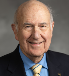 Gilbert S. Omenn合作论文数Department of Computational Medicine and Bioinformatics, Medical School, University of Michigan;School of Public Health, University of Michigan1
 Philip C. Andrews合作论文数Department of Biological Chemistry, University of Michigan Medical School;Department of Computational Medicine and Bioinformatics, University of Michigan;Department of Chemistry, University of Michigan1Sooryanarayana Varambally合作论文数1
 Anders Jimmy Ytterberg合作论文数1
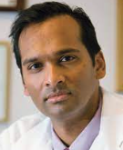 Arul M. Chinnaiyan合作论文数Rogel Cancer Center, Department of Pathology and Clinical Laboratories, University of Michigan;Michigan Center for Translational Pathology, University of Michigan;Howard Hughes Medical Institute1




