Biomarkers of ageing are defined as age-related changes in body function or composition that could serve as a measure of 'biological' age and predict the onset of age-related diseases and/or residual life expectancy. We conducted the MARK-AGE Study, a European population study (3300 subjects aged 35-74) to identify a powerful set of biomarkers of ageing. A total of 362 clinical-chemistry, genetic, cellular or molecular biomarkers were analysed for each subject. Using statistical models as well as machine learning we derived mathematical formulas for females and for males that yield a 'bioage score' of an individual, based on sets of 10 biomarkers for females and 10 for males. Collectively, these biomarkers model chronological age of our study population and, thus yield the 'biological' age of a certain person. 'Age difference' (defined as biological minus chronological age) should then identify biologically older or younger individuals. Using our set of biomarkers, subjects with Down Syndrome and smoking females are biologically older, whereas postmenopausal females taking hormone replacement therapy are biologically younger. Strikingly, our data reveal that age difference of MARK-AGE subjects, but not chronological age, is linearly correlated with levels of HDL, 25-hydroxy-Vitamin D, and CD3+ CD4+/CD45+ ratio in such a way that biologically younger subjects display values that are favourable to good health, whereas other markers such as glucose and HbA1c are correlated with chronological age, but not age difference. This dichotomy of correlations may point to different roles of such markers, that is, drivers of the ageing process versus bystanders of ageing.
Torquetenovirus (TTV) is a prevalent virus whose clinical significance remains unclear, potentially linked to immunosenescence. This study examines TTV species in relation to immune impairment, inflammation, and cellular stress response in aging. A subset of recruited age-stratified individuals (RASIG) from the MARK-AGE study was divided into three groups: Cohort 1 A (healthy young adults), Cohort 1B (older adults with mild immune decline), and Cohort 1 C (older adults with marked immune impairment). Analyses included TTV load, species diversity, lymphocyte subpopulations, inflammatory markers, Poly-(ADP-ribose) polymerase (PARP-1) expression/activity. Alpha- and beta-diversity analyses showed the highest TTV species diversity in Cohort 1 C, with significant cohort-dependent differences and partially cohort-specific clustering patterns. Increased TTV species number correlated with higher TTV load, elevated CMV IgG levels, and greater immune impairment risk. Specific TTV species were associated with CD4/CD8, and reduced T-cell receptor excision circles, suggesting impaired T-cell homeostasis. TTV viremia positively correlated with C-reactive protein (CRP) and α2-macroglobulin. PARP-1 expression and activity increased in individuals with higher TTV diversity, particularly in the presence of TTV9 and TTV20. TTV load and species diversity are associated with immunosenescence, inflammation, and PARP-1 activation suggesting their potential as biomarkers of age-related immune decline. Longitudinal studies are needed to clarify underlying mechanisms.
Cytomegalovirus (CMV) infection has been linked to accelerated biological aging, potentially increasing the risk of cardiovascular disease. DNA methylation of the gene Elongation Of Very Long Chain Fatty Acids-Like 2 (ELOVL2) is a molecular biomarker for aging, and its gene product is involved in polyunsaturated fatty acid synthesis, which impacts immune and inflammatory responses. This study, conducted in the MARK-AGE population, aimed to investigate the relationship between CMV infection and ELOVL2 methylation in adults aged 35-75, as well as the influence of CMV IgG levels on lipid metabolism, inflammation, DNA damage, and DNA repair. Our data revealed a higher prevalence of ischemic heart disease, atrial fibrillation, hypertension, and diabetes in CMV-positive individuals. CMV IgG levels were positively associated with ELOVL2 methylation at specific CpG sites and with increased expression of DNA methyltransferase-1 (DNMT1). CMV IgG was linked to lipid imbalances, such as increased BMI, VLDL-cholesterol, triglycerides, and HDL1-cholesterol. Additionally, ELOVL2 methylation was associated with systemic inflammation markers, lipid parameters and altered T-cell subsets. A negative correlation was observed between CMV IgG levels and both baseline DNA integrity and repair capacity. These results suggest that CMV infection might promote cardiovascular disease through ELOVL2 hypermethylation, lipid dysregulation, inflammation, and DNA damage.
Control of ribosome biogenesis is a critical aspect of the regulation of cell metabolism. As ribosomal genes (rDNA) are organized in repeated clusters on chromosomes 13, 14, 15, 21, and 22, trisomy of chromosome 21 confers an excess of rDNA copies to persons with Down syndrome (DS). Previous studies showed an alteration of ribosome biogenesis in children with DS, but the epigenetic regulation of rDNA genes has not been investigated in adults with DS so far. In this study, we used a targeted deep-sequencing approach to measure DNA methylation (DNAm) of rDNA units in whole blood from 69 adults with DS and 95 euploid controls. We further evaluated the expression of the precursor of ribosomal RNAs (RNA45S) in peripheral blood mononuclear cells (PBMCs) from the same subjects. We found that the rDNA promoter tends to be hypermethylated in DS concerning the control group. The analysis of epihaplotypes (the combination of methylated and unmethylated CpG sites along the same DNA molecule) showed a significantly lower intra-individual diversity in the DS group, which at the same time was characterized by a higher interindividual variability. Finally, we showed that RNA45S expression is lower in adults with DS. Collectively, our results suggest a rearrangement of the epigenetic profile of rDNA in DS, possibly to compensate for the extranumerary rDNA copies. Future studies should assess whether the regulation of ribosome biogenesis can contribute to the pathogenesis of DS and explain the clinical heterogeneity characteristic of the syndrome.
Ageing leaves characteristic traces in the DNA methylation make-up of the genome. However, the importance of DNA methylation in ageing remains unclear. The study of subtelomeric regions could give promising insights into this issue. Previously reported associations between susceptibility to age-related diseases and epigenetic instability at subtelomeres suggest that the DNA methylation profile of subtelomeres undergoes remodelling during ageing. In the present work, this hypothesis has been tested in the context of the European large-scale project MARK-AGE. In this cross-sectional study, we profiled the DNA methylation of chromosomes 5 and 21 subtelomeres, in more than 2000 age-stratified women and men recruited in eight European countries. The study included individuals from the general population as well as the offspring of nonagenarians and Down syndrome subjects, who served as putative models of delayed and accelerated ageing, respectively. Significant linear changes of subtelomeric DNA methylation with increasing age were detected in the general population, indicating that subtelomeric DNA methylation changes are typical signs of ageing. Data also show that, compared to the general population, the dynamics of age-related DNA methylation changes are attenuated in the offspring of centenarian, while they accelerate in Down syndrome individuals. This result suggests that subtelomeric DNA methylation changes reflect the rate of ageing progression. We next attempted to trace the age-related changes of subtelomeric methylation back to the influence of diverse variables associated with methylation variations in the population, including demographics, dietary/health habits and clinical parameters. Results indicate that the effects of age on subtelomeric DNA methylation are mostly independent of all other variables evaluated.
characterized by skewed epigenetic patterns, raising the possibility of therapeutically targeting epigenetic factors in this disease. Here we report that among different cancer types, epigenetic 38 factor TET1 is highly expressed in T-ALL and is crucial for human T-ALL cell growth in vivo . 39 Tet1 knockout mice and knockdown in human T-cells did not perturb normal T-cell proliferation, 40 indicating that TET1 expression is dispensable for normal T-cell growth. The promotion of 41 leukemic growth by TET1 was depending on its catalytic property to maintain global 5- 42 hydroxymethylcytosine (5hmC) marks, thereby regulating cell cycle, DNA repair genes and T- 43 ALL associated oncogenes. Furthermore, overexpression of the Tet1 catalytic domain was 44 sufficient to augment global 5hmC levels and leukemic growth of T-ALL cells in vivo . We 45 demonstrate that PARP enzymes, which are highly expressed in T-ALL patients, participate in 46 establishing H3K4me3 marks at the TET1 promoter and that PARP1 interacts with the TET1 47 protein. Importantly, the growth related role of TET1 in T-ALL could be antagonized by the 48 clinically approved PARP inhibitor Olaparib, which abrogated TET1 expression, induced loss of 49 5hmC marks and antagonized leukemic growth of T-ALL cells, opening a therapeutic avenue for 50 this disease. targeting epigenetic factors for treating this malignancy. Recent studies in healthy human CD4 naïve T-cells indicate that TET1 negatively regulates Th1/Th2 differentiation by suppressing the expression of pro-differentiation genes such as GATA3 , CD69 and IFNG in vitro growth promoting. In this study, we now demonstrate that in human T-ALL cells, high TET1 expression maintains global hydroxymethylome which positively regulates gene expression, safeguards genome integrity, and thereby promotes leukemic growth. Furthermore, our data indicate that the growth promoting activity of TET1 can be pharmacologically targeted via inhibition of PARPs, 82 which act as TET1 upstream regulators, opening a potential treatment modality for T-ALL patients.
T-cell acute lymphoblastic leukemia (T-ALL) is an aggressive hematological cancer characterized by skewed epigenetic patterns, raising the possibility of therapeutically targeting epigenetic factors in this disease. Here we report that among different cancer types, epigenetic factor TET1 is highly expressed in T-ALL and is crucial for human T-ALL cell growth in vivo. Knockout of TET1 in mice and knockdown in human T cell did not perturb normal T-cell proliferation, indicating that TET1 expression is dispensable for normal T-cell growth. The promotion of leukemic growth by TET1 was dependent on its catalytic property to maintain global 5-hydroxymethylcytosine (5hmC) marks, thereby regulate cell cycle, DNA repair genes, and T-ALL associated oncogenes. Furthermore, overexpression of the Tet1-catalytic domain was sufficient to augment global 5hmC levels and leukemic growth of T-ALL cells in vivo. We demonstrate that PARP enzymes, which are highly expressed in T-ALL patients, participate in establishing H3K4me3 marks at the TET1 promoter and that PARP1 interacts with the TET1 protein. Importantly, the growth related role of TET1 in T-ALL could be antagonized by the clinically approved PARP inhibitor Olaparib, which abrogated TET1 expression, induced loss of 5hmC marks, and antagonized leukemic growth of T-ALL cells, opening a therapeutic avenue for this disease.
T-cell acute lymphoblastic leukemia (T-ALL) is an aggressive blood cancer caused by the deregulation of key T-cell developmental pathways, including Notch signaling. Aberrant Notch signaling in T-ALL occurs by NOTCH1 gain-of-function mutations and by NOTCH3 overexpression. Although NOTCH3 is assumed as a Notch1 target, machinery driving its transcription in T-ALL is undefined in leukemia subsets lacking Notch1 activation. Here, we found that the binding of the intracellular Notch3 domain, as well as of the activated Notch1 fragment, to the NOTCH3 gene locus led to the recruitment of the H3K27 modifiers JMJD3 and p300, and it was required to preserve transcriptional permissive/active H3K27 marks and to sustain NOTCH3 gene expression levels. Consistently, pharmacological inhibition of JMJD3 by GSKJ4 treatment or of p300 by A-485 decreased the levels of expression of NOTCH3, NOTCH1 and of the Notch target genes DELTEX1 and c-Myc and abrogated cell viability in both Notch1- and Notch3-dependent T-cell contexts. Notably, re-introduction of exogenous Notch1, Notch3 as well as c-Myc partially rescued cells from anti-growth effects induced by either treatment. Overall our findings indicate JMJD3 and p300 as general Notch1 and Notch3 signaling co-activators in T-ALL and suggest further investigation on the potential therapeutic anti-leukemic efficacy of their enzymatic inhibition in Notch/c-Myc axis-related cancers and diseases.
Alu hypomethylation promotes genomic instability and is associated with aging and age-related diseases. Dietary factors affect global DNA methylation, leading to changes in genomic stability and gene expression with an impact on longevity and the risk of disease. This preliminary study aims to investigate the relationship between nutritional factors, such as circulating trace elements, lipids and antioxidants, and Alu methylation in elderly subjects and offspring of healthy nonagenarians. Alu DNA methylation was analyzed in sixty RASIG (randomly recruited age-stratified individuals from the general population) and thirty-two GO (GeHA offspring) enrolled in Italy in the framework of the MARK-AGE project. Factor analysis revealed a different clustering between Alu CpG1 and the other CpG sites. RASIG over 65 years showed lower Alu CpG1 methylation than those of GO subjects in the same age class. Moreover, Alu CpG1 methylation was associated with fruit and whole-grain bread consumption, LDL2-Cholesterol and plasma copper. The preserved Alu methylation status in GO, suggests Alu epigenetic changes as a potential marker of aging. Our preliminary investigation shows that Alu methylation may be affected by food rich in fibers and antioxidants, or circulating LDL subfractions and plasma copper.
DNA methylation is currently the most promising molecular marker for monitoring aging and predicting life expectancy. However, the mechanisms underlying age-related DNA methylation changes remain mostly undiscovered. Here we discuss the current knowledge of the dynamic nature of DNA epigenome landscape in mammals, and propose putative molecular mechanisms for aging-associated DNA epigenetic changes. Specifically, we describe age-related variations of methylcytosine and its oxidative derivatives in relation to the dynamics of chromatin structure, histone post-translational modifications and their modulators. Finally, we are proposing a conceptual framework that could explain the complex nature of the effects of age on DNA methylation patterns. This combines the accumulation of DNA methylation noise and also all of the predictable, site-specific DNA methylation changes. Gathering information in this area would pave the way for future investigation aimed at establishing a possible causative role of epigenetic mechanisms in aging.
Down syndrome (DS) is caused by the presence of part or an entire extra copy of chromosome 21, a phenomenon that can cause a wide spectrum of clinically defined phenotypes of the disease. Most of the clinical signs of DS are typical of the aging process including dysregulation of immune system. Beyond the causative genetic defect, DS persons display epigenetic alterations, particularly aberrant DNA methylation patterns that can contribute to the heterogeneity of the disease. In the present work, we investigated the levels of 5-hydroxymethylcytosine and of the Ten-eleven translocation dioxygenase enzymes, which are involved in DNA demethylation processes and are often deregulated in pathological conditions as well as in aging. Analyses were carried out on peripheral blood mononuclear cells of DS volunteers enrolled in the context of the MARK-AGE study, a large-scale cross-sectional population study with subjects representing the general population in eight European countries. We observed a decrease in 5-hydroxymethylcytosine, TET1, and other components of the DNA methylation/ demethylation machinery in DS subjects, indicating that aberrant DNA methylation patterns in DS, which may have consequences on the transcriptional status of immune cells, may be due to a global disturbance of methylation control in DS.
The process of aging culminates in a multitude of chronological irreversible changes at the cellular and molecular levels that push the organism toward a progressive loss of physiological integrity and increased susceptibility to disease. Mounting evidence indicates that aging trajectories can be closely tracked down by changes in the DNA methylation patterns over the entire lifespan. The tracing of underlying mechanisms is a promising research path to gain a deeper understanding of the molecular nature of aging. Here we discuss current knowledge about dynamics of DNA methylation in mammals and describe changes in DNA methylation occurring during aging. We explore mechanisms that could underpin aging-related DNA methylation changes and highlight models in which those changes can be understood in the context of the complex interplay between DNA methylation, chromatin states, and their modulators.
Metallothionein (MT) family are cysteine-rich proteins that regulate zinc (Zn) homeostasis and protect against oxidative damage. Studies in transgenic mice have shown that MT favorably influence longevity, although their role in human aging is not completely understood. Within the European multicenter study MARK-AGE, we analyzed MT induction after Zn treatment in peripheral blood mononuclear cells (PBMCs) and its relation with redox biomarkers in 2,936 age-stratified subjects (35-75 years) including the general population (RASIG), centenarian offspring (GO), and their spouses (SGO). We found that the lymphocyte capability to induce MT in response to Zn is not affected by aging. However, GO participants showed lower Zn-induced MT and increased basal expression of MT1A, MT1X, and ZnT-1 genes than RASIG subjects. Moreover, Zn-induced MT levels were found to be inversely related with oxidative stress markers (plasma protein carbonyls, 3-nitrotyrosine, and malondialdehyde) in the whole population, but not in GO subjects. In conclusion, our results support the hypothesis that the response to Zn is attenuated in PBMCs of centenarian offspring compared to the general population as a consequence of a tighter control of Zn homeostasis which is likely to provide them constant protection against stress stimuli over the whole lifespan.
AML1-ETO (AE) is the most commonly occurring fusion gene in AML and shows a distinct methylation pattern, the underlying mechanisms for which is poorly understood. TET1 dioxygenase regulates methylation patterns via conversion of 5-methylcytosine (5mC) to 5-hydroxymethylcytosine (5hmC), thereby regulating a variety of biological processes. However, the role of TET1 in AE+ AML cases is yet unexplored. Using qRT- PCR we observed that TET1 was significantly higher expressed in the majority of AE+ patients compared to other AML subtypes and CD34+ normal BM cells. This observation was consistent with published cDNA microarray and transcriptome data. Knockdown (KD) of TET1 using two shRNAs in AE+ AML cell lines impaired their cell growth and clonogenicity in vitro. KD of Tet1 in AE9a+ murine leukemic cell line inhibited its clonogenicity in vitro and delayed onset of leukemia in vivo. Tet1-ko mouse derived HSPCs transduced with AE9a showed impaired serial replating capacity compared to AE9a transduced Tet1-wt HSPCs in vitro. hMeDIP and MeDIP-seq performed on TET1 depleted KASUMI1 cells revealed 8,562 5hmC-enriched promoters (-5kbTSS) in the scrambled arm and a 50% decrease in 5hmC-enriched promoters in KD arm. In RNA-seq the genes associated with AML, Pleuripotency and WNT signaling were downregulated upon TET1 KD and over 200 of these genes exhibited loss of 5hmC on their promoters. MeDIPseq and RNA-seq data revealed a global decrease of promoter methylation and increased expression of myeloid differentiation associated genes. IP analysis confirmed that TET1 physically interacted with PARP1 in AE+ cell line. Oncogenic TET1 expression was antagonized by PARP inhibitor Olaparib in AE+ human leukemic cell lines, which induced reduction of H3K4me3 marks on the TET1 promoter and 5hmC levels in AML cell lines. Furthermore, Olaparib treatment decreased cell growth and clonogenicity of human and murine AE+ cell lines. In conclusion, our data indicate that aberrant TET1 expression contributes to the growth of AE+ AML by maintain 5hmC marks and that the PARP inhibitor olaparib can at least partially antagonize the oncogenic effect of TET in AML.
Epigenetic events include reversible modifications of DNA and histone tails driving chromatin organization and thus transcription. The epigenetic regulation is a highly integrated process underlying the plasticity of the genomic information both in the context of complex physiological and pathological processes. The global regulatory aspects of epigenetic events are largely unknown. PARylation and PARP1 are recently emerging as multi-level regulatory effectors that modulate the topology of chromatin by orchestrating very different processes. This review focuses in particular on the role of PARP1 in epigenetics, trying to build a comprehensive perspective of its involvement in the regulation of epigenetic modifications of histones and DNA, contextualizing it in the global organization of chromatin domains in the nucleus.
The Ten-eleven translocation 1 (TET1) enzyme catalyzes the oxidation of 5-methylcytosine (5mC) into 5-hydroxymethylcytosine (5hmC) and plays a key role in active DNA demethylation and regulation of gene expression. TET1 has been shown play a tumor supper function in B-cell malignancies however, its roles in T-cell acute lymphoblastic leukemia (T-ALL) is yet unknown. qRTPCR revealed that TET1 was significantly higher expressed in all human T-ALL cell lines and 95% of primary T-ALL patients compared to B-ALL cell lines, other primary leukemia types and healthy T-cells. This was further confirmed by cDNA microarray analysis on larger cohort of primary leukemia patient samples. Knockdown (KD) of TET1 using shRNAs in T-ALL cell lines adversely affected their cell growth and clonogenicity in vitro, and significantly reduced their leukemic engraftment potential in xenografts. Global analysis of 5hmC and 5mC marks using hMeDIP-seq and MeDIP-seq performed on TET1 depleted JURKAT cells revealed lower global 5hmC levels and increased 5mC levels (n=2). Promoters (-5kbTSS) demonstrating loss of 5hmC or gain of 5mC marks belonged to gene families involved in DNA repair, Cell cycle, T-cell development, Cancer and Wnt signaling. Changes in promoter 5hmC/5mC marks were paralleled by changes in expression of DNA repair and T-cell associated genes as measured by RNA-Seq, and protein levels as measured by western blot. Furthermore, KD of TET1 in T-ALL cell lines induced DNA damage and cell cycle arrest in the G2M phase. Aberrant expression of TET1 could be reverted by Olaparib, an inhibitor of DNA repair enzyme PARP, which induced a reduction of euchromatic marks on the TET1 promoter accompanied by decrease in TET1 mRNA and protein levels and global reduction of 5-hmC levels in T-ALL cell lines. Olaparib treatment adversely affected cell growth, clonogenicity and leukemic engraftment potential of T-ALL cell lines. In conclusion, these data demonstrate for the first time that TET1 plays an important oncogenic role in T-ALL. We also identify PARPs as upstream regulators of TET1 in T-ALL, opening the way to antagonize TET1 pharmacologically in this poor prognosis leukemia.
DNA methylation patterns are highly deregulated in human acute myeloid leukemia (AML) cases and stratify AML patient samples into different subgroup. AML1-ETO is the most commonly occurring fusion gene in AML and these AML cases exhibit an aberrant and distinct methylation pattern. So far, the underlying mechanisms for this are only poorly understood. The TET1 dioxygenase has recently emerged as an important epigenetic modifier: by catalyzing the conversion of 5-methylcytosine (5mC) to 5-hydroxymethylcytosine (5hmC) TET1 plays an important role in active demethylation, thereby regulating a variety of biological processes. It was linked to tumorigenesis based on the observation that its expression is frequently deregulated in solid cancer. However, the role of TET1 in AML1-ETO+ (AE+)human AML cases is yet unexplored. Using quantitative real time (qRT)- PCR we now show that AE+ AML is characterized by high and aberrant expression of TET1: the gene was significantly higher expressed in the majority of AE+ patients (n=7, p<0.01) compared to other AML subtypes such as inv(16) (n=11), PML-RARα+ (n=31), cytogenetically normal (CN)-AML patients (n=33) and CD34+ normal BM cells (n=4). This observation was consistent with published cDNA microarray data on large patient cohorts (Haferlach et al., JCO 2010, p<0.008 t-test, p<0.01 Anova) and recently published transcriptome data (TCGA) of AML patients. In contrast to TET1, TET2 and TET3 did not show significant higher expression in AE+ patients compared to other AML subtypes. In line with patient data, TET1 was highest expressed in the AE+ AML cell line KASUMI-1 and SKNO-1 compared to other AML cell lines (p<0.05 and n=3). Compared to normal CD34+ and myeloid (CD33+, CD15+ and CD14+) cells (n=3), TET1 was 10-fold and 16-fold higher expressed in AE+ patient samples (n= 7). Aberrant expression of TET1 in AE+ leukemic cells was associated with hypomethylation of its promoter and enrichment for H3K4me3 euchromatic marks at its promoter as determined by LC/MS and ChIP-qPCR respectively. Knockdown (KD) of TET1 mRNA using two short hairpin RNAs (shRNAs) in AE+ AML cell lines impaired their cell growth and clonogenicity by over 50% in vitro (n=3 and p<0.01). shRNA mediated depletion of TET1 did not impact the cell growth and clonogenicity of the TET1 negative cell line RAJI, ruling out off target effects of the shRNAs (n=3). In mice, KD of Tet1 in leukemic bone marrow cells expressing the truncated leukemogenic AML1-ETO9a (AE9a) fusion, dramatically inhibited cell growth (>60% compared to scrambled, n=3, p<0.01), clonogenicity (>50-70% reduction in primary CFCs, p<0.01, n=3) and importantly delayed onset of leukemia in vivo (median survival 35 days for scr vs 80 days for shRNA mice, n=4/arm, p<0.03). Tet1-knock-out c-kit+ hematopoietic stem and progenitor cells (HSPCs) transduced with AE9a showed reduced primary colony formation and impaired serial replanting capacity in vitro compared to AE9a transduced Tet1-wild-type HSPCs (>50% and >70%, respectively; p<0.001, n=3). Global analysis of 5hmC and 5mC levels using hMeDIP/MeDIP-seq performed on TET1 depleted KASUMI-1 cells revealed lower global 5hmC levels and increase in 5mC as compared to cells transduced with scrambled control (n=2). 3324 promoter regions lost 5hmC and gained 5mC upon TET1 depletion (-5kTSS, Fold enrichment cut off <2-fold, q-value<1e5). Recent studies have shown that PARP activity induces TET1 expression by regulating its promoter epigenetically. We could show that aberrant TET1 expression could be antagonized by the PARP inhibitor olaparib in AE+ leukemic cell lines. Furthermore, olaparib treatment decreased 5hmC levels and reduced cell growth and clonogenicity of human AE+ cell lines and of the murine AE9a+ leukemic cell line in vitro (n=3, p<0.01). In conclusion, our data indicates that aberrant TET1 expression contributes to the growth of AE+ AML by maintaining the 5-hydroxymethylome and that the PARP inhibitor olaparib can at least partially antagonize the oncogenic effect of TET in AML.
 Anna Reale合作论文数41
 Michele Zampieri合作论文数39Fabio Ciccarone合作论文数33Roberto Strom合作论文数22Tiziana Guastafierro合作论文数‡Department of Anatomy, Histology, Forensic Medicine and Orthopedics, Section of Histology and Embryology, Faculty of Pharmacy and Medicine, "Sapienza"University Rome19Giuseppe Zardo合作论文数Brain Tumor Research Center;Department of Neurological Surgery;medical Sciences Program, Bio;Department of Laboratory Medicine;Cancer Research Institute19Maria Giulia Bacalini合作论文数18Calabrese Roberta合作论文数17
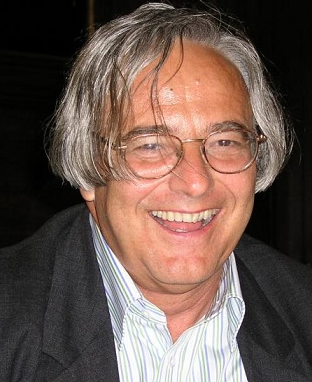 Claudio Franceschi合作论文数Faculty of Medicine, University of Bologna14
 Alexander Burkle合作论文数Department of Biology, University of Konstanz14
 Maria Moreno-Villanueva合作论文数Lehrstuhl Molekulare Toxikologie, Universität Konstanz13
 Paola Allegra合作论文数11M D'Erme合作论文数11
 Jordanka Zlatanova合作论文数University of Wyoming10
 Tilman Grune合作论文数Department of Molecular Toxicology, German Institute of Human Nutrition Potsdam-Rehbrucke;University of Potsdam;Faculty of Chemistry, University of Vienna10
 Miriam Capri合作论文数Department of Biomedical Sciences, University of Modena10Nicolle Breusing合作论文数9
 Marco Malavolta合作论文数Dipartimento di Scienze Cliniche e Molecolari, Università Politecnica delle Marche;Gerontological Research Department, INRCA Istituto Nazionale di Ricovero e Cura per Anziani9Martijn E.T. Dollé合作论文数9
 Ewa Sikora合作论文数9S Marenzi合作论文数8
 Eline Slagboom合作论文数Department of Biomedical Data Sciences, Leiden University Medical Center;Koninklijke Nederlandse Akademie van Wetenschappen8
 Raffaella Santoro合作论文数Biochemical Sciences “A. Rossi Fanelli” 2nd Institute of Internal Medicine, University of Rome “La Sapienza,”8
 Efstathios S Gonos合作论文数8Eugene Jansen合作论文数Centre for Health Protection, National Institute for Public Health and the Environment8M Hurme合作论文数7Elisabetta Valentini合作论文数7Antti Hervonen合作论文数7
 Viay Rawat合作论文数Inst Expt Canc Res, Univ Hosp Ulm7Christian Buske合作论文数7S Mastrantonio合作论文数6Alex Jose Pulikkottil合作论文数6Francesca Ciccarone合作论文数6
 Medhanie A. Mulaw合作论文数Clinical Cooperative Group “Leukemia, Helmholtz Zentrum Muenchen;Department of Medicine III, University of Munich-Grosshadern;Clinical Cooperative Group “Leukemia, University of Munich-Grosshadern6
 Marco Rispoli合作论文数6
 Hartmut Döhner合作论文数Klinik für Innere Medizin III, Zentrum für Innere Medizin, Universitätsklinikum Ulm6
 Michaela Feuring合作论文数Klinik für Innere Medizin III, Universitätsklinikum Ulm6
 Claudio Passananti合作论文数Istituto di Tecnologie Biomediche, CNR6
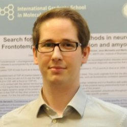 Fabian Mohr合作论文数Institute of Experimental Cancer Research, University Hospital of Ulm5
 Bamezai Shiva合作论文数Institute of Experimental Cancer Research, University Hospital Ulm5
 Klaus-Michael Debatin合作论文数Department of Pediatrics and Adolescent Medicine, Ulm University;Ulm University Medical Center;Universitätsklinikum Ulm5
 Robertina Giacconi合作论文数5Jorge Mejia合作论文数5
 Konstanze Döhner合作论文数Department of Internal Medicine III, Ulm University5
 Francesco Piacenza合作论文数Nutrigenomic and Immunosenescence Laboratory, INRCA5
 Angela Catizone合作论文数Department of Histology and Medical Embriology, “La Sapienza” University of Rome5Mariagrazia Perilli合作论文数Dipartimento di Scienze e Tecnologie Biomediche5
 Fabio Altieri合作论文数5
 Wolfgang Stuetz合作论文数Department of Biological Chemistry and Nutrition, University of Hohenheim5




