The dot tracking script takes timelapse images of fluorescent micropatterns and determines the frame-by-frame displacement of each fluorescent dot. From these displacement values, the traction force calculation script calculates the corresponding traction force values using the known material properties of the gel being imaged, as described in Equation 2 of the main text. It can also plot either traction or displacement maps, user’s choice. Final x and y displacement vectors (taken from dot displacement script and thresholded based on the chosen displacement threshold value) are saved to the variables “x_dis_final_thresh” and “y_dis_final_thresh,” and the magnitude of these vectors is saved to “magD.” Unbalanced traction force vectors are saved to “T1” and “T2,” while balanced traction force vectors (for static equilibrium) are saved to “feX” and “feY,” respectively. The magnitudes of the unbalanced and balanced traction force vectors are saved to “magT_UB” and “magT_B,” respectively.
Background:Dispersion presents both a challenge and a diagnostic opportunity in shear wave elastography (SWE).Shear Wave Rheometry(SWR) is an inversion technique for processing SWE data acquired using an acoustic radiation force impulse (ARFI) excitation. The main advantage of SWR is that it can characterize the shear properties of homogeneous soft media over a wide frequency range. Assumptions associated with SWR include tissue homogeneity, tissue isotropy, and axisymmetry of the ARFI excitation).Objective:Evaluate the validity of the SWR assumptions in ex vivo bovine liver.Approach:SWR was used to measure the shear properties of bovine liver tissue as function of frequency over a large frequency range. Assumptions associated with SWR (tissue homogeneity, tissue isotropy, and axisymmetry of the ARFI excitation) were evaluated through measurements performed at multiple locations and probe orientations. Measurements focused on quantities that would reveal violations of the assumptions.Main results:Measurements of shear properties were obtained over the 25-250 Hz range, and showed a 4-fold increase in shear storage modulus (from 1 to 4 kPa) and over a 10-fold increase in the loss modulus (from 0.2 to 3 kPa) over that decade-wide frequency range. Measurements under different conditions were highly repeatable, and model error was low in all cases.Significance and Conclusion:SWR depends on modeling the ARFI-induced shear wave as a full vector viscoelastic shear wave resulting from an axisymmetric source; it is agnostic to any specific rheological model. Despite this generality, the model makes three main simplifying assumptions. These results show that the modeling assumptions used in SWR are valid in bovine liver over a wide frequency band.
OBJECTIVE:Ultrasound elasticity imaging is a class of ultrasound techniques with applications that include the detection of malignancy in breast lesions. Although elasticity imaging traditionally assumes linear elasticity, the large strain elastic response of soft tissue is known to be nonlinear. This study evaluates the nonlinear response of breast lesions for the characterization of malignancy using force measurement and force-controlled compression during ultrasound imaging. METHODS:54 patients were recruited for this study. A custom force-instrumented compression device was used to apply a controlled force during ultrasound imaging. Motion tracking derived strain was averaged over lesion or background ROIs and matched with compression force. The resulting force-matched strain was used for subsequent analysis and curve fitting. RESULTS:Greater median differences between malignant and benign lesions were observed at higher compressional forces (p-value < 0.05 for compressional forces of 2-6N). Of three candidate functions, a power law function produced the best fit to the force-matched strain. A statistically significant difference in the scaling parameter of the power function between malignant and benign lesions was observed (p-value = 0.025). CONCLUSIONS:We observed a greater separation in average lesion strain between malignant and benign lesions at large compression forces and demonstrated the characterization of this nonlinear effect using a power law model. Using this model, we were able to differentiate between malignant and benign breast lesions. SIGNIFICANCE:With further development, the proposed method to utilize the nonlinear elastic response of breast tissue has the potential for improving non-invasive lesion characterization for potential malignancy.
• An example presented in the original is incorrect. • A new example is presented. • An extensive discussion of the example application is provided.
This study aims to gain mechanistic understanding of how aging-related changes in the microstructure of cortical bone drive mechanical consequences at the macroscale. To that end, cortical bone was modeled as a bundle of elastic-plastic, parallel fibers, which represented osteons and interstitial tissue, loaded in uniaxial tension. Distinct material properties were assigned to each fiber in either the osteon or interstitial fiber "families."Models representative of mature (20-60 yrs.) bone, and elderly (60+) bone were created by modeling aging via the following changes to the input parameters: (i) increasing porosity from 5% to 15%, (ii) increasing the ratio of the number of osteon fibers relative to interstitial fibers from 40% to 50%, and (iii) changing the fiber material properties from representing mature bone samples to representing elderly bone samples (i.e., increased strength and decreased toughness of interstitial fibers together with decreased toughness of osteon fibers). To understand the respective contributions of these changes, additional models isolating one or two of each of these were also created. From the computed stress-strain curve for the fiber bundle, the yield point (ey, & sigma;y), ultimate point (eu, & sigma; u), and toughness (UT) for the bundle as a whole were measured. We found that changes to all three input parameters were required for the model to capture the aging-related decline in cortical bone mechanical properties consistent with those previously reported in the literature. In both mature and elderly bundles, rupture of the interstitial fibers drove the initial loss of strength following the ultimate point. Plasticity and more gradual rupture of the osteons drove the remainder of the response. Both the onset and completion of interstitial fiber rupture occurred at lower strains in the elderly vs. mature case. These findings point to the importance of studying microstructural changes beyond porosity, such as the area fraction of osteons and the material properties of osteon and interstitial tissue, in order to further understanding of aging-related changes in bone.
Compression elastography allows the precise measurement of large deformations of soft tissue in vivo. From an image sequence showing tissue undergoing large deformation, an inverse problem for both the linear and nonlinear elastic moduli distributions can be solved. As part of a larger clinical study to evaluate nonlinear elastic modulus maps (NEMs) in breast cancer, we evaluate the repeatability of linear and nonlinear modulus maps from repeat measurements. Within the cohort of subjects scanned to date, 20 had repeat scans. These repeated scans were processed to evaluate NEM repeatability. In vivo data were acquired by a custom-built, digitally controlled, uniaxial compression device with force feedback from the pressure-plate. RF-data were acquired using plane-wave imaging, at a frame-rate of 200 Hz, with a ramp-and-hold compressive force of 8N, applied at 8N/sec. A 2D block-matching algorithm was used to obtain sample-level displacement fields which were then tracked at subsample resolution using 2D cross correlation. Linear and nonlinear elasticity parameters in a modified Veronda-Westmann model of tissue elasticity were estimated using an iterative optimization method. For the repeated scans, B-mode images, strain images, and linear and nonlinear elastic modulus maps are measured and compared. Results indicate that when images are acquired in the same region of tissue and sufficiently high strain is used to recover nonlinearity parameters, then the reconstructed modulus maps are consistent.
We study the vibration modes of a short section in the middle turn of the gerbil cochlea including both longitudinal and radial interstitial fluid spaces between the pillar cells (PC) and the sensory hair cells to determine the role of the interstitial fluid flow within the organ of corti (OoC). Three detailed finite element (FE) models of the cochlear short section (CSS) are studied. In model 1, the CSS is without fluids; model 2 includes the OoC fluid, but not the exterior scalae fluids; and model 3 is the CSS with both scalae and OoC fluids. We find that: (1) the fundamental mode shape of models 1 or 3 is similar to the classical basilar membrane (BM) bending mode that includes pivoting of the arch of corti, and hence determines the low frequency vibrational mode shape of the cochlea in the presence of the cochlear wave. (2) The fundamental mode shape of model 2 is characterized by a cross-sectional shape change similar to the passive response of the cochlea. This mode shape includes a tilting motion of the inner hair cell (IHC) region, a fluid motion within the tunnel of corti (ToC) in the radial direction and along the OoC, and a bulging motion of the reticular lamina (RL) above the outer hair cell (OHC). Each of these motions provides a plausible mode of excitation of the sensory hair cells. (3) The higher vibrational modes of model 1 are similar to the electrically evoked response within the OoC and suggests that the higher vibrational modes are responsible for the active response of the cochlea. We also observed that the fluid flow through the OoC interstitial space is significant, and the model comparison suggests that the OoC fluid contributes to the biphasic BM motion seen in electrical stimulation experiments. The effect of fluid viscosity on cilium deflection was assessed by performing a transient analysis to calculate the cilium shearing gain. The gain values are found to be within the range of experimentally measured values reported by Dallos et al. (1996, The Cochlea, Springer-Verlag, New York).
We consider an inverse problem where the forward problem is that of linear plane stress elasticity, or equivalently, that of linear heat/hydraulic conduction. We demonstrate that the linearized version of the saddle point problem obtained from the minimization problem inherits some stability from the forward elliptic problem. In particular, it is stable for the response variable and the Lagrange multiplier, but not for the material property field. This lack of stability implies that we are unable to prove optimal convergence with mesh refinement for the overall problem. We overcome this difficulty by adding to the saddle point problem a residual-based term that provides sufficient stability, and prove optimal convergence in an energy-like norm. We verify these estimates through simple numerical examples. We note that while we have considered a specific model for an inverse elliptic problem in this manuscript, similar ideas could be developed for a broad class of inverse elliptic problems.
We present an overview of the physical basis of quasi-static elastography. We discuss the deformation used, its measurement with ultrasonography, and its presentation as a strain image. We review the simplifying assumptions that go into the interpretation of strain images as reciprocal to elastic modulus. We end by discussing extensions of quasi-static elastography beyond applications to linear elastic modulus imaging.
A persistent challenge present in inverse or parameter estimation problems with interior data is how to deal with uncertainty in the boundary conditions employed in the forward or state model. In this work we focus on a linear plane stress inverse elasticity problem with measured displacement data where one component of the measured displacement field is known with considerably greater precision than the other. This situation is commonly encountered when the displacement field is measured using ultrasound or optical coherence tomography. We present a novel computational formulation in which no displacement or traction boundary conditions are assumed. The formulation results in coupling the state and adjoint equations, that are typically uncoupled when a well-posed state model is available. Two variants of residual-based stabilization are added. Our approach is applied to a simulated data set and experimental data from an ultrasound phantom.
Shear wave imaging techniques allow the evaluation of rigidity and viscosity of tissues locally within a material. From an inverse problem perspective, the approach is quite attractive insofar as it provides a densely sampled displacement field in the interior of the object from which to invert for material properties. We consider several challenges related to elastic wave inverse problems arising in acoustic radiation force imaging. First, we validate an axisymmetric viscoelasticity model suitable for some applications of acoustic radiation force imaging. Second, we consider reconstructing lateral displacement components from measured axial displacement components. Finally, we present a new variational formulation, the direct error in constitutive equation formulation, for inverse problems in time harmonic viscoelastic wave propagation with full-field data. The formulation relies on minimizing the error in the constitutive equation with a momentum equation constraint. Numerical results on model problems show that the formulation is capable of handling discontinuous and noisy strain fields and also converging with mesh refinement for continuous and discontinuous material property distributions. Applications to MRE and ARFI measured wave data are considered.
Ultrasound shear wave elastography uses shear waves to infer the viscoelastic properties of soft tissues. Since soft tissues tend to exhibit dispersive behavior, it is important to standardize the frequency at which measurements are reported. Shear wave pulses excited by acoustic radiation force in different materials, however, do not have the same frequency content, even if the radiation force pulse has precisely the same characteristics. Here, we present a simple model for shear wave pulse creation by acoustic radiation force. This model explains the frequency content of the acoustic radiation force pulse and can therefore be used to design radiation forcing parameters to obtain a desired frequency content in a given material. To derive our model, we use separation of time-scales to show that the shear wave pulse propagation may be treated as an initial value problem. The combination of the (particle) initial velocity distribution and the tissue mechanical properties determines the resulting frequency content of the shear wave pulse.
Dispersion, or the frequency dependence of mechanical parameters, is a primary confounding factor in elastography comparisons. We present a study of dispersion in tissue-mimicking gels over a wide frequency band using a combination of ultrasound shear wave elastography (SWE), and a novel torsional vibration rheometry which allows independent mechanical measurement of SWE samples. Frequency-dependent complex shear modulus was measured in homogeneous gelatin hydrogels of two different bloom strengths while controlling for confounding factors such as temperature, water content and material aging. Furthermore, both techniques measured the same physical samples, thereby eliminating possible variation caused by batch-to-batch gel variation, sample geometry differences and boundary artifacts. The wide-band measurement, from 1 to 1800 Hz, captured a 30%-50% increase in the storage modulus and a nearly linear increase with frequency of the loss modulus. The magnitude of the variation suggests that accounting for dispersion is essential for meaningful comparisons between SWE implementations.
SummaryThe propagation of waves in elastic solids at or near the incompressible limit is of interest in many current and emerging applications. Standard low‐order Galerkin finite element discretization struggles with both incompressibility and wave dispersion. Galerkin least squares stabilization is known to improve computational performance of each of these ingredients separately. A novel approach of combined pressure‐curl stabilization is presented, facilitating the use of continuous, equal‐order interpolation of displacements and pressure. The pressure stabilization parameter is determined by stability considerations, while the curl stabilization parameter is determined by dispersion considerations. The proposed pressure‐curl–stabilized scheme provides stable and accurate results on a variety of numerical tests for incompressible and nearly incompressible elastic waves computed with linear elements.
Mapping neuronal activity noninvasively is a key requirement for in vivo human neuroscience. Traditional functional magnetic resonance (MR) imaging, with a temporal response of seconds, cannot measure high-level cognitive processes evolving in tens of milliseconds. To advance neuroscience, imaging of fast neuronal processes is required. Here, we show in vivo imaging of fast neuronal processes at 100-ms time scales by quantifying brain biomechanics noninvasively with MR elastography. We show brain stiffness changes of ~10% in response to repetitive electric stimulation of a mouse hind paw over two orders of frequency from 0.1 to 10 Hz. We demonstrate in mice that regional patterns of stiffness modulation are synchronous with stimulus switching and evolve with frequency. For very fast stimuli (100 ms), mechanical changes are mainly located in the thalamus, the relay location for afferent cortical input. Our results demonstrate a new methodology for noninvasively tracking brain functional activity at high speed.
Compression elastography allows the precise measurement of large deformations of soft tissue in vivo. From a measured large deformation, an inverse problem for both the linear and nonlinear elastic moduli distributions can be solved. As part of a larger clinical study to evaluate NEMs in breast cancer, we evaluate the repeatability of linear and nonlinear modulus maps from repeat measurements. Within the cohort of 31 subjects scanned to date, several had repeated scans. These repeated scans were processed to evaluate NEM repeatability. In vivo data were acquired by a custom, digitally controlled, uniaxial compression device with force feedback. RF-data were acquired using plane wave imaging, at a frame-rate of 200 Hz, with a ramp-and-hold compressive force of 8N, applied at 8 N/s. A two-dimensional (2D) block-matching algorithm was used to obtain sample-level displacement fields which were then tracked at subsample resolution using 2D cross correlation. Linear and nonlinear elasticity parameters in the Blatz model of tissue elasticity are estimated using iterative optimization. Repeatability between both modes and elastic modulus maps is measured and compared. Preliminary results indicate that when images are acquired in the same region of tissue, the modulus maps are consistent. [Work supported by NIH R01CA195527.]
The ability of cells to maintain a constant level of cytoskeletal tension in response to external and internal disturbances is referred to as tensional homeostasis. It is essential for the normal physiological function of cells and tissues, and for protection against disease progression, including atherosclerosis and cancer. In previous studies, we defined tensional homeostasis as the ability of cells to maintain a consistent level of cytoskeletal tension with low temporal fluctuations. In those studies, we measured temporal fluctuations of cell-substrate traction forces in clusters of endothelial cells and of fibroblasts. We observed those temporal fluctuations to decrease with increasing cluster size in endothelial cells, but not in fibroblasts. We quantified temporal fluctuation, and thus homeostasis, through the coefficient of variation (CV) of the traction field; the lower the value of CV, the closer the cell is to the state of tensional homeostasis. This metric depends on correlation between individual traction forces. In this study, we analyzed the contribution of correlation between traction forces on traction field CV in clusters of endothelial cells and fibroblasts using experimental data that we had obtained previously. Results of our analysis showed that positive correlation between traction forces was detrimental to homeostasis, and that it was cell type-dependent.
Shear wave elastography is a non-invasive measurement technique to infer mechanical properties of soft tissue. A shear wave is propagated inside a region of interest. The medium perturbations caused by the wave are measured using an imaging modality, e.g., ultrasound, or NMR. The shear wave speed in the medium is thus inferred. Recent studies reported different shear wave speeds in pressurized soft tissue when measured in constrained versus unconstrained conditions, i.e., the volume of the body is free to change or not. Acoustoelasticity theory, which accounts for prestress in elastic materials, does not explain this phenomenon. We model this phenomenon by examining incremental waves in a pressurized poroelastic medium with incompressible phases. We derive a simple formula for the shear wave speed in an isotropic poroelastic medium, with the solid phase modeled by an arbitrary strain energy function. We discuss necessary constraints on the form of the strain energy function that are evident in this context. Interestingly, these constraints exclude all decoupled strain energy functions. We then specialize to a specific exponential model that yields shear wave speeds consistent with previously published measurements.
A simple but general solution of Navier's equation for axisymmetric shear wave propagation in a homogeneous isotropic viscoelastic medium is presented. It is well-suited for use as a forward model for some acoustic radiation force impulse based shear wave elastography applications because it does not require precise knowledge of the strength of the source, nor its spatial or temporal distribution. Instead, it depends on two assumptions: (1) the source distribution is axisymmetric and confined to a small region near the axis of symmetry, and (2) the propagation medium is isotropic and homogeneous. The model accounts for the vector polarization of shear waves and exactly represents geometric spreading of the shear wavefield, whether spherical, cylindrical, or neither. It makes no assumption about the frequency dependence of material parameters, i.e., it is material-model independent. Validation using measured shear wavefields excited by acoustic radiation force in a homogeneous gelatin sample show that the model accounts for well over 90% of the measured wavefield "energy." An optimal fit of the model to simulated shear wavefields with noise in a homogeneous viscoelastic medium enables estimation of both the shear storage modulus and shear wave attenuation to within 1%.
ABSTRACT The ability of cells to maintain a constant level of their cytoskeletal tension in response to external and internal disturbances is referred to as tensional homeostasis. It is essential for the normal physiological function of cells and tissues, and for protection against disease progression, including atherosclerosis and cancer. It has been shown recently that some cell types, such as endothelial cells, can maintain tensional homeostasis only when they form multicellular clusters, whereas other cell types, such as fibroblasts, do not require clustering for tensional homeostasis. For example, measurements of cell-extracellular matrix traction forces have shown that temporal fluctuations of the traction field in clusters of endothelial cells become progressively attenuated with increasing number of cells in the cluster, whereas in fibroblasts cell clustering does not influence traction field variability. Mechanisms that are responsible for these observations are largely unknown. In this study, a theoretical analysis and mathematical modeling have been applied to analyze experimental data obtained previously from traction microscopy measurements in order to investigate possible physical mechanisms that influence temporal variability of the traction field in multicellular forms. The focus of the analysis is on the contribution of dynamics and distribution of focal adhesion traction forces in conjunction with geometrical shape and size of multicellular clusters. Results of the analysis revealed that cluster size, magnitude and temporal fluctuations of focal adhesion traction forces have a major influence on traction field variability, whereas the influence of cluster shape appears to be minor.
 Assad A Oberai合作论文数Department of Aerospace and Mechanical Engineering, Viterbi School of Engineering, University of Southern California77
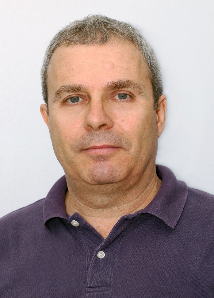 Isaac Harari合作论文数The Iby and Aladar Fleischman Faculty of Engineering, Tel Aviv University22
 Timothy J. Hall合作论文数Department of Medical Physics, School of Medicine and Public Health, University of Wisconsin–Madison;Department of Biomedical Engineering, University of Wisconsin–Madison18
 Jeff Bamber合作论文数Department of Physics, The Institute of Cancer Research and Royal Marsden NHS Foundation Trust14
 Olalekan Babaniyi合作论文数Boston Univ, Mech Engn, Boston, MA 02215 USA12Carlos E. Rivas合作论文数12Sevan Goenezen合作论文数Mechanical , Aerospace and Nuclear Engineering;Rensselaer Polytechnic Institute;Mechanical , Aerospace and Nuclear Engineering, Rensselaer Polytechnic Institute11Ricardo Leiderman合作论文数Department of Mechanical Engineering;Centro de Tecnologia;Cidade Universitaria Rio de Janeiro;Centro de Tecnologia|, Cidade Universitaria Rio de Janeiro9Elizabete Rodrigues Ferreira合作论文数8
 Ali Nadim合作论文数Keck Graduate Institute of Applied8Nachiket Gokhale合作论文数8
 Navid Nazari合作论文数Brigham & Womens Hosp, Dept Radiol, 75 Francis St, Boston, MA 02115 USA7
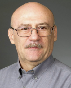 Dimitrije Stamenovic合作论文数Department of Biomedical Engineering, College of Engineering, Boston University7
 Jingfeng Jiang合作论文数Department of Biomedical Engineering, College of Engineering, Michigan Technological University7
 Arthur M. B. Braga合作论文数Department of Mechanical Engineering, Pontificia Universidade Católica do Rio de Janeiro7
 Michael Richards合作论文数7
 Sanjay S. Yengul合作论文数Department of Mechanical Engineering, Boston University6
 Michael Smith合作论文数6Uri Albocher合作论文数Faculty of Engineering, Tel Aviv University6Gearóid P. Berry合作论文数5
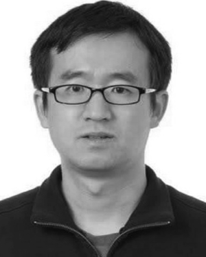 Yuqi Wang合作论文数Department of Medical Physics, University of Wisconsin, Madison, Wisconsin, USA5Nigel L. Bush合作论文数5
 Bruno Madore合作论文数Surgical Planning Laboratory, Department of Radiology, Brigham and Women's Hospital, Harvard Medical School5Gonzalo R. Feijoo合作论文数4jerome john cartmell合作论文数4D Thomas Seidl合作论文数4
 Daniel Gendin合作论文数Boston University4tengxiao liu合作论文数4
 Ralph Sinkus合作论文数School of Biomedical Engineering & Imaging Sciences, Faculty of Life Sciences & Medicine, King’s College London4
 Allan D. Pierce合作论文数College of Engineering, Boston University4
 Katharina Schregel合作论文数4Brian Harvey合作论文数4
 Dan Givoli合作论文数Faculty of Aerospace Engineering, Technion – Israel Institute of Technology4
 Palotai Miklós合作论文数Faculty of Medicine, University of Szeged4Crescenti Remo A合作论文数4Dord Jean Francois合作论文数4
 Kenneth R. Lutchen合作论文数Respiratory and Physiological Systems Identification Laboratory, College of Engineering, Boston University;Department of Biomedical Engineering, Boston University4
 Mohit Tyagi合作论文数TPPED, Bhabha Atomic Research Centre4Joseph P. Richter合作论文数4
 Yixiao Zhang合作论文数Sichuan University, Sichuan University4
 Samuel Patz合作论文数Department of Radiology, Harvard Medical School;Athinoula A. Martinos Center for Biomedical Imaging, Massachusetts General Hospital;Department of Radiology, Brigham & Womens Hospital;Center for Pulmonary Functional Imaging, Brigham & Womens Hospital4I Patlashenko合作论文数4george herrmann合作论文数3Steve Webb合作论文数3O Michael合作论文数3
 Daniel H. Goldman合作论文数3
 Rohit Nayak合作论文数Dept. of Elec. & Electron. Eng., Manipal Univ.;c;Dept. of Elec. & Electron. Eng., Manipal Univ.3
 Canović Elizabeth P合作论文数Department of Biomedical Engineering, Boston University3
 Robin Cleveland合作论文数Department of Engineering Science, Magdalen College, University of Oxford3




