Background The small nuclear RNA-activating complex polypeptide 4 (SNAPC4) encodes the largest scaffolding subunit of the SNAPc complex, essential for transcription of spliceosomal small nuclear RNAs (snRNAs) by RNA polymerase II and III. Although pathogenic variants in SNAPC4 have been associated with a rare neurodevelopmental disorder, the full phenotypic spectrum and underlying molecular mechanisms remain poorly defined. Methods We assembled an international cohort of 14 individuals harboring 18 distinct pathogenic SNAPC4 variants and one multi-exon deletion, three of which had been previously reported, including frameshift, splice-site, multi-exon deletions, and missense alleles. Detailed clinical and neuroimaging data were integrated with in silico structural and protein–protein interaction analyses to assess the impact of these variants on SNAPc architecture and function. Results SNAPC4 -related disease emerges as a highly heterogeneous clinical continuum rather than a uniform progressive disorder. Core features included global developmental delay and microcephaly, with variable trajectories ranging from static cognitive impairment to adult-onset progressive motor dysfunction, alongside neurobehavioral phenotypes such as autism and ADHD. Neuroimaging revealed abnormalities suggestive of impaired neurogenesis and gliogenesis, including lissencephaly-pachygyria and hypomyelinating leukodystrophy. Structural modeling supports a unifying loss-of-function (LoF) mechanism, whereby variants destabilize the SNAPc complex or disrupt key interfaces required for DNA binding and RNA polymerase recruitment, consistent with impaired SNAPc function and transcriptional dysregulation. Conclusion These findings redefine SNAPC4 -related disease as a stage-dependent neurodevelopmental spliceosomopathy, linking disruption of SNAPC4 to impaired SNAPc function and transcriptional dysregulation, with direct implications for diagnosis and variant interpretation.
The TP53 gene encodes the well-known p53 tumor suppressor protein, which plays a crucial role in preventing cancer development. Germline TP53 variants cause Li-Fraumeni Syndrome (LFS), an autosomal dominant disorder associated with early-onset cancers, including breast cancer, brain tumors, leukemias, bone cancers, and soft tissue sarcomas. Here, we described a germline TP53 variant c.671A>C, located at the penultimate nucleotide of exon 6 and predicted to result in the missense substitution p.E224A. The variant was identified in a 2-year-old child with retroperitoneal rhabdomyosarcoma and with a strong family history suggestive of LFS. Functional assays in yeast and human cells demonstrated wild type-like activity of the protein p.E224A; however, in silico splicing analysis indicated potential splice defects (e.g., SpliceAI score = 0.77). Given this discrepancy, we further investigated this variant using a minigene approach, demonstrating that it causes the skipping of exon 6, likely resulting in a frameshift and the introduction of a premature stop codon. These findings supported the classification of the TP53 germline variant c.671A>C (p.E224A) as likely pathogenic, providing a definitive molecular diagnosis for family counselling. Additionally, the present results sheds light on how certain predicted TP53 missense variants can be linked to disease mechanisms through RNA splicing disruption.
Horizontal gaze palsy with progressive scoliosis (HGPPS) is a rare autosomal recessive disorder caused by biallelic ROBO3 variants, characterized by congenital horizontal gaze restriction, early-onset scoliosis, and distinctive hindbrain malformations. We report two pediatric Patients and integrate current evidence to expand the ROBO3 variant spectrum and refine phenotypic delineation. Detailed clinical evaluation was combined with whole-spine radiography and high-resolution brain MRI with diffusion tensor imaging. Trio-based exome sequencing identified three ROBO3 variants, interpreted according to ACMG/AMP criteria. A novel splice-region variant was evaluated using SpliceAI and Pangolin, while AlphaFold2 modeling and ThermoMPNN, DDMut, SIFT, PolyPhen-2, and ClinPred were used to assess a missense variant affecting the Ig-like domain. Both patients exhibited congenital horizontal gaze palsy, early-onset scoliosis, and the characteristic HGPPS hindbrain triad. Patient #1 carried compound heterozygous variants p.(Arg703Pro) and c.2073+4A > G, while Patient #2 harbored the homozygous p.(Arg245Trp) variant. Literature review confirmed a uniform neuroimaging signature despite variable clinical severity. These findings expand the molecular landscape of HGPPS and underscore the importance of early neuroimaging recognition and orthopedic surveillance in the absence of robust genotype-phenotype correlations.
BackgroundDiGeorge syndrome is a rare genetic disorder with variable phenotypic and immunologic features associated to an increased risk of malignancy. Hemophagocytic lymphohistiocytosis (HLH) is a rare hyperinflammatory condition that can be triggered by infectious complications in immunodeficient patients like those with DGS.CaseWe report the case of a 20-year-old female with intellectual and motor disabilities who was diagnosed with non-familial HLH at age of 12 months and underwent allogeneic hematopoietic stem cell transplantation due to early disease reactivation. Despite resolution of HLH and post-transplant GVHD, the patient’s psychomotor delay persisted, and dysmorphic features became more pronounced. A an array-CGH on her fibroblasts was performed and identified a partial de novo deletion on chromosome 22, consistent with DiGeorge Syndrome (DGS). At 15 years, imaging performed because of persistent and worsening back pain, revealed an intraneural/intraradicular atypical neurofibroma which was partially resected leading to symptoms disappearance.ConclusionThis is the first report of a case of DGS with early onset HLH and late occurrence of a neoplasm of uncertain biological potential. Careful clinical monitoring is essential due to the variability in clinical manifestations and level of immune alteration.
BACKGROUND/OBJECTIVES:Neural tube defects (NTDs) are congenital malformations arising when the neural tube (NT), precursor of the brain and spine, fails to properly close during neurulation. Etiology is multifactorial, with environmental and genetic factors variably contributing on a case-by-case basis. Molecular genetic studies of murine NTD genes have been precious in the identification of predisposing NTD genes in humans, highlighting the peculiar role of the planar cell polarity (PCP) pathway in a fraction of human NTD patients. METHODS:Seventy-eight patients with NTD treated at a pediatric tertiary care center were selected for genetic analysis. A custom next-generation sequencing (NGS) panel of 29 genes encoding for components of the core PCP pathway or for family members and paralogs of proteins (including SHROOM and GRHL) underlying NTDs in well-known animal models was used to re-sequence patients with NTD. A gene-burden analysis was also performed to assess potential enrichment of rare damaging variants in the NTD cohort compared to ethnically matched controls. RESULTS:Thirty-nine of 78 individuals (50%) presented with at least one putatively damaging rare variant, most of which (87%) were missense substitutions. Rare variants of GRHL1 and WNT5A, and among gene families GRHL and SHROOM, were significantly enriched in the patients' cohort compared to controls. CONCLUSION:This study supports the involvement of human orthologues of mouse genes in human NTD phenotypes. Further re-sequencing or, even better, whole-exome sequencing of a large group of cases will give the clues for a better understanding of NTD etiology, ameliorating the clinical management of patients and their families.
Introduction:Arteriovenous cerebral high-flow shunts include the vein of Galen aneurysmal malformation (VGAM) and vein of Galen dilatation, which are considered secondary to arteriovenous malformations or arteriovenous fistulas. These entities are often sporadic but are found in association with variants of the RASA1 and EPHB4 genes (capillary malformation-arteriovenous malformation, CMAVM; OMIM #608354) or ACVRL1, ENG, and SMAD4 genes (hereditary hemorrhagic telangiectasia, HHT; OMIM #187300). The clinical phenotypes associated with these conditions are highly variable, with incomplete penetrance and mostly dependent on the hemodynamic consequences (including heart failure and cerebral hemorrhage) or management complications rather than anatomical vascular variations per se. The present study aimed to genetically characterize a cohort of 29 patients affected by arteriovenous cerebral high-flow shunts who were treated at a pediatric referral center. Methods:The genetic techniques employed include next-generation sequencing, multiplex ligation-dependent probe amplification, and whole-exome sequencing. Results:Of the 29 patients, 11 cases were found to have variants in genes associated with vascular functions, five cases received a genetic diagnosis, one case presented with a variant of uncertain significance in the EPHB4 gene, and five cases showed variants in novel genes possibly linked with cerebrovascular disorders. Discussion:We provide extensive case descriptions and attempt to infer the genotype-phenotype correlations; variants in all of the known genes associated with arteriovenous cerebral shunts were reported in VGAM patients, while cutaneous angiomas were specific to RASA1 mutations. The genotypic and phenotypic descriptions of the affected individuals may thus have relevant implications in terms of better pathophysiological understanding, genotype-phenotype correlations, treatment strategies, and outcomes.
We report the first case of pleural mesothelioma (PM) occurring in a child affected by NF2-related schwannomatosis (NF2-SWN) and without any history of environmental exposure to asbestos. Mesothelioma is a rare secondary tumor in brain cancer patients and the association with NF2-SWN has been described only in a few anecdotal cases and never in the pediatric field. NF2-SWN is an autosomal dominant disease caused by inactivating germline mutations of the NF2 tumor suppressor gene, one of the most common mutations associated with human primary mesothelioma too. By MLPA assay, array-CGH analysis, and NGS on blood and tumor DNA, we determined the mutation profile of this rare NF2-driven PM and we identified several atypical chromosomal aberrations in tumor cells, suggesting a different genomic signature between pediatric and adult mesothelioma.
OBJECTIVES:Chiari malformation type 1 (CMI) is defined by the herniation of cerebellar tonsils of 5 mm or more, with possible neurological consequences, including compression of the neural tissue and/or anomalies in cerebral spinal fluid circulation. The etiology of CMI is not fully elucidated, with both genetic and environmental factors being involved. Several genes and pathways involved in bone development are pointed out like genes of the WNT, FGF, and BMP signaling pathways. More recently, the crucial role played by chromatin remodeling genes in the pathogenesis of CMI has increasingly emerged. METHODS:In this paper, we discuss a familial case of CMI and a single patient, harboring variants in chromatin remodeling genes, identified by whole exome sequencing. RESULTS:The first is a family with three affected members and one sibling with a cerebellar tonsil herniation of < 5 mm. The three CMI patients harbor a heterozygous missense variant in the SETD2 gene, whose truncating variants are responsible for Luscan-Lumish syndrome. A second variant in HP1BP3, a gene not previously associated with human pathology, with evidence of skeletal anomalies in mice models, was found in the three patients and also in the girl with a herniation of < 5 mm. The second case is a proband with a de novo variant in KMT2A, associated with Wiedemann-Steiner syndrome, in which anomalies of the craniocervical junction are described. DISCUSSION:We highlight the importance of chromatin remodeling genes in both isolated and syndromic CMI and suggest the potential role of HP1BP3 as a possible modifier gene in CMI pathogenesis, even if this association needs to be further clarified.
Asthma is a complex respiratory condition characterized by chronic airway inflammation and variable expiratory airflow limitation, affecting millions globally. Among athletes, particularly those competing at elite levels, the prevalence of respiratory conditions is notably heightened, varying between 20% and 70% across specific sports. Exercise-induced bronchoconstriction (EIB) is a common issue among athletes, impacting their performance and well-being. The prevalence rates vary based on the sport, training environment, and genetics. Exercise is a known trigger for asthma, but paradoxically, it can also improve pulmonary function and alleviate EIB severity. However, athletes' asthma phenotypes differ, leading to varied responses to medications and challenges in management. The unique aspects in athletes include heightened airway sensitivity, allergen, pollutant exposure, and temperature variations. This review addresses EIB in athletes, focusing on pathogenesis, diagnosis, and treatment. The pathogenesis of EIB involves complex interactions between physiological and environmental factors. Airway dehydration and cooling are key mechanisms, leading to osmotic and thermal theories. Airway inflammation and hyper-responsiveness are common factors. Elite athletes often exhibit distinct inflammatory responses and heightened airway sensitivity, influenced by sport type, training, and environment. Swimming and certain sports pose higher EIB risks, with chlorine exposure in pools being a notable factor. Immune responses, lung function changes, and individual variations contribute to EIB in athletes. Diagnosing EIB in athletes requires objective testing, as baseline lung function tests can yield normal results. Both EIB with asthma (EIBA) and without asthma (EIBwA) must be considered. Exercise and indirect bronchoprovocation tests provide reliable diagnoses. In athletes, exercise tests offer effectiveness in diagnosing EIB. Spirometry and bronchodilation tests are standard approaches, but the diagnostic emphasis is shifting toward provocation tests. Despite its challenges, achieving an optimal diagnosis of EIA constitutes the cornerstone for effective management, leading to improved performance, reduced risk of complications, and enhanced quality of life. The management of EIB in athletes aligns with the general principles for symptom control, prevention, and reducing complications. Non-pharmacological approaches, including trigger avoidance and warming up, are essential. Inhaled corticosteroids (ICS) are the cornerstone of asthma therapy in athletes. Short-acting beta agonists (SABA) are discouraged as sole treatments. Leukotriene receptor antagonists (LTRA) and mast cell stabilizing agents (MCSA) are potential options. Optimal management improves the athletes' quality of life and allows them to pursue competitive sports effectively.
Legius syndrome, commonly referred to as SPRED1-related neurofibromatosis type 1-like syndrome, is a rare autosomal dominant disorder characterized by café-au-lait macules, freckling, lipomas, macrocephaly, and heterogeneous neurodevelopmental manifestations, including a different degree of learning difficulties. Although a partial clinical overlap exists with neurofibromatosis type 1 (NF1), Legius syndrome is distinguished by its genetic etiology and the absence of neurofibromas, indicating an inherent lack of tumor risk. The SPRED1 gene encodes the Sprouty-related protein with an EVH1 domain 1 (SPRED1), a negative regulator of the RAS-MAPK signaling pathway with a crucial role in cellular growth and development. Despite various genetic variants and genomic deletions associated with Legius syndrome, the full genetic spectrum of this condition remains elusive. In this study, we investigated the underlying genetic etiology in a cohort of patients presenting with typical manifestations of Legius syndrome using a custom Next Generation Sequencing (NGS) panel and Multiplex Ligation-Dependent Probe Amplification (MLPA) for NF1 and SPRED1. We identified 12 novel SPRED1 damaging variants segregating with the phenotype in all families. These rare variants affect conserved residues of the protein and are predicted damaging according to in silico tools. No clear genotype-phenotype correlations could be observed in the current cohort and previously reported patients, underscoring the heterogeneous genotype spectrum of this condition. Our findings expand the understanding of SPRED1 variants causing Legius syndrome and underscore the importance of comprehensively characterizing the genetic landscape of this disorder. Despite the absence of clear genotype-phenotype correlations, elucidating the genetic etiology of Legius syndrome is pertinent for facilitating accurate diagnosis, genetic counseling, and therapeutic interventions.
Pediatric low-grade gliomas (pLGG) frequently harbor MAPK-pathway mutations that determine poor response to standard chemotherapy (sCt). Targeted agents against BRAF-V600E mutation (MAPK-i) are emerging as molecularly driven treatments. Few studies have compared sCt with MAPK-i. We aimed to compare response and progression-free survival (PFS) of unresectable brain pLGG treated with sCt or MAPK-i. MAPK-i were used in first line or at relapse. We conducted a single-center observational retrospective study of all brain pLGG consecutively diagnosed and treated at Gaslini Children’s Hospital between January 2008 and December 2021. Treatment was assessed using RAPNO criteria at 6, 12-18 months, and at last follow-up. MRI studies were reviewed by two experienced neuroradiologists. Seventy-one patients were enrolled: 66 received sCt and 10 MAPK-i (5 in first line, 5 after sCt failure). All patients who received MAPK-i were BRAF-V600E mutated. Groups were homogeneous for tumor location and dissemination. sCt group was enriched for pilocytic astrocytoma, MAPK-I group for ganglioglioma. sCt patients were younger. SCt lasted 12-18 months, MAPK-i were continuously given. Tumor size reduction was 19.5% at 6 months and 40.5% at 12-18 months for sCt group; 48% at 6 months and 43.5% at 12-18 months for MAPK-i group. SCt determined at least stable disease in 74.2% of patients; 60% of responders reached the best response at 6 months, 40% at 12-18 months. 5y-PFS was 40.3% (median time for progression: 2.5 years). MAPK-i determined at least stable disease in all patients at first evaluation with long-lasting response (median time of follow-up: 5.1 years). In our study sCt was effective in inducing response but prolonged disease control is not always achieved. BRAF-V600E mutated patients upon MAPK-i start gained a rapid disease control with long-lasting response, also if they have been pretreated. Acknowledgement: this study was supported by ARTUCEBA ONLUS.
The occurrence of an abdominal tumor invading the spinal canal and causing symptoms of epidural compression is rare in an infant, and exceptional at birth. Peripheral neuroblastic tumors are by far the most common cause. Emergency chemotherapy is commonly curative, though permanent sequelae are possible. Although other malignancies may be involved, no case of rhabdoid tumors at birth has been reported. We describe the case of a neonate who presented symptoms of spinal epidural compression at birth secondary to a rhabdoid tumor. As expected with this highly malignant tumor, the patient experienced a rapidly progressive clinical course and died within three months of diagnosis.
Background: Infantile myofibromatosis (IM) is a rare disorder characterized by benign tumors in the skin, subcutaneous tissue, muscle, and occasionally viscera. IM can be hereditary due to PDGFRB or NOTCH3 variants. Treatment is mainly conservative or surgical. Combination regimens have been used in case of disseminated disease. Observation: We present relapsed disease of IM 11 years after diagnosis in a 2-year-old child initially treated by microscopically complete resection. A new heterozygous c.1687G>A (p.Glu563Lys) mutation in the PDGFRB gene was identified (considered likely pathogenic). Conclusions: In association with initial treatment, genetic testing is crucial for tailored clinical practice and follow-up in patients diagnosed with IM.
In MAPK-altered pediatric low-grade glioma (pLGG), immune infiltration may be involved in treatment response. It is debated whether tumor immune cell infiltration is due to expansion of endogenous resident microglia or to the active recruitment of bone marrow-derived microglial progenitors from the bloodstream. This study aimed to investigate the relation between neuroradiological features of MAPK-altered pLGG and treatment response to MAPK-inhibitors (MAPKi). We conducted a retrospective analysis of all patients with unresectable BRAF-V600E mutated brain pLGG, radiotherapy naïve, treated with MAPKi at our institution since 2015. MRI studies were reviewed by two experienced neuroradiologist and tumor response assessed according to RAPNO criteria. 12 patients were enrolled (9 female). Median age at treatment start was 6.2 years (range 2.7–14.3 years). Four were pilocytic astrocytomas, 6 gangliogliomas, and 2 desmoplastic infantile gangliogliomas. Five patients received MAPKi as first line, 7 following other chemotherapy lines. Median follow-up was 4.1 years (range 2.5–8.7 years). Tumor response was associated with contrast enhancement (CE): 9 out of 10 patients with CE at treatment onset had at least partial response and subsequent CE resolution, while both 2 out of 2 patients without CE showed stable disease. A 4-year old contrast enhancing-ganglioglioma who reached partial response and CE resolution with MAPKi, presented disease progression four days after treatment withdrawal; treatment re-start determined tumor and CE reduction after only 7 days. We hypothesize that brain-blood barrier breakage is induced by tumor infiltration of bloodstream immune cells, and that this trafficking is stimulated by tumor cell secreted products and determined by microglial progenitors shift from the bloodstream to the tumor site. MAPKi treatment may incite blood-brain barrier reconstitution as it reduces the trafficking between tumor site and bloodstream. This work was supported by grant number NET-2018-12366666 NeuroArtP3 from the Italian Ministry of Health.
Immune dysregulation in Inborn Errors of Immunity (IEI) shows a broad phenotype, including autoimmune disorders, benign lymphoproliferation, and malignancies, driven by an increasing number of implicated genes. Recent findings suggest that childhood cancer survivors (CCSs) may exhibit immunological abnormalities potentially linked to an underlying IEI, along with a well-known increased risk of subsequent malignancies due to prior cancer treatments. We describe a patient with two composite heterozygous pathogenic variants in the interleukin-2-inducible T-cell kinase (ITK) gene and a history of multiple tumors, including recurrent Epstein-Barr virus (EBV)-related nodular sclerosis and Hodgkin's lymphoma (NSHL), associated with unresponsive multiple hand warts, immune thrombocytopenia, and an impaired immunological profile (CD4+ lymphocytopenia, memory B-cell deficiency, reduction in regulatory T-cells, and B-cell- and T-cell-activated profiles). In our case, ITK-related immune dysregulation and prior exposure to oncological treatments seem to have simultaneously intervened in the same individual, leading to the development of a unique clinical profile. It is essential to raise awareness of the two-way association between immune dysregulation disorders and multiple tumors.
PurposePathogenic variants of FIG4 generate enlarged lysosomes and neurological and developmental disorders. To identify additional genes regulating lysosomal volume, we carried out a genome-wide activation screen to detect suppression of enlarged lysosomes in FIG4-/- cells.MethodsThe CRISPR-a gene activation screen utilized sgRNAs from the promoters of protein coding genes. Fluorescence-activated cell sorting separated cells with correction of the enlarged lysosomes from uncorrected cells. Patient variants of SLC12A9 were identified by exome or genome sequencing and studied by segregation analysis and clinical characterization.ResultsOverexpression of SLC12A9, a solute co-transporter, corrected lysosomal swelling in FIG4-/- cells. SLC12A9 (NP_064631.2) co-localized with LAMP2 at the lysosome membrane. Biallelic variants of SLC12A9 were identified in three unrelated probands with neurodevelopmental disorders. Common features included intellectual disability, skeletal and brain structural abnormalities, congenital heart defects and hypopigmented hair. Patient 1 was homozygous for nonsense variant p.(Arg615*); patient 2 was compound heterozygous for p.(Ser109Lysfs*20) and a large deletion, and proband 3 was compound heterozygous for p.(Glu290Glyfs*36) and p.(Asn552Lys). Fibroblasts from proband 1 contained enlarged lysosomes that were corrected by wildtype SLC12A9 cDNA. Patient variant p.(Asn552Lys) failed to correct the lysosomal defect.ConclusionsImpaired function of SLC12A9 results in enlarged lysosomes and a recessive disorder with a recognizable neurodevelopmental phenotype.
 Valeria Capra合作论文数U.O. Neurochirurgia, Istituto G. Gaslini91
 Elisa Merello合作论文数39
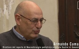 Armando Cama合作论文数Department of Pediatric Neuroradiology, G. Gaslini Children’s Research Hospital37
 Marco Pavanello合作论文数Department of Neurosurgery, Giannina Gaslini Institute28
 Gianluca Piatelli合作论文数Department of Neurosurgery, Giannina Gaslini Institute23
 Zoha Kibar合作论文数22
 Federico Zara合作论文数University of Genoa22Marzia Ognibene合作论文数Laboratory of Molecular Biology, G. Gaslini Institute18E Merello合作论文数15
 Marcello Scala合作论文数Dept Neurosci Rehabil Ophthalmol Genet Maternal &, Univ Genoa15
 Maria Grazia Calevo合作论文数Istituto Giannina Gaslini;Giannina Gaslini Hospital15
 Alessandro Raso合作论文数Neurosurgery Unit, IRCCS G. Gaslini Hospital15A Moroni合作论文数13
 Mariasavina Severino合作论文数12Claudia Milanaccio合作论文数12Andrea Rossi合作论文数11
 Gianluca Piccolo合作论文数Pediatric Neurology and Muscular Diseases Unit, IRCCS Giannina Gaslini Institute11
 Andrea Accogli合作论文数10
 Samantha Mascelli合作论文数9
 Antonio Verrico合作论文数9Iacomino Michele合作论文数9
 Maria Luisa Garre合作论文数Istituto Giannina Gaslini9Tortora Domenico合作论文数Department of Radiology, University Hospital of Chieti;Chieti;Department of Radiology, University Hospital of Chieti8Marco Di Duca合作论文数8Ferruccio Romano合作论文数7Allache Redouane合作论文数7
 Pasquale Striano合作论文数Center of Experimental and Translational Myology, Dipartimento Di Neuroscienze, Riabilitazione, Oftalmologia, Genetica E Scienze Materno-Infantili, Università Degli Studi Di Genova;IRCCS 'G. Gaslini'7
 Paola Rogliani合作论文数Department of Internal Medicine, University of Rome ‘Tor Vergata’7Maria Cristina Diana合作论文数6Cristina Chelleri合作论文数6
 Marco Crocco合作论文数Dept Neurosci Rehabil Ophthalmol Genet Maternal &, Univ Genoa6Simona Baldassari合作论文数6Stefano Calzolari合作论文数6
 Richard Finnell (理查德·菲纳尔)合作论文数Center for Precision Environmental Health, Baylor College of Medicine;Department of Nutritional Sciences, University of Texas;Dell Children's Medical Center;Department of Chemistry and Biochemistry, University of Texas6Francesca Madia合作论文数6Gabriele Gaggero合作论文数6P Negrin合作论文数6G Testa合作论文数5
 Stacey Cherny合作论文数Tel Aviv University5
 Kshitij Mankad合作论文数Bupa Cromwell Hospital5Robert Milan Porsch合作论文数Department of Psychiatry, University of Hong Kong5Vincenzo Salpietro合作论文数5
 Philippe Gros合作论文数Department of Biochemistry, School of Biomedical Sciences, McGill University;McGill University Research Centre on Complex Traits5
 Pak Chung Sham (沈伯松)合作论文数Department of Psychiatry, School of Clinical Medicine, Li Ka Shing Faculty of Medicine, The University of Hong Kong;State Key Laboratory of Brain and Cognitive Sciences, The University of Hong Kong5Monica Traverso合作论文数5Sara Guerrisi合作论文数4S Mascelli合作论文数4Antonia Ramaglia合作论文数4L Arata合作论文数4




