A 58-year-old woman was admitted to the hospital 3 weeks after lumbar surgery because of diplopia and fever. Left leg pain and a rash had preceded the fever and diplopia. A diagnosis was made.
BACKGROUND AND PURPOSE:A scout accelerated motion estimation and reduction (SAMER) framework has been developed for efficient retrospective motion correction. The goal of this study was to perform an initial evaluation of SAMER in a series of clinical brain MR imaging examinations. MATERIALS AND METHODS:Ninety-seven patients who underwent MR imaging in the inpatient and emergency department settings were included in the study. SAMER motion correction was retrospectively applied to an accelerated T1-weighted MPRAGE sequence that was included in brain MR imaging examinations performed with and without contrast. Two blinded neuroradiologists graded images with and without SAMER motion correction on a 5-tier motion severity scale (none = 1, minimal = 2, mild = 3, moderate = 4, severe = 5). RESULTS:The median SAMER reconstruction time was 1 minute 47 seconds. SAMER motion correction significantly improved overall motion grades across all examinations (P < .005). Motion artifacts were reduced in 28% of cases, unchanged in 64% of cases, and increased in 8% of cases. SAMER improved motion grades in 100% of moderate motion cases and 75% of severe motion cases. Sixty-nine percent of nondiagnostic motion cases (grades 4 and 5) were considered diagnostic after SAMER motion correction. For cases with minimal or no motion, SAMER had negligible impact on the overall motion grade. For cases with mild, moderate, and severe motion, SAMER improved the motion grade by an average of 0.3 (SD, 0.5), 1.1 (SD, 0.3), and 1.1 (SD, 0.8) grades, respectively. CONCLUSIONS:SAMER improved the diagnostic image quality of clinical brain MR imaging examinations with motion artifacts. The improvement was most pronounced for cases with moderate or severe motion.
BACKGROUND AND PURPOSE:Patients infected with the Severe Acute Respiratory Syndrome coronavirus 2 (SARS-CoV-2) can develop a spectrum of neurological disorders, including a leukoencephalopathy of variable severity. Our aim was to characterize imaging, lab, and clinical correlates of severe coronavirus disease 2019 (COVID-19) leukoencephalopathy, which may provide insight into the SARS-CoV-2 pathophysiology. MATERIALS AND METHODS:Twenty-seven consecutive patients positive for SARS-CoV-2 who had brain MR imaging following intensive care unit admission were included. Seven (7/27, 26%) developed an unusual pattern of "leukoencephalopathy with reduced diffusivity" on diffusion-weighted MR imaging. The remaining patients did not exhibit this pattern. Clinical and laboratory indices, as well as neuroimaging findings, were compared between groups. RESULTS:The reduced-diffusivity group had a significantly higher body mass index (36 versus 28 kg/m2, P < .01). Patients with reduced diffusivity trended toward more frequent acute renal failure (7/7, 100% versus 9/20, 45%; P = .06) and lower estimated glomerular filtration rate values (49 versus 85 mL/min; P = .06) at the time of MRI. Patients with reduced diffusivity also showed lesser mean values of the lowest hemoglobin levels (8.1 versus 10.2 g/dL, P < .05) and higher serum sodium levels (147 versus 139 mmol/L, P = .04) within 24 hours before MR imaging. The reduced-diffusivity group showed a striking and highly reproducible distribution of confluent, predominantly symmetric, supratentorial, and middle cerebellar peduncular white matter lesions (P < .001). CONCLUSIONS:Our findings highlight notable correlations between severe COVID-19 leukoencephalopathy with reduced diffusivity and obesity, acute renal failure, mild hypernatremia, anemia, and an unusual brain MR imaging white matter lesion distribution pattern. Together, these observations may shed light on possible SARS-CoV-2 pathophysiologic mechanisms associated with leukoencephalopathy, including borderzone ischemic changes, electrolyte transport disturbances, and silent hypoxia in the setting of the known cytokine storm syndrome that accompanies severe COVID-19.
We would like to commend Bulut et al[1][1] for their investigation of brain MR imaging findings that could potentially be useful in discriminating pediatric-onset neuromyelitis optica spectrum disorder (NMOSD) from acute disseminated encephalomyelitis (ADEM). These 2 entities exist within the
Background: Biliary tract carcinoma (BTC) is a fatal malignancy with a late clinical presentation. Novel molecular markers for early diagnosis are urgently needed. The purpose of this study was to evaluate the diagnostic and prognostic value of promoter hypermethylation of SHOX2 and SEPT9 in BTC. Methods: Relative DNA methylation of SHOX2 and SEPT9 was quantified in tumor specimens and matched normal adjacent tissue (NAT) from 71 BTC patients, as well as in plasma samples from an independent prospective cohort of 20 cholangiocarcinoma patients and 100 control patients. Receiver operating characteristic (ROC) curve analyses were performed to probe the diagnostic ability of both methylation markers. DNA methylation was correlated to clinicopathological and survival data. Results: SHOX2 methylation was significantly higher in tumor tissue than in NAT (p < 0.001) and correctly identified 71% of BTC specimens with 100% specificity (AUC = 0.918; 95% CI 0.865-0.971). Elevated DNA methylation levels were also found in plasma derived from cholangiocarcinoma patients. SHOX2 and SEPT9 methylation as a marker panel achieved a sensitivity of 45% and a specificity of 99% in differentiating between samples from patients with and without cholangiocarcinoma (AUC = 0.752; 95% CI 0.631-0.873). Conclusion: SHOX2 and SEPT9 are frequently methylated in biliary tract cancers. Promoter hypermethylation of SHOX2 and SEPT9 may therefore serve as a biomarker supporting diagnosis finding and therapy monitoring in clinical specimens.
A 63-year-old man presented with confusion and weakness 5 months after he had undergone allogeneic stem-cell transplantation for chronic lymphocytic leukemia. MRI did not show evidence of acute brain abnormalities. Diagnostic tests were performed.
A 61-year-old man with a history of stroke and alcohol-use disorder presented with grandiosity, impulsivity, and decreased sleep. An evaluation was notable for an expansive affect, emotional lability, and impaired memory. A diagnosis was made.
A total of 53 patients underwent MR imaging consisting of 5 basic fast precontrast sequences plus standard and accelerated versions of the same postcontrast T1WI sequences. Two neuroradiologists assessed the image quality and the final diagnosis for each set of postcontrast sequences and compared their performances. The 10-minute brain MR imaging protocol with contrast was comparable in diagnostic performance with the standard protocol in an inpatient motion-prone population, with the additional benefits of reducing acquisition times and image artifacts.BACKGROUND AND PURPOSE: The development of new MR imaging scanners with stronger gradients and improvement in coil technology, allied with emerging fast imaging techniques, has allowed a substantial reduction in MR imaging scan times. Our goal was to develop a 10-minute gadolinium-enhanced brain MR imaging protocol with accelerated sequences and to evaluate its diagnostic performance compared with the standard clinical protocol.MATERIALS AND METHODS: Fifty-three patients referred for brain MR imaging with contrast were scanned with a 3T scanner. Each MR image consisted of 5 basic fast precontrast sequences plus standard and accelerated versions of the same postcontrast T1WI sequences. Two neuroradiologists assessed the image quality and the final diagnosis for each set of postcontrast sequences and compared their performances.RESULTS: The acquisition time of the combined accelerated pre- and postcontrast sequences was 10 minutes and 15 seconds; and of the fast postcontrast sequences, 3 minutes and 36 seconds, 46% of the standard sequences. The 10-minute postcontrast axial T1WI had fewer image artifacts (P < .001) and better overall diagnostic quality (P < .001). Although the 10-minute MPRAGE sequence showed a tendency to have more artifacts than the standard sequence (P = .08), the overall diagnostic quality was similar (P = .66). Moreover, there was no statistically significant difference in the diagnostic performance between the protocols. The sensitivity, specificity, and accuracy values for the 10-minute protocol were 100.0%, 88.9%, and 98.1%.CONCLUSIONS: The 10-minute brain MR imaging protocol with contrast is comparable in diagnostic performance with the standard protocol in an inpatient motion-prone population, with the additional benefits of reducing acquisition times and image artifacts.
Biliary tract carcinoma (BTC) is a fatal malignancy which aggressiveness contrasts sharply with its relatively mild and late clinical presentation. Novel molecular markers for early diagnosis and precise treatment are urgently needed. The purpose of this study was to evaluate the diagnostic and prognostic value of promoter hypermethylation of the SHOX2 and SEPT9 gene loci in BTC.
Einleitung/Ziel: Gallengangskarzinome (GGK) weisen trotz intensiver Forschung immer noch eine schlechte Prognose auf. Die einzige kurative Therapiemöglichkeit ist eine radikale chirurgische Resektion in einem frühen Tumorstadium. Eine frühzeitige Diagnose stellt aufgrund des späten Auftretens von Symptomen jedoch eine Herausforderung dar. Neue molekulare Biomarker zur Diagnoseverbesserung sowie Prognoseeinschätzung werden daher dringend benötigt. Ziel dieser Studie ist die Rolle der DNA-Methylierung von SHOX2 und SEPT9 als potentielle Biomarker im GGK zu evaluieren.
Magnetic resonance sequences that take advantage of susceptibility effects to demonstrate pathology are powerful and sensitive aids for diagnostic imaging. An important distinction should be highlighted at this point. Although the term susceptibility-weighted imaging (SWI) has been used in the past to refer to T2*-weighted gradient recall echo (GRE) techniques, the more recent convention is to reserve this term for a distinct new sequence utilizing both magnitude and phase information. The bulk of the stroke-related research discussed in this chapter relates to conventional T2*-weighted GRE sequences; susceptibility sequences and SWI are discussed in Ch. 10. In particular, one of the key applications of susceptibility sequences is the identification of hemorrhage and blood products.
A 59-year-old man was admitted to this hospital with fever and the subacute onset of progressive neurologic symptoms that included multiple cranial-nerve palsies. Magnetic resonance imaging of the brain and spine showed leptomeningeal enhancement. Analysis of the cerebrospinal fluid disclosed a lymphocytic pleocytosis and elevated protein and glucose levels. Respiratory failure developed, requiring intubation and mechanical ventilation. The result of a diagnostic test was received.
A 59-year-old man was admitted to this hospital in late summer because of back pain, weakness in the right arm, and cranial-nerve palsies. The patient was in his usual state of health until the first week in July, 5 weeks before admission, when fevers led to body temperatures up to 39.4°C and neck stiffness developed. He saw his primary care physician, who noted thrombocytopenia; a viral illness was diagnosed, and antibiotics were administered. The fevers resolved within 5 days after they started, and the platelet count returned to normal within 2 weeks. However, the discomfort in his neck gradually worsened. One week before admission, numbness of the right hand developed, followed by weakness of the right hand and neck during the next 3 days. Four days before admission, the patient was evaluated by an orthopedist. Magnetic resonance imaging (MRI) of the neck 2 days later revealed degenerative changes of the spine at multiple levels from C3 to T2, with a broad-based central herniation at the C3–C4 level and mild protrusions at other cervical levels. An electrocardiogram was normal. Ibuprofen, methocarbamol, and oxycodone–acetaminophen were administered. The right arm became weaker, and the patient reported that the neck pain became sharp and more severe (9 on a scale of 1 to 10, with 1 being the least severe), radiating down the spine. The day before admission, left ptosis and diplopia developed. On the day of admission, the patient was seen at another hospital. He did not have fever, headache, nausea, vomiting, or a skin rash. The blood pressure was 156/85 mm Hg, the pulse 101 beats per minute, the respiration 20 breaths per minute, and the temperature 36.1°C. The oxygen saturation was 95% while the patient was breathing ambient air. Gaze was directed to the right, and strength in the right hand was decreased. Hydromorphone was administered intravenously for pain control. Computed tomographic (CT) scanning of the head showed no abnormalities suggestive of acute disease. He was transferred to the emergency department of this hospital. Diabetes mellitus had been diagnosed 1 year earlier and was managed with glipizide (5 mg daily). A lumbar laminectomy had been performed in the past. He Case 11-2007: A 59-Year-Old Man with Neck Pain, Weakness in the Arms, and Cranial-Nerve Palsies
Part I: Fundamentals of Acute Ischemic Stroke.- Ischemic Stroke: Basic Pathophysiology and Neuroprotective Strategies. Causes of Ischemic Stroke.- Part II: Imaging of Acute Ischemic Stroke.- Unenhanced Computed Tomography.-Stroke CT Angiography (CTA).-CT Perfusion (CTP).-Conventional MRI and MR Angiography of Stroke.-Diffusion MR of Acute Stroke.-Perfusion MRI of Acute Stroke.-The Clinical Ischemic Penumbra.- Part III: Intervention in Acute Ischemic Stroke.- Clinical Management of Acute Stroke.-Intravenous Thrombolysis.- Imaging for Endovascular Stroke Therapy.-Endovascular Approaches to Acute Stroke.
 Michael Lev合作论文数Harvard Medical School;Massachusetts General Hospital5
 Ramon Gonzalez合作论文数Division of Neuroradiology, Department of Radiology, Massachusetts General Hospital, Mass General Brigham;Mass General Cancer Center, Massachusetts General Hospital, Mass General Brigham;Harvard Medical School, Harvard University5
 A Semaan合作论文数3WJ Koroshetz合作论文数3
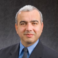 Otto Rapalino合作论文数Harvard Medical School;Division of Neuroradiology, Massachusetts General Hospital3
 Vittorio Branchi合作论文数University Hospital Bonn3
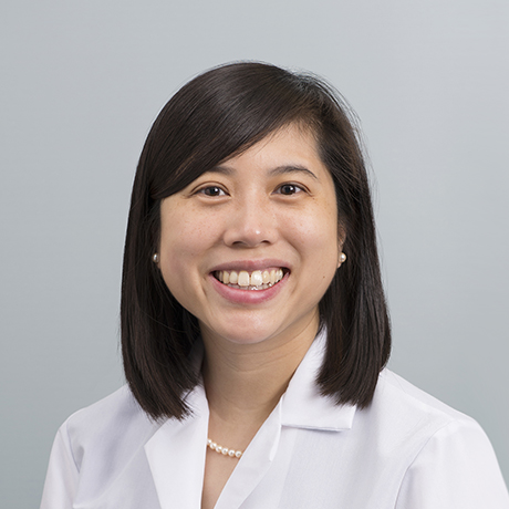 Susie Huang合作论文数Division of Neuroradiology, Massachusetts General Hospital;Harvard Medical School;Athinoula A. Martinos Center3
 Jörg Kalff合作论文数Department of Surgery, Medical Faculty, University of Bonn3
 Hanno Matthaei合作论文数Rheinische Friedrich-Wilhelms-Universität Bonn3
 Dimo Dietrich合作论文数Epigenomics AG3Nico Schäfer合作论文数3
 Joshua Hirsch合作论文数Massachusetts General Hospital2
 Allen Steere合作论文数Medicine Department, Harvard Medical School;Rheumatology Unit, Mass General Research Institute, Massachusetts General Hospital2
 Conklin John合作论文数2
 Philipp Lingohr合作论文数2
 Glen Kristiansen合作论文数Institute of Pathology, University Hospital Bonn2
 E Tessa Hedley-Whyte合作论文数Harvard Medical School, Massachusetts General Hospital2
 Scott Plotkin合作论文数The Stephen E. and Catherine Pappas Center for Neuro-Oncology, Massachusetts General Hospital, Harvard Medical School;Family Center for Neurofibromatosis, Massachusetts General Hospital, Harvard Medical School2
 David M. Greer合作论文数Chobanian & Avedisian School of Medicine, Boston University;Boston Medical Center2
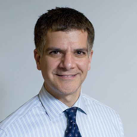 Robert Hasserjian合作论文数Department of Pathology, Massachusetts General Hospital;Harvard Medical School;Dana-Farber / Harvard Cancer Center2
 Aneesh B Singhal合作论文数Massachusetts General Hospital2G Sorensen合作论文数1A. Tabari合作论文数1J M Romero合作论文数1
 Brendan Kelley合作论文数1Keith Heberlein合作论文数1W-C Lo合作论文数1BR ROSEN合作论文数1Richard C. Cabot合作论文数1D N Splitthoff合作论文数1St Traweek合作论文数1Rd Adams合作论文数1BC Lucey合作论文数1R Gilberto Gonzalez合作论文数1steven thibodeau合作论文数1Jo-Anne O. Shepard合作论文数1Rajiv Gupta合作论文数1Stuart Pomerantz合作论文数1
 Ali Pourvaziri合作论文数Massachusetts Gen Hosp, Dept Radiol, 55 Fruit St,White 270, Boston, MA 02114 USA1
 Adrian Jaramillo-Cardoso合作论文数Harvard Medical School, Massachusetts General Hospital1G Gonzalez合作论文数1
 Daniel J Boulter合作论文数Wexner Med Ctr, Ohio State Univ1GA Rordorf合作论文数1Komal Manzoor合作论文数Harvard Med Sch, Boston, MA USA1B Wang合作论文数1Jacob E Lemieux合作论文数Broad Inst, Cambridge, MA 02142 USA1R. Gilberto Gonzalez合作论文数1
 Daniel Polak合作论文数A. A. Martinos Center for Biomedical Imaging, Massachusetts General Hospital1
 John Henson合作论文数1




