Background and purposeWe systematically reviewed available evidence for reports of neurological signs and symptoms in patients with COVID‐19 to identify cases with severe acute respiratory syndrome coronavirus (SARS‐CoV)‐2 infection or immune‐mediated reaction in the nervous system.MethodsWe followed PRISMA guidelines and used the MEDLINE, EMBASE, Google Scholar, MedRxiv and ChinaXiv databases to search for articles on COVID‐19 and nervous system involvement that were published from 1 January to 24 April 2020. Data on design, sample size, neurological assessment and related work‐up were extracted. Biases were assessed with the Newcastle–Ottawa scale.ResultsWe analysed 27 publications on potential neuroinvasive or parainfectious neurological complications of COVID‐19. The reports focused on smell and taste (n = 5) and evaluation of neurological symptoms and signs in cohorts (n = 5). There were cases of Guillain‐Barré syndrome/Miller‐Fisher syndrome/cranial neuropathy (seven cases), meningitis/encephalitis (nine cases) and various other conditions (five cases). The number of patients with examination of cerebrospinal fluid and, in particular, SARS‐CoV‐2 polymerase chain reaction was negligible. Two had a positive SARS‐CoV‐2 polymerase chain reaction examination of cerebrospinal fluid specimen. Study of potential parenchymal involvement with magnetic resonance imaging was rare. Only four reports received a rating of the highest quality standards.ConclusionsThis systematic review failed to establish comprehensive insights into nervous system manifestations of COVID‐19 beyond immune‐mediated complications in the aftermath of respiratory symptoms. The authors therefore provide guidance for more careful clinical, diagnostic and epidemiological studies to characterize the manifestations and burden of neurological disease caused by SARS‐CoV‐2 on behalf of the Infectious Disease Panel of the European Academy of Neurology.
Background and purposeAlthough the main clinical features of COVID‐19 infection are pulmonary, several associated neurological signs, symptoms and diseases are emerging. The incidence and characteristics of neurological complications are unclear. For this reason, the European Academy of Neurology (EAN) core COVID‐19 Task Force initiated a survey on neurological symptoms observed in patients with COVID‐19 infection.MethodsA 17‐question online survey was made available on the EAN website and distributed to EAN members and other worldwide physicians starting on 9 April 2020.ResultsBy 27 April 2020, proper data were collected from 2343 responders (out of 4199), of whom 82.0% were neurologists, mostly from Europe. Most responders (74.7%) consulted patients with COVID‐19 mainly in emergency rooms and in COVID‐19 units. The majority (67.0%) had evaluated fewer than 10 patients with neurological manifestations of COVID‐19 (neuro COVID‐19). The most frequently reported neurological findings were headache (61.9%), myalgia (50.4%), anosmia (49.2%), ageusia (39.8%), impaired consciousness (29.3%) and psychomotor agitation (26.7%). Encephalopathy and acute cerebrovascular disorders were reported at 21.0%. Neurological manifestations were generally interpreted as being possibly related to COVID‐19; they were most commonly recognized in patients with multiple general symptoms and occurred at any time during infection.ConclusionNeurologists are currently and actively involved in the management of neurological issues related to the COVID‐19 pandemic. This survey justifies setting up a prospective registry to better capture the prevalence of patients with neuro COVID‐19, neurological disease characteristics and the contribution of neurological manifestations to outcome.
BACKGROUND AND PURPOSE:The recent SARS-CoV-2 pandemic has posed multiple challenges to the practice of clinical neurology including recognition of emerging neurological complications and management of coexistent neurological diseases. In a fast-evolving pandemic, evidence-based studies are lacking in many areas. This paper presents European Academy of Neurology (EAN) expert consensus statements to guide neurologists caring for patients with COVID-19. METHODS:A refined Delphi methodology was applied. In round 1, statements were provided by EAN scientific panels (SPs). In round 2, these statements were circulated to SP members not involved in writing them, asking for agreement/disagreement. Items with agreement >70% were retained for round 3, in which SP co-chairs rated importance on a five-point Likert scale. Results were graded by importance and reported as consensus statements. RESULTS:In round one, 70 statements were provided by 23 SPs. In round two, 259/1061 SP member responses were received. Fifty-nine statements obtained >70% agreement and were retained. In round three, responses were received from 55 co-chairs of 29 SPs. Whilst general recommendations related to prevention of COVID-19 transmission had high levels of agreement and importance, opinion was more varied concerning statements related to therapy. CONCLUSION:This is the first structured consensus statement on good clinical practice in patients with neurological disease during the COVID-19 pandemic that provides immediate guidance for neurologists. In this fast-evolving pandemic, a rapid response using refined Delphi methodology is possible, but guidance may be subject to change as further evidence emerges.
European Journal of NeurologyVolume 27, Issue 9 p. e31-e32 Letters To The Editor The need for neurologists in the care of COVID-19 patients J. Sellner, Corresponding Author J. Sellner [email protected] orcid.org/0000-0001-8749-5533 Department of Neurology, Landesklinikum Mistelbach-Gänserndorf, Mistelbach, Austria Department of Neurology, Christian Doppler Medical Center, Paracelsus Medical University, Salzburg, Austria Department of Neurology, Klinikum rechts der Isar, Technische Universität München, München, Germany Correspondence: J. Sellner, Department of Neurology, Landesklinikum Mistelbach-Gänserndorf, Liechtensteinstr. 67, 2130 Mistelbach, Austria (tel.: +43 2572 9004-11550; fax: +43 2572 9004-49332; e-mail: [email protected]).Search for more papers by this authorP. Taba, P. Taba orcid.org/0000-0002-5439-1022 Department of Neurology and Neurosurgery, University of Tartu, Tartu, Estonia Neurology Clinic, Tartu University Hospital, Tartu, EstoniaSearch for more papers by this authorS. Öztürk, S. Öztürk Department of Neurology, Faculty of Medicine, Selcuk University, Konya, TurkeySearch for more papers by this authorR. Helbok, R. Helbok Neurological Intensive Care Unit, Department of Neurology, Medical University of Innsbruck, Innsbruck, AustriaSearch for more papers by this author J. Sellner, Corresponding Author J. Sellner [email protected] orcid.org/0000-0001-8749-5533 Department of Neurology, Landesklinikum Mistelbach-Gänserndorf, Mistelbach, Austria Department of Neurology, Christian Doppler Medical Center, Paracelsus Medical University, Salzburg, Austria Department of Neurology, Klinikum rechts der Isar, Technische Universität München, München, Germany Correspondence: J. Sellner, Department of Neurology, Landesklinikum Mistelbach-Gänserndorf, Liechtensteinstr. 67, 2130 Mistelbach, Austria (tel.: +43 2572 9004-11550; fax: +43 2572 9004-49332; e-mail: [email protected]).Search for more papers by this authorP. Taba, P. Taba orcid.org/0000-0002-5439-1022 Department of Neurology and Neurosurgery, University of Tartu, Tartu, Estonia Neurology Clinic, Tartu University Hospital, Tartu, EstoniaSearch for more papers by this authorS. Öztürk, S. Öztürk Department of Neurology, Faculty of Medicine, Selcuk University, Konya, TurkeySearch for more papers by this authorR. Helbok, R. Helbok Neurological Intensive Care Unit, Department of Neurology, Medical University of Innsbruck, Innsbruck, AustriaSearch for more papers by this author First published: 23 April 2020 https://doi.org/10.1111/ene.14257Citations: 34Read the full textAboutPDF ToolsRequest permissionExport citationAdd to favoritesTrack citation ShareShare Give accessShare full text accessShare full-text accessPlease review our Terms and Conditions of Use and check box below to share full-text version of article.I have read and accept the Wiley Online Library Terms and Conditions of UseShareable LinkUse the link below to share a full-text version of this article with your friends and colleagues. Learn more.Copy URL Share a linkShare onEmailFacebookTwitterLinkedInRedditWechat No abstract is available for this article. References 1Wu Z, McGoogan JM. Characteristics of and important lessons from the coronavirus disease 2019 (COVID-19) outbreak in China: summary of a report of 72314 cases from the Chinese Center for Disease Control and Prevention. JAMA 2020; 323: 1239. 10.1001/jama.2020.2648 CASPubMedWeb of Science®Google Scholar 2Huang C, Wang Y, Li X, et al. Clinical features of patients infected with 2019 novel coronavirus in Wuhan, China. Lancet 2020; 395: 497–506. 10.1016/S0140-6736(20)30183-5 CASPubMedWeb of Science®Google Scholar 3Wang HY, Li XL, Yan ZR, Sun XP, Han J, Zhang BW. Potential neurological symptoms of COVID-19. Ther Adv Neurol Dis. 2020; 13. https://doi.org/10.1177/1756286420917830. 10.1177/1756286420917830 Web of Science®Google Scholar 4Mao L, Wang M, Chen S, et al. Neurological manifestations of hospitalized patients with COVID-19 in Wuhan, China: a retrospective case series study. medRxiv. 2020doi: 10.1101/2020.02.22.20026500doi. Google Scholar 5Desforges M, Le Coupanec A, Dubeau P, et al. Human coronaviruses and other respiratory viruses: underestimated opportunistic pathogens of the central nervous system? Viruses 2019; 12: 14. 10.3390/v12010014 PubMedGoogle Scholar 6Baig AM, Khaleeq A, Ali U, Syeda H. Evidence of the COVID-19 virus targeting the CNS: tissue distribution, host–virus interaction, and proposed neurotropic mechanisms. ACS Chem Neurosci 2020; 11: 995–998. 10.1021/acschemneuro.0c00122 CASPubMedWeb of Science®Google Scholar 7Netland J, Meyerholz DK, Moore S, Cassell M, Perlman S. Severe acute respiratory syndrome coronavirus infection causes neuronal death in the absence of encephalitis in mice transgenic for human ACE2. J Virol 2008; 82: 7264–7275. 10.1128/JVI.00737-08 CASPubMedWeb of Science®Google Scholar 8Giacomelli A, Pezzati L, Conti F, et al. Self-reported olfactory and taste disorders in SARS-CoV-2 patients: a cross-sectional study. Clin Infect Dis 2020; 71: 889–890. 10.1093/cid/ciaa330 CASPubMedWeb of Science®Google Scholar 9 The Encephalitis Society UE. Coronavirus statement. https://wwwencephalitisinfo/blog/coronavirus. 2020. Google Scholar 10Poyiadji N, Shahin G, Noujaim D, Stone M, Patel S, Griffith B. COVID-19-associated acute hemorrhagic necrotizing encephalopathy: CT and MRI features. Radiology 2020; 201187. Google Scholar 11Bernstein KE, Khan Z, Giani JF, Cao DY, Bernstein EA, Shen XZ. Angiotensin-converting enzyme in innate and adaptive immunity. Nat Rev Nephrol 2018; 14: 325–336. 10.1038/nrneph.2018.15 CASPubMedWeb of Science®Google Scholar 12Li YC, Bai WZ, Hashikawa T. The neuroinvasive potential of SARS-CoV2 may play a role in the respiratory failure of COVID-19 patients. J Med Virol 2020; 92: 552–555. 10.1002/jmv.25728 CASPubMedWeb of Science®Google Scholar 13Duffin J. Functional organization of respiratory neurones: a brief review of current questions and speculations. Exp Physiol 2004; 89: 517–529. 10.1113/expphysiol.2004.028027 CASPubMedWeb of Science®Google Scholar 14Bohmwald K, Galvez NMS, Rios M, Kalergis AM. Neurologic alterations due to respiratory virus infections. Front Cell Neurosci 2018; 12: 386. 10.3389/fncel.2018.00386 CASPubMedWeb of Science®Google Scholar 15 Hospital Reggio Emilia. Frontline Report: COVID-19 and Impact on Neurology Unit at Reggio Emilia Hospital. https://wwweanpagesorg/2020/03/31/frontline-report-covid-19-impact-on-neurology-unit-at-reggio-emilio-hospital/. 2020. Google Scholar 16Madjid M, Safavi-Naeini P, Solomon SD, Vardeny O. Potential effects of coronaviruses on the cardiovascular system. A Review. JAMA Cardiol 2020; 5: 831–840. 10.1001/jamacardio.2020.1286 Web of Science®Google Scholar Citing Literature Volume27, Issue9September 2020Pages e31-e32 ReferencesRelatedInformation
Background and aims: Next to characteristic motor triad of Parkinson’s Disease (PD) due to loss of nigrostriatal neurons, more symptoms associated with non-neuronal tissues are emerging. Little is known about the molecular alterations underlying dermatologic issues or epidemiologic associations like increased incidence of melanoma in PD. The aim is to give an overview of the altered gene expression profiles of PD skin and blood using the novel method of RNA Sequencing. Networks of different genes are analyzed to map affected pathways that contribute to pathomolecular mechanism of PD in the periphery. Methods: Whole transcriptomic profiling of 12+12 idiopathic PD patients’ and matched controls’ skin biopsies and venous whole blood was performed with highthroughput RNA-sequencing analysis. Followingly, pathway analysis of differentially changed gene expressions was performed. The results were validated using RT-qPCR. Results: PD skin RNA-Seq resulted in a large collection of over 1000 differentially expressed genes, among which a clear pattern of global downregulation appeared. In blood, the differential changes were more subtle, blood being a heterogenous tissue. Pathways associated with mitochondrial metabolism and protein degradation by the ubiquitin-proteasome system were dysregulated in both. Conclusion: The concordance of these results with previous gene expression profiling studies demonstrate that the molecular alterations in PD leading to neurodegeneration in the CNS are systemic and manifest also in peripheral tissues. Major affected pathways include dysfunction in protein metabolism, mitochondrial dysfunction and impaired immune system. Homeostatic imbalance in the skin can lead to increased susceptibility to mutagenic hazards and provide a possible molecular link between melanoma and PD.
Acta Neurologica ScandinavicaVolume 138, Issue 3 p. 266-266 LETTER TO THE EDITOR Response to the letter by Scorza et al L. Kadastik-Eerme, L. Kadastik-Eerme liiskadastikeerme@gmail.com orcid.org/0000-0002-9788-9402 Department of Neurology and Neurosurgery, University of Tartu, Tartu, EstoniaSearch for more papers by this authorN. Taba, N. Taba Estonian Genome Centre, Institute of Genomics, University of Tartu, Tartu, EstoniaSearch for more papers by this authorT. Asser, T. Asser Department of Neurology and Neurosurgery, University of Tartu, Tartu, EstoniaSearch for more papers by this authorP. Taba, P. Taba Department of Neurology and Neurosurgery, University of Tartu, Tartu, EstoniaSearch for more papers by this author L. Kadastik-Eerme, L. Kadastik-Eerme liiskadastikeerme@gmail.com orcid.org/0000-0002-9788-9402 Department of Neurology and Neurosurgery, University of Tartu, Tartu, EstoniaSearch for more papers by this authorN. Taba, N. Taba Estonian Genome Centre, Institute of Genomics, University of Tartu, Tartu, EstoniaSearch for more papers by this authorT. Asser, T. Asser Department of Neurology and Neurosurgery, University of Tartu, Tartu, EstoniaSearch for more papers by this authorP. Taba, P. Taba Department of Neurology and Neurosurgery, University of Tartu, Tartu, EstoniaSearch for more papers by this author First published: 21 June 2018 https://doi.org/10.1111/ane.12970Read the full textAboutPDF ToolsRequest permissionExport citationAdd to favoritesTrack citation ShareShare Give accessShare full text accessShare full-text accessPlease review our Terms and Conditions of Use and check box below to share full-text version of article.I have read and accept the Wiley Online Library Terms and Conditions of UseShareable LinkUse the link below to share a full-text version of this article with your friends and colleagues. Learn more.Copy URL Share a linkShare onFacebookTwitterLinkedInRedditWechat No abstract is available for this article. Volume138, Issue3September 2018Pages 266-266 RelatedInformation
A previous epidemiological study of Parkinson's disease (PD) in the county of Tartu, Estonia, found an adjusted prevalence rate of 152/100 000 persons. We aimed to determine PD prevalence almost 20 years later, as well as evaluate any dynamic changes in disease frequency compared to the first study.
BACKGROUND AND PURPOSE:Tick-borne encephalitis (TBE) is an infection of the central nervous system (CNS) caused by tick-borne encephalitis virus (TBEV) and transmitted by ticks, with a variety of clinical manifestations. The incidence of TBE in Europe is increasing due to an extended season of the infection and the enlargement of endemic areas. Our objectives are to provide recommendations on the prevention, diagnosis and management of TBE, based on evidence or consensus decisions. METHODS:For systematic evaluation, the literature was searched from 1970 to 2015 (including early online publications of 2016), and recommendations were based on evidence or consensus decisions of the Task Force when evidence-based data were not available. RECOMMENDATIONS:Vaccination against TBE is recommended for all age groups above 1 year in highly endemic areas (≥5 cases/100 000/year), but also for individuals at risk in areas with a lower incidence. Travellers to endemic areas should be vaccinated if their visits will include extensive outdoor activities. Post-exposure prophylaxis after a tick bite is not recommended. A case of TBE is defined by the presence of clinical signs of meningitis, meningoencephalitis or meningoencephalomyelitis with cerebrospinal fluid (CSF) pleocytosis (>5 × 10⁶ cells/l) and the presence of specific TBEV serum immunoglobulin M (IgM) and IgG antibodies, CSF IgM antibodies or TBEV IgG seroconversion. TBEV-specific polymerase chain reaction in blood is diagnostic in the first viremic phase but it is not sensitive in the second phase of TBE with clinical manifestations of CNS inflammation. Lumbar puncture should be performed in all patients with suspected CNS infection unless there are contraindications. Imaging of the brain and spinal cord has a low sensitivity and a low specificity, but it is useful for differential diagnosis. No effective antiviral or immunomodulating therapy is available for TBE; therefore the treatment is symptomatic. Patients with a potentially life threatening meningoencephalitis or meningoencephalomyelitis should be admitted to an intensive care unit. In the case of brain oedema, analgosedation should be deepened; osmotherapy and corticosteroids are not routinely recommended. If intracranial pressure is increased, therapeutic hypothermia or decompressive craniectomy might be considered. Seizures should be treated as any other symptomatic epileptic seizures. CONCLUSIONS:Tick-borne encephalitis is a viral CNS infection that may result in long-term neurological sequelae. Since its incidence in Europe is increasing due to broadening of endemic areas and prolongation of the tick activity season, the health burden of TBE is enlarging. There is no effective antiviral treatment for TBE, but the disease may be effectively prevented by vaccination.
 Liis Kadastik-Eerme合作论文数Department of Neurology and Neurosurgery, University of Tartu17
 Toomas Asser合作论文数Institute of Clinical Medicine, Faculty of Medicine, University of Tartu;Department of Neurology and Neurosurgery, University of Tartu;Division of Biology, Geology and Chemistry, Estonian Academy of Sciences16
 Toomsoo Toomas合作论文数East Tallinn Central Hospital7
 K. Rallmann合作论文数6M. Muldmaa合作论文数6Sikk K合作论文数6
 Sulev Koks合作论文数Personalised Medicine Centre, Health Futures Institute, Murdoch University6Sulev Haldre合作论文数6
 Nele Taba合作论文数University of Tartu5
 Daniela Berg合作论文数Department of Neurology, Christian-Albrechts-Universitat zu Kiel5
 Sten-Magnus Aquilonius合作论文数5
 Anette Schrag合作论文数Queen Square Institute of Neurology, University College London4Liepelt-Scarfone Inga合作论文数4
 Werner Poewe合作论文数Department of Neurology, Medical University Innsbruck4
 Jonas Bergquist合作论文数Department of Chemistry, Uppsala University;Riding Institution, Uppsala University4Rosenthal Marika合作论文数4
 Johann Sellner合作论文数Department of Neurology, Technische Universität München4
 Dag Nyholm合作论文数Department of Neuroscience, Neurology, Uppsala University4Ülle Krikmann合作论文数4Klaus Seppi合作论文数4C. Rodriguez-Blazquez合作论文数3I. Rubanovits合作论文数3S. Seeman合作论文数3V. Datieva合作论文数3F. Cormier合作论文数3I. Aviles Olmos合作论文数3A. Cervantes Arriaga合作论文数3P Martínez-Martín合作论文数Department of Neurology, University Hospital of Getafe3Andres Asser合作论文数3Eve Benchetrit合作论文数3N. Garretto合作论文数3E. Reiter合作论文数3
 Henry Moore合作论文数University of Miami - Miller School of Medicine3
 Lille Kurvits合作论文数Dept Neurol & Neurosurg, Univ Tartu3
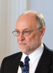 Mati Paasuke合作论文数Inst Exercise Biol & Physiotherapy, Univ Tartu3
 Zuzana Gdovinova合作论文数Department of Neurology, Faculty of Medicine, Pavol Jozef Šafárik University3Mendoza Rodríguez A合作论文数3
 Mangone Graziella合作论文数'Sapienza' University, Tor 'Vergata' University3Vladimír Haň合作论文数Department of Neurology, Košice Faculty of Medicine, Pavol Jozef Šafárik University3
 Milan Grofik合作论文数Jessenius Faculty of Medicine, Comenius University3
 Michal Minar合作论文数3
 Matej Skorvanek合作论文数Faculty of Medicine, Safarik University;Dept. of Neurology, University Hospital of L. Pasteur;Graduate School Kosice Institute for Society and Health, Safarik University;Safarik University, Safarik University3
 Pool Allan-Hermann合作论文数Department of Molecular and Cell Biology and Helen Wills Neuroscience Institute, University of California3Barbara C. Tilley合作论文数3
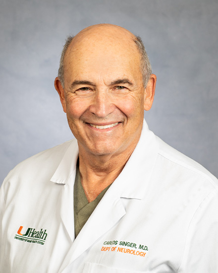 Carlos Singer合作论文数Department of Neurology, Miller School of Medicine, University of Miami3Aschermann Zsuzsanna合作论文数3Ene Reimann合作论文数Department of Physiology;University of Tartu;Department of Physiology, University of Tartu3Tomoko Arakaki合作论文数3
 Peter Valkovic合作论文数2nd Department of Neurology, Faculty of Medicine, Comenius University Bratislava3




