Amyloid-beta (A beta) and human islet amyloid polypeptide (hIAPP) aggregate to form amyloid fibrils that deposit in tissues and are associated with Alzheimer's disease (AD) and type II diabetes (T2D), respectively. Individuals with T2D have an increased risk of developing AD, and conversely, AD patients have an increased risk of developing T2D. Evidence suggests that this link between AD and T2D might originate from a structural similarity between aggregates of A beta and hIAPP. Using the cryoEM method microelectron diffraction, we determined the atomic structures of 11-residue segments from both A beta and hIAPP, termed A beta (24-34) WT and hIAPP(19-29) S20G, with 64% sequence similarity. We observed a high degree of structural similarity between their backbone atoms (0.96-angstrom root mean square deviation). Moreover, fibrils of these segments induced amyloid formation through self- and cross-seeding. Furthermore, inhibitors designed for one segment showed cross-efficacy for full-length A beta and hIAPP and reduced cytotoxicity of both proteins, although by apparently blocking different cytotoxic mechanisms. The similarity of the atomic structures of A beta (24-34) WT and hIAPP(19-29) S20G offers a molecular model for cross-seeding between A beta and hIAPP.
The cryoEM method micro-electron diffraction (MicroED) utilizes 3D crystals only a few hundred nanometers thick for atomic structure determination, thus shrinking the crystal size limits set by synchrotron micro-focus beamlines. MicroED is ideal for 3D nanocrystals, such as those formed from amyloid protein segments. Amyloid proteins form fibers that are the hallmarks of a class of more than 25 amyloid diseases, ranging from Parkinson's disease to Type II Diabetes (T2D). Although full-length amyloid proteins have so far been resistant to crystallization, select protein segments that form the spines of amyloid fibrils do form such tiny 3D crystals. Gaining knowledge of the atomic structures of these protein segments is critical to our understanding of amyloid fibril structures that contribute to disease onset and progression. Here, we use MicroED to determine the structures of two 11-residue segments from Islet Amyloid Polypeptide (IAPP) at 1.4 Å and 2.0 Å resolution, respectively. Amyloid fibrils of IAPP are found in approximately 90% of T2D patients and the formation of these fibrils is tightly linked to pancreatic β-cell death and insulin dependence. The two segments overlap in sequence by 5 residues. Their structures add to the range of polymorphism observed previously in crystal structures of shorter IAPP segments. One segment, 15-25WT, forms an unusual arrangement of single, out-of-register β-sheets. The second segment, 19-29S20G, contains the familial S20G mutation that leads to early-onset T2D. It forms pairs of β-sheets mated by a dry interface that share structural features with and are similarly cytotoxic to full-length IAPP fibrils. Thus, this structure may serve as a model for the toxic spine of IAPP aggregates. These findings, facilitated by MicroED, give us insight into disease-relevant structures that can be utilized for structure-based design of therapeutics.
hIAPP fibrils are associated with Type-II Diabetes, but the link of hIAPP structure to islet cell death remains elusive. Here we observe that hIAPP fibrils are cytotoxic to cultured pancreatic β-cells, leading us to determine the structure and cytotoxicity of protein segments composing the amyloid spine of hIAPP. Using the cryoEM method MicroED, we discover that one segment, 19–29 S20G, forms pairs of β-sheets mated by a dry interface that share structural features with and are similarly cytotoxic to full-length hIAPP fibrils. In contrast, a second segment, 15–25 WT, forms non-toxic labile β-sheets. These segments possess different structures and cytotoxic effects, however, both can seed full-length hIAPP, and cause hIAPP to take on the cytotoxic and structural features of that segment. These results suggest that protein segment structures represent polymorphs of their parent protein and that segment 19–29 S20G may serve as a model for the toxic spine of hIAPP.
Monocarboxylate transporter 1 (MCT1) inhibition is thought to block tumor growth through disruption of lactate transport and glycolysis. Here, we show MCT1 inhibition impairs proliferation of glycolytic breast cancer cells co-expressing MCT1 and MCT4 via disruption of pyruvate rather than lactate export. MCT1 expression is elevated in glycolytic breast tumors, and high MCT1 expression predicts poor prognosis in breast and lung cancer patients. Acute MCT1 inhibition reduces pyruvate export but does not consistently alter lactate transport or glycolytic flux in breast cancer cells that co-express MCT1 and MCT4. Despite the lack of glycolysis impairment, MCT1 loss-of-function decreases breast cancer cell proliferation and blocks growth of mammary fat pad xenograft tumors. Our data suggest MCT1 expression is elevated in glycolytic cancers to promote pyruvate export that when inhibited, enhances oxidative metabolism and reduces proliferation. This study presents an alternative molecular consequence of MCT1 inhibitors, further supporting their use as anti-cancer therapeutics.
In order to evaluate potential therapeutic targets for treatment of amyloidoses such as Alzheimer's disease (AD), it is essential to determine the structures of toxic amyloid oligomers. However, for the amyloid β-protein peptide (Aβ), thought to be the seminal neuropathogenetic agent in AD, its fast aggregation kinetics and the rapid equilibrium dynamics among oligomers of different size pose significant experimental challenges. Here we use ion-mobility mass spectrometry, in combination with electron microscopy, atomic force microscopy, and computational modeling, to test the hypothesis that Aβ peptides can form oligomeric structures resembling cylindrins and β-barrels. These structures are hypothesized to cause neuronal injury and death through perturbation of plasma membrane integrity. We show that hexamers of C-terminal Aβ fragments, including Aβ(24-34), Aβ(25-35) and Aβ(26-36), have collision cross sections similar to those of cylindrins. We also show that linking two identical fragments head-to-tail using diglycine increases the proportion of cylindrin-sized oligomers. In addition, we find that larger oligomers of these fragments may adopt β-barrel structures and that β-barrels can be formed by folding an out-of-register β-sheet, a common type of structure found in amyloid proteins.
In order to evaluate potential therapeutic targets for treatment of amyloidoses such as Alzheimers disease (AD), it is essential to determine the structures of toxic amyloid oligomers. However, for the amyloid beta-protein peptide (A beta), thought to be the seminal neuropathogenetic agent in AD, its fast aggregation kinetics and the rapid equilibrium dynamics among oligomers of different size pose significant experimental challenges. Here we use ion-mobility mass spectrometry, in combination with electron microscopy, atomic force microscopy, and computational modeling, to test the hypothesis that A beta peptides can form oligomeric structures resembling cylindrins and beta-barrels. These structures are hypothesized to cause neuronal injury and death through perturbation of plasma membrane integrity. We show that hexamers of C-terminal A beta fragments, including A beta(24-34), A beta(25-35) and A beta(26-36), have collision cross sections similar to those of cylindrins. We also show that linking two identical fragments head-to-tail using diglycine increases the proportion of cylindrin-sized oligomers. In addition, we find that larger oligomers of these fragments may adopt beta-barrel structures and that beta-barrels can be formed by folding an out-of-register beta-sheet, a common type of structure found in amyloid proteins.
Specific docking interactions between mitogen-activated protein kinases (MAPKs), their regulators, and their downstream substrates, are crucial for efficient and accurate signal transmission. To identify novel substrates of the c-Jun N-terminal kinase (JNK) family of MAPKs, we searched the human genome for proteins that contained (1), a predicted JNK-docking site (D-site); and (2), a cluster of putative JNK target phosphosites located close to the D-site. Here we describe a novel JNK substrate that emerged from this analysis, the functionally uncharacterized protein smoothelin-like 2 (SMTNL2). SMTNL2 protein bound with high-affinity to multiple MAPKs including JNK1-3 and ERK2; furthermore, the identity of conserved amino acids in the predicted docking site (residues 180-193) was necessary for this high-affinity binding. In addition, purified full-length SMTNL2 protein was phosphorylated by JNK1-3 in vitro, and this required the integrity of the D-site. Using mass spectrometry and mutagenesis, we identified four D-site-dependent phosphoacceptor sites in close proximity to the docking site, at S217, S241, T236 and T239. A short peptide comprised of the SMTNL2 D-site inhibited JNK-mediated phosphorylation of the ATF2 transcription factor, showing that SMTNL2 can compete with other substrates for JNK binding. Moreover, when transfected into HEK293 cells, SMTNL2 was phosphorylated by endogenous JNK in a D-site dependent manner, on the same residues identified in vitro. SMTNL2 protein was expressed in many mammalian tissues, with a notably high expression in skeletal muscle. Consistent with the hypothesis that SMTNL2 has a function in skeletal muscle, SMTNL2 protein expression was strongly induced during the transition from myoblasts to myotubes in differentiating C2C12 cells.
The major malaria vector in Sub-Saharan Africa is the Anopheles gambiae mosquito. This species is a key target of malaria control measures. Mosquitoes find humans primarily through olfaction, yet the molecular mechanisms associated with host-seeking behavior remain largely unknown. To further understand the functionality of A. gambiae odorant binding protein 1 (AgamOBP1), we combined in silico protein structure modeling and site-directed mutagenesis to generate 16 AgamOBP1 protein analogues containing single point mutations of interest. Circular dichroism (CD) and ligand-binding assays provided data necessary to probe the effects of the point mutations on ligand binding and the overall structure of AgamOBP1. Far-UV CD spectra of mutated AgamOBP1 variants displayed both substantial decreases to ordered α-helix structure (up to22%) and increases to disordered α-helix structure(up to 15%) with only minimal changes in random coil (unordered) structure. In mutations Y54A, Y122A and W114Q, aromatic side chain removal from the binding site significantly reduced N-phenyl-1-naphthylamine binding. Several non-aromatic mutations (L15T, L19T, L58T, L58Y, M84Q, M84K, H111A, Y122A and L124T) elicited changes to protein conformation with subsequent effects on ligand binding. This study provides empirical evidence for the in silico predicted functions of specific amino acids in AgamOBP1 folding and ligand binding characteristics.
 David Eisenberg合作论文数Department of Chemistry and Biochemistry, College of Physical Sciences, University of California, Los Angeles5
 Eric Y. Hayden合作论文数3Sarah Quan合作论文数3
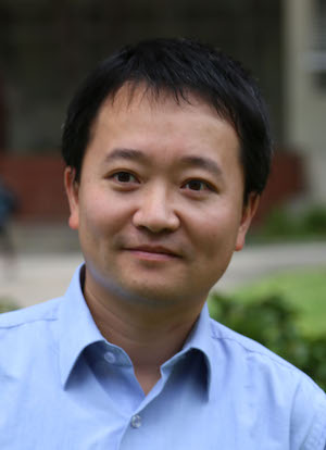 Lin Jiang合作论文数Molecular Biology Institute, University of California, Los Angeles3
 Tamir Gonen合作论文数Department of Biological Chemistry, University of California, Los Angeles3
 Duilio Cascio合作论文数Department of Biological Chemistry, University of California, Los Angeles3
 Jose A. Rodriguez合作论文数Biochemistry Division, Department of Chemistry and Biochemistry, College of Physical Sciences, University of California, Los Angeles;Institute for Genomics and Proteomics, Department of Energy Office of Science, University of California, Los Angeles;California NanoSystems Institute, University of California, Los Angeles;Molecular Biology Institute, University of California, Los Angeles;NSF-STROBE Science and Technology Center3
 David B. Teplow合作论文数Brain Research Institute, University of California, Los Angeles;Biopolymer Laboratory, David Geffen School of Medicine, University of California, Los Angeles3LaPointe Nichole E合作论文数3Michael Sawaya合作论文数Molecular Biology Institute, University of California, Los Angeles;Institute for Genomics and Proteomics, US Department of Office Energy of Science3Do Thanh D合作论文数3Shi Dan合作论文数3
 Mike Bowers合作论文数Department of Chemistry & Biochemistry, University of California, Santa Barbara3Rebecca Nelson合作论文数3
 Stuart Feinstein合作论文数University of California Santa Barbara3
 Charles G. Glabe合作论文数School of Biological Sciences, University of California, Irvine;Alzheimer's Disease Research Center, University of California, Irvine2
 Nannenga Brent L合作论文数Chem Engn, Arizona State Univ2
 Hattne Johan合作论文数Lawrence Berkeley Natl Lab, Univ Calif Berkeley2Oskarsson Marie E合作论文数2
 Francis E. Reyes合作论文数Department of Chemistry and Biochemistry, University of Colorado at Boulder2Sarah L Griner合作论文数2Gunilla T. Westermark合作论文数Dept Med Cell Biol, Uppsala Univ2
 Stephan Philipp合作论文数2Vishal R Patel合作论文数1Mohammed Alavi合作论文数1
 Paul Seidler合作论文数1Susan E. Critchlow合作论文数1Ji H. Lee合作论文数1Thomas C Whisenant合作论文数1
 Robyn Kaake合作论文数1Kevin Murray合作论文数1William Gordon合作论文数School of Medicine, University of California1
 Michael Zeller合作论文数1
 Steven Dubinett合作论文数Molecular Biology Institute, University of California, Los Angeles1
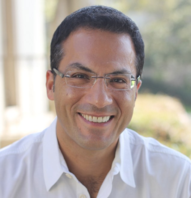 Siavash K. Kurdistani合作论文数Department of Biological Chemistry, David Geffen School of Medicine, University of California1
 Ingo Mellinghoff合作论文数Pharmacology, Weill Cornell Graduate School of Medical Sciences, Cornell University1
 Lorena Saelices合作论文数Departamento de Genética, Universidad de Sevilla1Lee Bardwell合作论文数1
 Thireou Trias N合作论文数1David Chia合作论文数1
 Elizabeth Gordon合作论文数1Wen Gu合作论文数1
 Steve Horvath合作论文数Department of Biostatistics, School of Public Health, University of California Los Angeles;Institutes of Science, Altos Labs1
 Nick Graham合作论文数Mork Family Department of Chemical Engineering and Materials Science, Viterbi School of Engineering, University of Southern California1
 Pierre Baldi合作论文数Department of Information and Computer Science, School of Information and Computer Sciences, University of California, Irvine;Center for Machine Learning and Intelligent Systems, Bren School of Information and Computer Science, University of California, Irvine;Mohamed bin Zayed University of Artificial Intelligence1
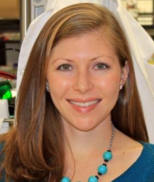 Heather Christofk合作论文数Jonsson Comprehensive Cancer Center, University of California Los Angeles;Department of Biological Chemistry, University of California Los Angeles1
 Iman Saramipoor Behbahan合作论文数Department of Internal Medicine, University of California Davis Medical Center1
 Sara A. Hurvitz合作论文数Clinical Research Division, Fred Hutchinson Cancer Center;Division of Hematology and Oncology, Department of Medicine, School of Medicine, University of Washington1
 Joan-Emma Shea合作论文数Department of Chemistry and Biochemistry, University of California, Santa Barbara1




