The lipid bilayer is a dynamic environment that consists of a mixture of lipids with different properties that regulate the function of membrane proteins; these lipids are either annular, masking the protein hydrophobic surface, or specific lipids, essential for protein function. In this study, using tandem mass spectrometry, we have identified specific lipids associated with the Escherichia coli ABC transporter McjD, which translocates the antibacterial peptide MccJ25. Using non-denaturing mass spectrometry, we show that McjD in complex with MccJ25 survives the gas phase. Partial delipidation of McjD resulted in reduced ATPase activity and thermostability as shown by circular dichroism, both of which could be restored upon addition of defined E. coli lipids. We have resolved a phosphatidylglycerol lipid associated with McjD at 3.4 Å resolution, whereas molecular dynamic simulations carried out in different lipid environments assessed the binding of specific lipids to McjD. Combined, our data show a synergistic effect of zwitterionic and negatively charged lipids on the activity of McjD; the zwitterionic lipids provide structural stability to McjD, whereas the negatively charged lipids are essential for its function.
Ionizing radiation is a universal tool in tumor therapy but may also cause secondary cancers or cell invasiveness. These negative side effects could be causally related to the human-intermediate-conductance Ca2+-activated-K+-channel (hIK), which is activated by X-ray irradiation and affects cell proliferation and migration. To analyze the signaling cascade downstream of ionizing radiation we use genetically encoded reporters for H2O2 (HyPer) and for the dominant redox-buffer glutathione (Grx1-roGFP2) to monitor with high spatial and temporal resolution, radiation-triggered excursions of H2O2 in A549 and HEK293 cells. The data show that challenging cells with ≥1 Gy X-rays or with UV-A laser micro-irradiation causes a rapid rise of H2O2 in the nucleus and in the cytosol. This rise, which is determined by the rate of H2O2 production and glutathione-buffering, is sufficient for triggering a signaling cascade that involves an elevation of cytosolic Ca2+ and eventually an activation of hIK channels.
To understand the impact of ionizing irradiation from diagnostics and radiotherapy on cells, we examined K(+) channel activity before and immediately after exposing cells to X-rays. Already, low dose in the cGy range caused in adenocarcinoma A549 cells within minutes a hyperpolarization following activation of the human intermediate-conductance Ca(2+)-activated K(+) channel (hIK). The response was specific for cells, which functionally expressed hIK channels and in which hIK activity was low before irradiation. HEK293 cells, which do not respond to X-ray irradiation, accordingly develop a sensitivity to this stress after heterologous expression of hIK channels. The data suggest that hIK activation involves a Ca(2+)-mediated signaling cascade because channel activation is suppressed by a strong cytosolic Ca(2+) buffer. The finding that an elevation of H2O2 causes an increase in the concentration of cytosolic Ca(2+) suggests that radicals, which emerge early in response to irradiation, trigger this Ca(2+) signaling cascade. Inhibition of hIK channels by specific blockers clotrimazole and TRAM-34 slowed cell proliferation and migration in "wound" scratch assays; ionizing irradiation, in turn, stimulated the latter process presumably via its activation of the hIK channels. These data stress an indirect radiosensitivity of hIK channels with an impact on cell differentiation.
The viral channel KcvNTS belongs to the smallest K(+) channels known so far. A monomer of a functional homotetramer contains only 82 amino acids. As a consequence of the small size the protein is almost fully submerged into the membrane. This suggests that the channel is presumably sensitive to its lipid environment. Here we perform a comparative analysis for the function of the channel protein embedded in three different membrane environments. 1. Single-channel currents of KcvNTS were recorded with the patch clamp method on the plasma membrane of HEK293 cells. 2. They were also measured after reconstitution of recombinant channel protein into classical planar lipid bilayers and 3. into horizontal bilayers derived from giant unilamellar vesicles (GUVs). The recombinant channel protein was either expressed and purified from Pichia pastoris or from a cell-free expression system; for the latter a new approach with nanolipoprotein particles was used. The data show that single-channel activity can be recorded under all experimental conditions. The main functional features of the channel like a large single-channel conductance (80pS), high open-probability (>50%) and the approximate duration of open and closed dwell times are maintained in all experimental systems. An apparent difference between the approaches was only observed with respect to the unitary conductance, which was ca. 35% lower in HEK293 cells than in the other systems. The reason for this might be explained by the fact that the channel is tagged by GFP when expressed in HEK293 cells. Collectively the data demonstrate that the small viral channel exhibits a robust function in different experimental systems. This justifies an extrapolation of functional data from these systems to the potential performance of the channel in the virus/host interaction. This article is part of a Special Issue entitled: Viral Membrane Proteins-Channels for Cellular Networking.
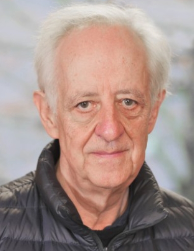 Gerhard Thiel合作论文数Department of Biosciences, University of Milan3
 Anna Moroni合作论文数Department of Biosciences, University of Milan3
 Bastian Roth合作论文数2Christine S. Gibhardt合作论文数2Claudia Fournier合作论文数2Indra Schroeder合作论文数2Sebastian Fuck合作论文数1
 Christian Braun合作论文数1Manuela Gebhardt合作论文数1
 Cristina Arrigoni合作论文数1
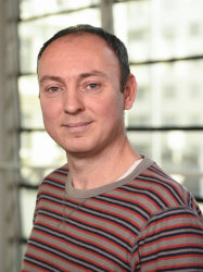 Konstantinos Beis合作论文数Bacterial Pathogenesis, Department of Life Sciences, Faculty of Natural Sciences, Imperial College London1
 Severine Zirah合作论文数1
 Rohanah Hussain合作论文数1
 Hassanul G Choudhury合作论文数1Henkes Leonhard M合作论文数1
 Peter Tieleman合作论文数Department of Biological Sciences, Faculty of Science, University of Calgary;Centre for Molecular Simulation, Faculty of Science, University of Calgary1Shahid Mehmood合作论文数Physical and Theoretical Chemistry Laboratory, University of Oxford1Lachnit Christine合作论文数1
 Valentina Corradi合作论文数University of Exeter1
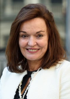 Carol Robinson合作论文数Department of Chemistry, University of Oxford;Kavli Institute for Nanoscience Discovery, University of Oxford;OMass Therapeutics1Danny Axford合作论文数1Stefan M. Kast合作论文数1
 Sylvie Rebuffat合作论文数1
 Burkhard Jakob合作论文数1




