Background and Importance Tegmen defects associated with cerebrospinal fluid (CSF) leaks are a rare pathology that can result in severe complications if left untreated. There is no universal optimal surgical algorithm for repair, although the most common techniques are the middle fossa craniotomy (traditionally 25 cm(2) in area), the transmastoid approach, or both. Here, we describe successful use of a keyhole mini-craniotomy, only 6 cm(2) in area, without mastoidectomy or days of lumbar drainage. Clinical Presentation Three patients presented with right-sided CSF otorrhea and hearing loss, with varying sizes of tegmen defects and associated encephaloceles. Keyhole craniotomies measuring 3 x 2 cm were used to perform a multilayer repair comprising an intradural collagen dural substitute, extradural fascial graft, extradural collagen dural substitute, fibrin sealant, and sometimes bony reconstruction using partial thickness craniotomy grafting. All patients were discharged on postoperative day 1 or 2, with no recurrence of symptoms at 6 months. Conclusion The keyhole craniotomy approach does not sacrifice the extent of operative access for this pathology. This minimally invasive approach can likely be used more often without need for concomitant mastoidectomy, ultimately enabling shorter hospital stays and more rapid recovery.
Raman spectroscopy is a photonic modality defined as the inelastic backscattering of excitation coherent laser light. It is particularly beneficial for rapid tissue diagnosis in sensitive intraoperative environments like those involving the brain, due to its nonionizing potential, point-scanning capability, and highly-specific spectral fingerprint signatures that can characterize tissue pathology [1]. While Raman scattering is an inherently weak process, Surface-Enhanced Raman Spectroscopy (SERS), which is based on the use of metal nanostructure surfaces to amplify Raman signals, has become a compelling method for achieving highly specific Raman spectra with detection sensitivity comparable to conventional modalities such as fluorescence [2]. A unique plasmonics-active nanoplatform, SERS gold nanostars (GNS) have previously been designed in our group to accumulate preferentially in brain tumors [2]. Raman detection, when combined with machine learning and robotics, stands to enhance the diagnosis of ambiguous tissue during tumor resection surgery, with the potential to improve extent-of-resection and rapidly reconstruct the dynamic surgical field. Here we demonstrate preliminary results from the use of a SERS-based robotics platform to efficiently recreate a tumor embedded in healthy tissue, which is modeled here as a GNS-infused phantom. Transfer learning, specifically through use of the open-source RRUFF mineral database, is employed here to address the dearth of collected biomedical Raman data [3].
Background: One of the most devastating complications associated with vestibular schwannoma surgery is facial nerve (FN) injury and subsequent loss of function. In order to preserve FN function surgeons have previously utilized a monopolar stimulator to assess nerve function throughout surgery. This process is time-consuming and not continuous. One potential solution to this involves the utilization of the blink reflex where FN function can be stimulated through motor potentials and monitored continuously and autonomously. Despite the potential benefit of measuring the Blink reflex, it is currently unknown if subtle changes in EMG response will be detectable by neuromonitoring technicians and the degree to which the blink reflex can predict FN function. Therefore, this study seeks to implement a machine learning model to detect key features of the blink reflex response that can predict FN function vestibular schwannoma surgery.
Ten patients undergoing surgical resection for spinal tumors were selected. Samples of tumor, muscle, and bone were resected, de-identified by the treating surgeon, and then scanned with the TumorID technology ex vivo. This study investigates whether TumorID technology is able to differentiate three different human clinical fresh tissue specimens: spine tumor, normal muscle, and normal bone. The TumorID technology utilizes a 405 nm excitation laser to target endogenous fluorophores, thereby allowing for the detection of tissue based on emission spectra. Metabolic profiles of tumor and healthy tissue vary, namely NADH (bound and free emission peak, respectively: 487 nm, 501 nm) and FAD (emission peak: 544) are endogenous fluorophores with distinct concentrations in tumor and healthy tissue. Emission spectra analyzed consisted of 74 scans of spine tumor, 150 scans of healthy normal bone, and 111 scans of healthy normal muscle. An excitation wavelength of 405 nm was used to obtain emission spectra from tissue as previously described. Emission spectra consisted of approximately 1400 wavelength intensity pairs between 450 and 750 nm. Kruskal–Wallis tests were conducted comparing AUC distributions for each treatment group, α = 0.05. Spectral signatures varied amongst the three different tissue types. All pairwise comparisons among tissues for Free NADH were statistically significant (Tumor vs. Muscle: p = 0.0006, Tumor vs. Bone: p < 0.0001, Bone vs. Muscle: p = 0.0357). The overall comparison of tissues for FAD (506.5–581.5 nm) was also statistically significant (p < 0.0001), with two pairwise comparisons being statistically significant (Tumor vs. Muscle: p < 0.0001, Tumor vs. Bone: p = 0.0045, Bone vs. Muscle: p = 0.249). These statistically significant differences were maintained when stratifying tumor into metastatic carcinoma (N = 57) and meningioma (N = 17). TumorID differentiates tumor tissue from normal bone and normal muscle providing further clinical evidence of its efficacy as a tissue identification tool. Future studies should evaluate TumorID’s ability to serve as an adjunctive tool for intraoperative assessment of surgical margins and surgical decision-making.
Key points We developed a culture model of a human olfactory ensheathing cell tumor. Cultured organoids resemble normal ensheathing cells. Assays suggest that this model provides a tool for studying the roles of these glial cells in the maintenance of the peripheral olfactory system.
Background: Despite robust EMG monitoring efforts during surgery, complications are somewhat common with around a 40% chance of facial nerve sequelae following surgery. This research will explore the feasibility of utilizing large-scale EMG data to determine the prognostic value of blink reflex data in the preservation of facial nerve function.
In robotic laser surgery, shape prediction of an one-shot ablation crater is an important problem for minimizing errant overcutting of healthy tissue during the course of pathological tissue resection and precise tumor removal. Since it is difficult to physically model the laser-tissue interaction due to the variety of optical tissue properties, complicated process of heat transfer, and uncertainty about the chemical reaction, we propose a 3D crater prediction model based on an entirely data-driven method without any assumptions of laser settings and tissue properties. Based on the crater prediction model, we formulate a novel robotic laser planning problem to determine the optimal laser incident configuration, which aims to create a crater that aligns with the surface target (e.g. tumor, pathological tissue). To solve the one-shot ablation crater prediction problem, we model the 3D geometric relation between the tissue surface and the laser energy profile as a non-linear regression problem that can be represented by a single-layer perceptron (SLP) network. The SLP network is encoded in a novel kinematic model to predict the shape of the post-ablation crater with an arbitrary laser input. To estimate the SLP network parameters, we formulate a dataset of one-shot laser-phantom craters reconstructed by the optical coherence tomography (OCT) B-scan images. To verify the method. The learned crater prediction model is applied to solve a simplified robotic laser planning problem modelled as a surface alignment error minimization problem. The initial results report about $(91.2\pm 3.0)\%$ 3D-crater-Intersection-over-Union (3D-crater-IoU) for the 3D crater prediction and an average of about 98.0% success rate for the simulated surface alignment experiments.
In neurosurgical procedures maximizing the resection of tumor tissue while avoiding healthy tissue is of paramount importance and a difficult task due to many factors, such as surrounding eloquent brain. Swiftly identifying tumor tissue for removal could increase surgical outcomes. The TumorID is a laser-induced fluorescence spectroscopy device that utilizes endogenous fluorophores such as NADH and FAD to detect tumor regions. With the goal of creating an endoscopic tool for intraoperative tumor detection in mind, a study of the TumorID was conducted to assess how the angle of incidence (AoI) affects the collected spectral response of the scanned tumor. For this study, flat and convex NADH/FAD gellan gum phantoms were scanned at various AoI (a range of 36 degrees) to observe the spectral behavior. Results showed that spectral signature did not change significantly across flat and convex phantoms, and the Area under Curve (AUC) values calculated for each spectrum had a standard deviation of 0.02 and 0.01 for flat and convex phantoms, respectively. Therefore, the study showed that AoI will affect the intensity of the spectral response, but the peaks representative of the endogenous fluorophores are still observable and similar. Future work includes conducting an AoI study with a longer working-distance lens, then incorporating said lens to design an endoscopic, intraoperative tumor detection device for minimally invasive surgery, with first applications in endonasal endoscopic approaches for pituitary tumors.
Background: Near real-time intraoperative identification of tumor tissue is needed, as contemporary methods such as intraoperative MRI are expensive, time intensive, and only available at the most advanced care facilities. The TumorID is a low cost, portable, non-contact laser induced fluorescence spectroscopy device designed by our research group for intraoperative use. Importantly, the TumorID uses autofluorescence, meaning no additional fluorophores are given to the patient thereby avoiding the associated risks and timeline constraints. By using an excitation wavelength of 405 nm the TumorID targets differences in NADH and FAD noted by the Warburg effect. Providing the surgical team with increased knowledge about tumor location could potentially increase resection, decrease surgical time, and therefore increase patient outcomes. This study shows the TumorID's applicability in identifying differences using a logistic regression model in recently resected Pituitary Adenoma with an endonasal approach.
Automated laser surgery with sensor fusion is an important problem in medical robotics since it requires precise control of mirrors used to steer the laser systems. The propagation of the laser beam should satisfy the geometric constraints of the surgical site but the relation between the number of mirrors and the design of the optical path remains an unsolved problem. Furthermore, different types of surgery (e.g. endoscopic vs open surgery) can require different optical designs with varying number of mirrors to successfully steer the laser beam to the tissue. A generalized method for controlling the laser beam in such systems remains an open research question. This paper proposes an analytical model for a laser-based surgical system with an arbitrary number of mirrors, which is referred as an "N-mirror" robotic system. This system consists of three laser inputs to transmit the laser beam to the tissue surface through N number of mirrors, which can achieve surface scanning, tissue resection and tissue classification separately. For sensor information alignment, the forward and inverse kinematics of the N-mirror robot system are derived and used to calculate the mirror angles for laser steering at the target surface. We propose a system calibration method to determine the laser input configuration that is required in the kinematic modelling. We conduct simulation experiments for a simulated 3-mirror system of an actual robotic laser platform and a 6-mirror simulated robot, both with 3-laser inputs. The simulation experiments for system calibration show results of maximum position offset smaller than 0.127 mm and maximum angle offset smaller than 0.05° for the optimal laser input predictions.
This paper focuses on a research problem of robotic controlled laser orientation to minimize errant overcutting of healthy tissue during the course of pathological tissue resection. Laser scalpels have been widely used in surgery to remove pathological tissue targets such as tumors or other lesions. However, different laser orientations can create various tissue ablation cavities, and incorrect incident angles can cause over-irradiation of healthy tissue that should not be ablated. This work aims to formulate an optimization problem to find the optimal laser orientation in order to minimize the possibility of excessive laser-induced tissue ablation. We first develop a 3D data-driven geometric model to predict the shape of the tissue cavity after a single laser ablation. Modelling the target and non-target tissue region by an obstacle boundary, the determination of an optimal orientation is converted to a collision-minimization problem. The goal of this optimization formulation is maintaining the ablated contour distance from the obstacle boundary, which is solved by Projected gradient descent. Simulation experiments were conducted and the results validated the proposed method with conditions of various obstacle shapes and different initial incident angles.
Because contemporary intraoperative tumor detection modalities, such as intraoperative MRI, are not ubiquitously available and can disrupt surgical workflow, there is an imperative for an accessible diagnostic device that can meet the surgeon's needs in identifying tissue types. The objective of this paper is to determine the efficacy of a novel non-contact tumor detection device for metastatic melanoma boundary identification in a tissue-mimicking phantom, evaluate the identification of metastatic melanoma boundaries in ex vivo mouse brain tissue, and find the error associated with identifying this boundary. To validate the spatial and fluorescence resolution of the device, tissue-mimicking phantoms were created with modifiable optical properties. Phantom tissue provided ground truth measurements for fluorophore concentration differences with respect to spatial dimensions. Modeling metastatic disease, ex vivo melanoma brain metastases were evaluated to detect differences in fluorescence between healthy and neoplastic tissue. This analysis includes determining required-to-observe fluorescence differences in tissue. H&E staining confirmed tumor presence in mouse tissue samples. The device detected a difference in normalized average fluorescence intensity in all three phantoms. There were differences in fluorescence with the presence and absence of melanin. The estimated tumor boundary of all tissue phantoms was within 0.30 mm of the ground truth tumor boundary for all boundaries. Likewise, when applied to the melanoma-bearing brains from ex vivo mice, a difference in normalized fluorescence intensity was successfully detected. The potential prediction window for the tumor boundary location is less than 1.5 mm for all ex vivo mouse brain tumors boundaries. We present a non-contact, laser-induced fluorescence device that can identify tumor boundaries based on changes in laser-induced fluorescence emission intensity. The device can identify phantom ground truth tumor boundaries within 0.30 mm using instantaneous rate of change of normalized fluorescence emission intensity and can detect endogenous fluorescence differences in melanoma brain metastases in ex vivo mouse tissue.
Background Instrument oversupply drives cost in the operating room (OR). We review previously reported methodologies for surgical instrument reduction and report a pilot methodology for optimizing instrument supply via ethnographic instrument tracking of thumb carpometacarpal (CMC) arthroplasties. Additionally, we report a cost analysis of instrument oversupply and potential savings of tray optimization methods. Methods Instrument utilization was tracked over 8 CMC arthroplasties conducted by 2 surgeons at an ambulatory surgery center of a large academic hospital. An optimized supply methodology was designed. A cost analysis was conducted using health-system-specific data and previously published research. Results After tracking instrument use in 8 CMC arthroplasties, a cumulative total of 59 out of the 120 instruments in the Hand & Foot (H&F) tray were used in at least 1 case. Two instruments were used in all cases, and another 20 instruments were used in at least 50% of the cases. Using a reduced tray with 59 instruments, potential cost savings for tray reduction in 60 cases were estimated to be $2086 without peel-packing and $2356 with peel-packing. The estimated cost savings were lower than those reported in literature due to a reduced scope and exclusion of OR time cost in the analysis. Conclusions Instrument oversupply drives cost at our institution’s ambulatory surgery center. Ethnography is a cost-effective method to track instrument utilization and determine optimal tray composition for small services but is not scalable to large health systems. The time and cost required to observe sufficient surgeries to enable supply reduction to motivate the need for more efficient methods to determine instrument utility.
Objectives This article describes a novel technique implementing the use of a tympanostomy t-tube to provide long-term marsupialization of small Rathke's cleft cysts (RCCs). Design A retrospective review of electronic medical records was performed to collect demographic and clinical data on a series of four patients. Setting Academic medical center. Participants Four female patients (mean age of 34 years) underwent transsphenoidal endoscopic endonasal surgery for RCC. All four patients presented with headaches. Mean cyst size was 7 mm. Two of the four surgeries were revisions for RCC recurrence. Main Outcome Measures Symptom resolution after surgery, duration of follow-up, and feasibility of the proposed technique. Results Tympanostomy t-tube was used to marsupialize small RCCs (< 10 mm) for four patients. Three patients remained symptom-free with endoscopy and imaging showing patent t-tubes at 21 months' (range 20-24 months) follow-up. One patient experienced severe migraines immediately after surgery. Migraines were relieved after t-tube was removed 6 weeks after surgery. Conclusion Tympanostomy t-tubes placed via an endoscopic endonasal approach can provide long-term marsupialization for small RCCs.
Abstract Objective Surgical instrument oversupply drives cost, confusion, and workload in the operating room. With an estimated 78%–87% of instruments being unused, many health systems have recognized the need for supply refinement. By manually recording instrument use and tasking surgeons to review instrument trays, previous quality improvement initiatives have achieved an average 52% reduction in supply. While demonstrating the degree of instrument oversupply, previous methods for identifying required instruments are qualitative, expensive, lack scalability and sustainability, and are prone to human error. In this work, we aim to develop and evaluate an automated system for measuring surgical instrument use. Materials and Methods We present the first system to our knowledge that automates the collection of real-time instrument use data with radio-frequency identification (RFID). Over 15 breast surgeries, 10 carpometacarpal (CMC) arthroplasties, and 4 craniotomies, instrument use was tracked by both a trained observer manually recording instrument use and the RFID system. Results The average Cohen’s Kappa agreement between the system and the observer was 0.81 (near perfect agreement), and the system enabled a supply reduction of 50.8% in breast and orthopedic surgery. Over 10 monitored breast surgeries and 1 CMC arthroplasty with reduced trays, no eliminated instruments were requested, and both trays continue to be used as the supplied standard. Setup time in breast surgery decreased from 23 min to 17 min with the reduced supply. Conclusion The RFID system presented herein achieves a novel data stream that enables accurate instrument supply optimization.
Objective To assess olfactory outcomes as measured by an olfactory-specific quality of life (QOL) questionnaire in patients undergoing EESBS for sellar lesions. Design Retrospective case series. Setting Tertiary academic medical center. Participants In total, 36 patients undergoing EESBS for lesions limited to the sella were evaluated. Main Outcome Measures The following were performed before and three months after surgery: 22-Item Sinonasal Outcomes Test (SNOT-22), University of Pennsylvania Smell Identification Test (UPSIT), and the Assessment of Self-reported Olfactory Functioning (ASOF), which has three domains: subjective olfactory capability scale (SOC), smell-related problems (SRP), and olfactory-related quality of life (ORQ). Results Median age at surgery was 52.5 years, with a median tumor size of 1.8 cm (range: 0.2 to 3.9 cm). Pre- and postoperative median scores were 35 [34, 36.2] and 34.5 [32, 36] for UPSIT, 21 [7.5, 33.5] and 21.5 [6.8, 35.7] for SNOT-22, 10 [9, 10] and 9 [8, 10] for ASOF-SOC, 5 [4.8, 5] and 4.5 [4, 5] for ASOF-SRP, and 5 [5, 5] and 5 [4.5, 5] for ASOF-ORQ. There was no significant change in the two of the three domains of the ASOF. Correlation between ASOF and UPSIT scores were weak. Older age and larger tumor size were associated with worsened olfaction after surgery. Conclusions Patients did not experience significant changes in olfactory-specific QOL three months after EESBS, as measured by two domains of the ASOF. The ASOF may serve as a useful adjunctive tool for assessing olfaction after surgery. The lack of correlation between UPSIT and ASOF suggests the need for more research in subjective olfactory-related quality of life after surgery.
Objective: Create a device that improves the identification and extent of resection at the interface between healthy and tumor tissue; ultimately, using this device would improve surgical outcomes for patients and increase survival. Methods: We have created a contactless tumor removal system that utilizes endogenous fluorescence feedback to inform a laser ablation system to execute autonomous removal of phantom tumor tissue. Results: This completely non-contact surgical system is capable of resecting the tumor boundary of a tissue phantom with an average root mean square error (RMSE) of approximately 1.55 mm and an average max error of approximately 2.15 mm. There is no difference in the performance of the system when changing the size of the internal tumor from 7.5-12.5 mm in diameter. Discussion: Future research steps include creating a more intelligent spectral search strategy to increase the density of points around the resection boundary, and to develop a more sophisticated classifier to predict pathologic diagnosis and tissue subtypes located regionally around the tumor boundaries. We envision this device being used to resect the boundaries of tumors identified by exogenously delivered tumor-labeling fluorophores, such as fluorescein or 5-ALA, in addition to approaches relying on autofluorescence of endogenous fluorophores.
Abstract INTRODUCTION Vaccination with dendritic cells (DCs) fares poorly in primary and recurrent glioblastoma (GBM). Moreover, GBM vaccine trials are often underpowered due to limited sample size. METHODS To address these limitations, we conducted three sequential clinical trials utilizing Cytomegalovirus (CMV)-specific DC vaccines in patients with primary GBM. Autologous DCs were generated and electroporated with mRNA encoding for the CMV protein pp65. Serial vaccination was given throughout adjuvant temozolomide cycles, and 111Indium radiolabeling was implemented to assess migration efficiency of DC vaccines. Patients were followed for median overall survival (mOS) and OS. RESULTS Our initial study was the phase II ATTAC study (NCT00639639; total n=12) with 6 patients randomized to vaccine site preconditioning with tetanus-diphtheria (Td) toxoid. This led to an expanded cohort trial (ATTAC-GM; NCT00639639) of 11 patients receiving CMV DC vaccines containing granulocyte-macrophage colony-stimulating factor (GM-CSF). Follow-up data from ATTAC and ATTAC-GM revealed 5-year OS rates of 33.3% (mOS 38.3 months; CI95 17.5-undefined) and 36.4% (mOS 37.7 months; CI95 18.2-109.1), respectively. ATTAC additionally revealed a significant increase in DC migration to draining lymph nodes following Td preconditioning (P=0.049). Increased DC migration was associated with OS (Cox proportional hazards model, HR=0.820, P=0.023). Td-mediated increased migration has been recapitulated in our larger confirmatory trial ELEVATE (NCT02366728) of 43 patients randomized to preconditioning (Wilcoxon rank sum, Td n=24, unpulsed DC n=19; 24h, P=0.031 and 48h, P=0.0195). In ELEVATE, median follow-up of 42.2 months revealed significantly longer OS in patients randomized to Td (P=0.026). The 3-year OS for Td-treated patients in ELEVATE was 34% (CI95 19-63%) compared to 6% given unpulsed DCs (CI95 1-42%). CONCLUSION We report reproducibility of our findings across three sequential clinical trials using CMV pp65 DCs. Despite their small numbers, these successive trials demonstrate consistent survival outcomes, thus supporting the efficacy of CMV DC vaccine therapy in GBM.
Introduction: Estimating the risk of extended length of stay (LOS) or non-routine discharge disposition is helpful in surgical decision-making for patients with brain metastases (BM). In 2020, an online calculator was introduced by Khalafallah et al. that stratified the risk of patients with brain tumors based on poor surgical outcomes. We applied the calculator to our population of BM patients to determine its generalizability and validity. Methods: We included BM patients who underwent a cranial procedure between 2015 and 2018 at a single academic institution. Patient age, race, marital status, admission status, KPS score, and medical co-morbidities (5-point modified frailty index (mFI-5)) were included in the analysis. We calculated the areas under the Receiver Operating Characteristics (ROC) curves to determine the validity of the model proposed in predicting extended LOS (>7 days) and need for specialty care at discharge (non-routine discharge disposition). Results: We analyzed 244 patients (mean age 61.2 years (SD 11.1), 57.0% female, and 78.1% Caucasian). The areas under the ROC curves were 0.8427 and 0.8422 for extended LOS and non-routine discharge disposition, suggesting high accuracy of the models for these outcomes. However, the (mFI-5) was not a significant predictor of either outcome in our multivariate analyses. Conclusions: We validated Khalafallah et al.’s predictive models of extended LOS and non-routine discharge disposition in our patient population, which included a broader range of surgical procedures. Further investigation of this model could clarify how the type of neurosurgical procedure influences outcomes, the role of the mFI-5, and its overall generalizability.
With the increasing availability of new image registration approaches, an unbiased evaluation is becoming more needed so that clinicians can choose the most suitable approaches for their applications. Current evaluations typically use landmarks in manually annotated datasets. As a result, the quality of annotations is crucial for unbiased comparisons. Even though most data providers claim to have quality control over their datasets, an objective third-party screening can be reassuring for intended users. In this study, we use the variogram to screen the manually annotated landmarks in two datasets used to benchmark registration in image-guided neurosurgeries. The variogram provides an intuitive 2D representation of the spatial characteristics of annotated landmarks. Using variograms, we identified potentially problematic cases and had them examined by experienced radiologists. We found that (1) a small number of annotations may have fiducial localization errors; (2) the landmark distribution for some cases is not ideal to offer fair comparisons. If unresolved, both findings could incur bias in registration evaluation.
 Weston Ross合作论文数13
 Guangshen Ma合作论文数Duke Univ, Durham, NC 27708 USA7
 Ali R. Zomorodi合作论文数Department of Neurosurgery, Duke University School of Medicine6
 David W Jang合作论文数Department of Head and Neck Surgery and Communication Sciences, Duke University School of Medicine6
 Ralph Abi Hachem合作论文数Department of Head and Neck Surgery and Communication Sciences, Duke University School of Medicine6
 Peter Edward Fecci合作论文数Center for Brain and Spine Metastasis, Duke University;Clinical Science Departments, Pratt School of Engineering, Duke University;Department of Pathology, Pratt School of Engineering, Duke University;Duke Cancer Institute5
 Tanner Zachem合作论文数4
 Westin Hill合作论文数4
 Matthew Tucker合作论文数4
 Pierre E. Dupont合作论文数Boston Children's Hospital4C. Rory Goodwin合作论文数Department of Neurosurgery, Johns Hopkins Hospital4
 Andrew H. C. Gosline合作论文数Centre of Intelligent Machines, McGill University4Harrison Hockenberry合作论文数3
 Kent K. Yamamoto合作论文数3Jihad Abdelgadir合作论文数Department of Neurosurgery, Duke University Medical Center3Ian Hill合作论文数Department of Mechanical Engineering and Material Science, Duke University, Durham, NC, 27708, USA3
 Pankaj K. Agarwalla合作论文数Department of Neurosurgery, New Jersey Medical School3
 Allan H. Friedman合作论文数Duke University Medical Center3
 William E. Butler合作论文数Massachusetts General Hospital3
 Evan J. Butler合作论文数3
 Brian V. Nahed合作论文数Department of Neurosurgery, Massachusetts General Hospital and Harvard Medical School3Edward R Smith合作论文数2Suzanna Joseph合作论文数2Ravi Prakash合作论文数2Elliot Le合作论文数2Richard T. Chen合作论文数2Steven Cook合作论文数2
 Daniel Sexton合作论文数Computational Neurobiology Laboratory, Salk Institute for Biological Studies2Marcus Zachariah合作论文数Wexner Medical Center, The Ohio State University2Jimmy C Yang合作论文数Department of Neurosurgery, Emory University School of Medicine;Department of Neurosurgery, Ohio State University;Emory University School of Medicine, Ohio State University2Joshua K Helmkamp合作论文数2Tracy Truong合作论文数2Lyndon Chan合作论文数Div Rhinol & Endoscop Skull Base Surg, Duke Univ2
 Ren Odion合作论文数Fitzpatrick Inst Photon, Duke Univ2
 Brian P Walcott合作论文数Dept Neurol Surg, Univ Calif San Francisco2
 Nandan Lad合作论文数Department of Neurosurgery, Duke University School of Medicine2
 Margaret Johnson合作论文数Department of Neurosurgery, Duke University School of Medicine2
 Jordan Komisarow合作论文数Department of Neurosurgery, Duke University School of Medicine2
 Katherine Barnett Peters合作论文数Department of Neurology, Duke University School of Medicine;Duke Cancer Institute2
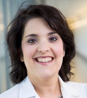 Annick Desjardins合作论文数Department of Neurosurgery, School of Medicine, Duke University;The Preston Robert Tisch Brain Tumor Center, Duke University;Duke Cancer Institute2
 Bradley John Goldstein合作论文数Department of Head and Neck Surgery & Communication Sciences, School of Medicine, Duke University2
 John Howard Sampson合作论文数School of Medicine, Duke University2
 Maragatha Kuchibhatla合作论文数Department of Biostatistics and Bioinformatics, Duke University School of Medicine2
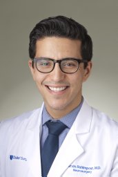 Rahimpour Shervin合作论文数Surgical Neurology Branch, National Institute of Neurological Disorders and Stroke2
 Pakawat Chongsathidkiet合作论文数Department of Surgery, Duke University Medical Center2
 Christopher Stapleton合作论文数Wang Ambulatory Care Center, Massachusetts General Hospital;Harvard Medical School2
 Brian P Mann合作论文数Department of Mechanical Engineering and Materials Science, Duke University2
 Pedro J. Del Nido合作论文数Department of Cardiac Surgery, Boston Children's Hospital;Harvard Medical School2
 James Emmett Herndon合作论文数Department of Biostatistics and Bioinformatics, School of Medicine, Duke University2




