IntroductionSex and gender are independently important in health and disease but have been incompletely explored in neurology. This is in part contributed to by the pre-existing male bias in scientific literature that results in fewer females being included in clinical research and the often interchangeable use of sex and gender in the literature. This scoping review intends to identify the advances as well as under-explored aspects of this field to provide a road map for future research. This paper outlines the methods for a scoping review of published, peer-reviewed literature on sex and gender differences in four subspecialty areas of neurology: demyelination, stroke, epilepsy and headache.Methods and analysisA detailed search strategy will be used to search five databases pertaining only to sex differences. Specific inclusion and exclusion criteria will be applied to capture relevant literature published from 2014 to 2020. Data will be collected and synthesised to provide an overview of information retrieved, a narrative synthesis of each subspecialty area and map of results.Ethics and disseminationResearch ethics board approval was not required for this study. This study will aid in mapping recent trends in sex differences in four major neurological conditions and will help identify areas for further research. A manuscript will be compiled for publication and presentations of findings.Registration detailsThe final protocol is registered with the Open Science Framework (https://osf.io/n937x/).
Zebrafish have made significant contributions to our understanding of the vertebrate brain and the neural basis of behavior, earning a place as one of the most widely used model organisms in neuroscience. Their appeal arises from the marriage of low cost, early life transparency, and ease of genetic manipulation with a behavioral repertoire that becomes more sophisticated as animals transition from larvae to adults. To further enhance the use of adult zebrafish, we created the first fully segmented three-dimensional digital adult zebrafish brain atlas (AZBA). AZBA was built by combining tissue clearing, light-sheet fluorescence microscopy, and three-dimensional image registration of nuclear and antibody stains. These images were used to guide segmentation of the atlas into over 200 neuroanatomical regions comprising the entirety of the adult zebrafish brain. As an open source, online (azba.wayne.edu), updatable digital resource, AZBA will significantly enhance the use of adult zebrafish in furthering our understanding of vertebrate brain function in both health and disease.
AbstractAddition of new neurons to the dentate gyrus might change the activity of neural circuitry in the areas which the hippocampus projects to. The size of the hippocampus and the number of adult newborn granule cells are decreased by unpredictable chronic mild stress (UCMS). Additionally, one of the notable effects of chronic stress is the induction of ΔFosB, an unusually stable transcription factor which accumulates over time in several brain areas. This accumulation has been observed in many animal models of depression and it could have a protective role against stress, but no studies so far have explored how a specific increase in neurogenesis might regulate the induction and which brain networks might be predominately affected.We attempted to investigate the role of increasing adult hippocampal neurogenesis on stress-related behavior and the functional brain circuitry involved in mice exposed to UCMS. We usediBaxmice, in which the pro-apoptotic geneBaxcan be selectively ablated in neural stem cells, therefore inducibly enhancing survival of newborn neurons after tamoxifen administration. The animals were exposed to UCMS for 9 weeks and treated with tamoxifen in week 3 after the beginning of UCMS. In week 8, they were submitted to a battery of behavioral tests to assess depressive-like and anxiety-like behavior. In week 9, blood was collected to assess basal corticosterone levels, and the animals were sacrificed and their brain collected for ΔFosB immunohistochemistry. Brain-wide maps of ΔFosB expression were constructed and graph theoretical analyses were used to study the changes in brain networks after stress.UCMS induced negative correlations between the lateral entorhinal cortex and both the hippocampal structures and the nucleus accumbens in the VEH-treated mice, which were not present in other groups. Ranking nodes by degree reveals a strong thalamic-cortical signature in both non-stress (NS) groups. Exposure to UCMS seems to induce activity in thalamic areas and cerebral nuclei, with a different signature in the UCMS TAM group, which seems to completely “disengage” the neocortex and has most of its nodes with the most connections in the thalamic areas.
Memory is coded by patterns of neural activity in distinct circuits. Therefore, it should be possible to reverse engineer a memory by artificially creating these patterns of activity in the absence of a sensory experience. In olfactory conditioning, an odor conditioned stimulus (CS) is paired with an unconditioned stimulus (US; for example, a footshock), and the resulting CS-US association guides future behavior. Here we replaced the odor CS with optogenetic stimulation of a specific olfactory glomerulus and the US with optogenetic stimulation of distinct inputs into the ventral tegmental area that mediate either aversion or reward. In doing so, we created a fully artificial memory in mice. Similarly to a natural memory, this artificial memory depended on CS-US contingency during training, and the conditioned response was specific to the CS and reflected the US valence. Moreover, both real and implanted memories engaged overlapping brain circuits and depended on basolateral amygdala activity for expression.
The 2018 Annual General Meeting (AGM) and Young Investigators’ Forum (YIF) of the Canadian Society of Clinician Investigators (CSCI) and Clinician Investigator Trainee Association of Canada/Association des Cliniciens-Chercheurs en Formation du Canada (CITAC/ACCFC) was held in Toronto, Ontario on November 19–20, 2018, in conjunction with the University of Toronto Clinician Investigator Program Research Day. The theme for the meeting was “Prepare for Success—Things to Master Now for Clinician Scientists in Training”; with lectures and workshops that were designed to provide knowledge and hands-on skills to navigate life as a clinician investigator. The opening remarks were by Jason Berman (President of CSCI), Josh Abraham (President of CITAC/ACCFC) and Nicola Jones (University of Toronto Clinician Investigator Symposium Chair). The keynote speakers were Dr. Ruth Ann Marrie (University of Manitoba), who received the Distinguished Scientist Award, Dr. Davinder Jassal (University of Manitoba), who received the CSCI-RCPSC Henry Friesen Award, and Dr. Aleixo Muise (University of Toronto), who received the Joe Doupe Young Investigator Award. Dr. Minna Woo (University of Toronto), Canada Research Chair in Diabetes Signal Transduction, delivered the keynote lecture “From Onion Cells to Single Cell Seq—A Constant Change in Lenses: A perspective of an evolving clinician scientist”. The workshops, focusing on career development for clinician-scientists, were hosted by Drs. Robert Chen, Stephen Juvet, Lorraine Kalia, Phyllis Billia, Neil Goldenberg, Nicola Jones, Srdjanaa Filipovic, Jason Berman, Josh Abraham, Melanie Szweras, Joseph Ferenbok and Uri Tabori. The AGM also included presentations from clinician investigator trainees from across the country, and these abstracts are summarized in this review. Over 80 abstracts were showcased at this year’s meeting during the poster session, with six outstanding abstracts selected for oral presentations during the President’s Forum.
The generation of myelin-forming oligodendrocytes persists throughout life and is regulated by neural activity. Here we tested whether experience-driven changes in oligodendrogenesis are important for memory consolidation. We found that water maze learning promotes oligodendrogenesis and de novo myelination in the cortex and associated white matter tracts. Preventing these learning-induced increases in oligodendrogenesis without affecting existing oligodendrocytes impaired memory consolidation of water maze, as well as contextual fear, memories. These results suggest that de novo myelination tunes activated circuits, promoting coordinated activity that is important for memory consolidation. Consistent with this, contextual fear learning increased the coupling of hippocampal sharp wave ripples and cortical spindles, and these learning-induced increases in ripple-spindle coupling were blocked when oligodendrogenesis was suppressed. Our results identify a non-neuronal formof plasticity that remodels hippocampal-cortical networks following learning and is required for memory consolidation.
Peripheral innervation plays an important role in regulating tissue repair and regeneration. Here we provide evidence that injured peripheral nerves provide a reservoir of mesenchymal precursor cells that can directly contribute to murine digit tip regeneration and skin repair. In particular, using single-cell RNA sequencing and lineage tracing, we identify transcriptionally distinct mesenchymal cell populations within the control and injured adult nerve, including neural crest-derived cells in the endoneurium with characteristics of mesenchymal precursor cells. Culture and transplantation studies show that these nerve-derived mesenchymal cells have the potential to differentiate into non-nerve lineages. Moreover, following digit tip amputation, neural crest-derived nerve mesenchymal cells contribute to the regenerative blastema and, ultimately, to the regenerated bone. Similarly, neural crest-derived nerve mesenchymal cells contribute to the dermis during skin wound healing. These findings support a model where peripheral nerves directly contribute mesenchymal precursor cells to promote repair and regeneration of injured mammalian tissues.
The 2017 Annual General Meeting of the Canadian Society of Clinician Investigators (CSCI) and Clinician Investigator Trainee Association of Canada/Association des Cliniciens-Chercheurs en Formation du Canada (CITAC/ACCFC) was a national Annual General Meeting (AGM) held in Toronto, Ontario November 20-22, 2017, in conjunction with the University of Toronto Clinician Investigator Program Research Day. The theme for this year's meeting was "Roll up your sleeves-How to manage your physician scientist career", emphasizing lectures and workshops that were designed to provide tools for being proactive and successful in career planning. The keynote speakers were Dr. Rod McInnes (McGill University and Canadian Institutes of Health Research Acting President), who was the Distinguished Scientist Award recipient, Dr. David Goltzman (McGill University), who was the 2017 Henry Friesen Award recipient, Dr. Gillian Hawker (University of Toronto), Dr. Mike Sapieha (Université de Montréal), who was the 2017 Joe Doupe Award recipient, and Dr. Alex MacKenzie (Children's Hospital of Eastern Ontario Research Institute, University of Ottawa). The workshops, focusing on career development for clinician scientists, were hosted by Dr. Lisa Robinson, Dr. Nicola Jones, Kevin Vuong, Fran Brunelle, Dr. Jason Berman and Dr. Alan Underhill. Further to this, the Young Investigators' Forum encompasses presentations from scientist-clinician trainees from across the country. All scientific abstracts are summarized in this review. There were over 100 abstracts showcased at this year's meeting during the highlighted poster sessions, with six outstanding abstracts selected for oral presentations during the President's Forum.
Arc (activity-regulated cytoskeleton-associated protein) is an immediate early gene that may be used to label recently active neurons. Arc is transcribed following neuronal activity, and its mRNA is then rapidly transported to dendrites. This feature allows nuclear-localized Arc mRNA to define ensembles of recently active neurons in systems or circuit neuroscience. However, typical in situ hybridization techniques severely constrain the thickness of the tissue specimen (typically 20-µm brain slices). Here, we describe a protocol for visualizing intranuclear Arc mRNA in large (4 × 4 × 3 mm) volumes of intact mouse brain tissue. We combined a tissue clearing protocol (iDISCO+) with an advanced in situ hybridization technique (hybridization chain reaction [HCR]) to detect nuclear-localized Arc mRNA in whole, intact brain regions without the need for brain sectioning or reconstruction. We successfully applied this protocol to image ensembles of neurons of the basolateral amygdala in mice that are active following the recall of a conditioned fear memory. © 2018 by John Wiley & Sons, Inc.
Adipose tissue is an important metabolic organ with high plasticity and is responsive to environmental stimuli and nutrient status. As such, various techniques have been developed to study the morphology and biology of adipose tissue. However, conventional visualization methods are limited to studying the tissue in 2D sections, failing to capture the 3D architecture of the whole organ. Here we present whole-mount staining, an immunohistochemistry method that preserves intact adipose tissue morphology with minimal processing steps. Hence, the structures of adipocytes and other cellular components are maintained without distortion, achieving the most representative 3D visualization of the tissue. In addition, whole-mount staining can be combined with lineage tracing methods to determine cell fate decisions. However, this technique has some limitations to providing accurate information regarding deeper parts of adipose tissue. To overcome this limitation, whole-mount staining can be further combined with tissue clearing techniques to remove the opaqueness of tissue and allow for complete visualization of entire adipose tissue anatomy using light-sheet fluorescent microscopy. Therefore, a higher resolution and more accurate representation of adipose tissue structures can be captured with the combination of these techniques.
BACKGROUND:Combined MD/PhD programs provide a structured path for physician-scientist training, but assessment of their success within Canada is limited by a lack of quantitative data. We collected outcomes data for graduates of Canadian MD/PhD programs. METHODS:We developed and implemented a Web-based survey consisting of 41 questions designed to collect outcomes data for Canadian MD/PhD program alumni from 8 Canadian universities who had graduated before September 2015. Respondents were categorized into 2 groups according to whether they had or had not completed all training. RESULTS:Of the 186 eligible alumni of MD/PhD programs, 139 (74.7%) completed the survey. A total of 136/138 respondents (98.6%) had completed or were currently completing residency training, and 66/80 (82%) had completed at least 1 postgraduate fellowship. Most (58 [83%]) of the 70 respondents who had completed all training were appointed as faculty at academic institutions, and 37 (53%) had been principal investigators on at least 1 recent funded project. Among the 58 respondents appointed at academic institutions, 44/57 (77%) dedicated at least 20% of their time to research, and 25/57 (44%) dedicated at least 50% to research. During their combined degree, 102/136 respondents (75.0%) published 3 or more first-author papers, and 133/136 (97.8%) matched with their first choice of specialty. The median length of physician-scientist training was 13.5 years. Most respondents graduated with debt despite having been supported by Canadian Institutes of Health Research MD/PhD studentships. INTERPRETATION:Most Canadian MD/PhD program alumni pursued careers consistent with their physician-scientist training, which indicates that these programs are meeting their primary objective. Nevertheless, our findings highlight that a minority of these positions are research intensive; this finding warrants further study. Our data provide a baseline for future monitoring of the output of Canadian MD/PhD programs.
Combined MD/PhD programs provide a structured path for physician-scientist training, but assessment of their success within Canada is limited by a lack of quantitative data. We collected outcomes data for graduates of Canadian MD/PhD programs.We developed and implemented a Web-based survey consisting of 41 questions designed to collect outcomes data for Canadian MD/PhD program alumni from 8 Canadian universities who had graduated before September 2015. Respondents were categorized into 2 groups according to whether they had or had not completed all training.Of the 186 eligible alumni of MD/PhD programs, 139 (74.7%) completed the survey. A total of 136/138 respondents (98.6%) had completed or were currently completing residency training, and 66/80 (82%) had completed at least 1 postgraduate fellowship. Most (58 [83%]) of the 70 respondents who had completed all training were appointed as faculty at academic institutions, and 37 (53%) had been principal investigators on at least 1 recent funded project. Among the 58 respondents appointed at academic institutions, 44/57 (77%) dedicated at least 20% of their time to research, and 25/57 (44%) dedicated at least 50% to research. During their combined degree, 102/136 respondents (75.0%) published 3 or more first-author papers, and 133/136 (97.8%) matched with their first choice of specialty. The median length of physician-scientist training was 13.5 years. Most respondents graduated with debt despite having been supported by Canadian Institutes of Health Research MD/PhD studentships.Most Canadian MD/PhD program alumni pursued careers consistent with their physician-scientist training, which indicates that these programs are meeting their primary objective. Nevertheless, our findings highlight that a minority of these positions are research intensive; this finding warrants further study. Our data provide a baseline for future monitoring of the output of Canadian MD/PhD programs.
Behavior depends on coordinated activity across multiple brain regions. Within such networks, highly connected hub regions are assumed to disproportionately influence behavioral output, although this hypothesis has not been systematically evaluated. Previously, by mapping brain-wide expression of the activity-regulated gene c-fos, we identified a network of brain regions co-activated by fear memory. To test the hypothesis that hub regions are more important for network function, here, we simulated node deletion in silico in this behaviorally defined functional network. Removal of high degree nodes produced the greatest network disruption (e.g., reduction in global efficiency). To test these predictions in vivo, we examined the impact of post-training chemogenetic silencing of different network nodes on fear memory consolidation. In a series of independent experiments encompassing 25% of network nodes (i.e., 21/84 brain regions), we found that node degree accurately predicted observed deficits in memory consolidation, with silencing of highly connected hubs producing the largest impairments.
The 2016 Annual General Meeting of the Canadian Society of Clinician Investigators (CSCI) and Clinician Investigator Trainee Association of Canada/Association des Cliniciens-Chercheurs en Formation du Canada (CITAC/ACCFC) was a national conference held in Toronto November 21-23, 2016, in conjunction with The University of Toronto Clinician Investigator Program Research Day. The theme for this year's meeting was "Mapping Your Career as a Clinician-Scientist"; emphasizing essential skills for developing a fruitful career as clinician-scientist. The meeting featured an opening presentation by Dr. Alan Underhill, Dr. Nicola Jones and Alexandra Kuzyk. The keynote speakers were Dr. Nada Jabado (McGill University), who discussed the association between cancer and histones, Dr. Norman Rosenblum (University of Toronto), who addressed the career path and the "calling" of the Clinician Scientist, Dr. Martin Schmeing (McGill University), who was the 2016 Joe Doupe Award recipient, and Dr. Linda Rabeneck (Cancer Care Ontario and University of Toronto), who received the Friends of CIHR lectureship. The workshops, focusing on career development for clinician scientists, were hosted by Drs. Alan Underhill, Nicola Jones, Lynn Raymond, Michael Schlossmacher and Norman Rosenblum, as well as University of Toronto communication specialists, Caitlin Johannesson and Suzanne Gold. In addition, the Young Investigators' Forum included presentations from clinician investigator trainees from across the country. The research topics were diverse and comprehensive: from basic sciences to clinical practice; from epidemiology to medical engineering. All scientific abstracts are summarized in this review. Over 70 abstracts were showcased at this year's meeting during two poster sessions, with six outstanding abstracts selected for oral presentations during the President's Forum.
Clinician-scientists are physicians with training in both clinical medicine and research that enables them to occupy a unique niche as specialists in basic and translational biomedical research. While there is widespread acknowledgement of the importance of clinician-scientists in today's landscape of evidence-based medical practice, training of clinician-scientists in Canada has been on the decline, with fewer opportunities to obtain funding. With the increasing length of training and lower financial compensation, fewer medical graduates are choosing to pursue such a career. MD-PhD programs, in which trainees receive both medical and research training, have the potential to be an important tool in training the next generation of clinician-scientists; however, MD-PhD trainees in Canada face barriers that include an increase in medical school tuition and a decrease in the amount of financial support. We examined the available data on MD-PhD training in Canada and identified a lack of oversight, a lack of funding and poor mentorship as barriers experienced by MD-PhD trainees. Specific recommendations are provided to begin the process of addressing these challenges, starting with the establishment of an overseeing national body that would track long-term outcome data for MD-PhD trainees. This national body could then function to implement best practices from individual programs across the country and to provide further mentorship and support for early-career physician-scientists. MD-PhD programs have the potential to address Canada's growing shortage of clinician-scientists, and strengthening MD-PhD programs will help to effect positive change.
The 2015 Annual General Meeting of The Canadian Society of Clinician Investigators (CSCI) and Clinician Investigator Trainee Association of Canada/Association des Cliniciens-Chercheurs en Formation du Canada (CITAC/ACCFC) was held in Toronto November 23-25, 2015, in conjunction with The University of Toronto Clinician Investigator Program Research Day. The theme for this year’s meeting was “It takes a village” and the focus was the various support systems necessary to train a successful clinician scientist. The meeting featured an opening presentation by Dr. Vincent Dumez and workshops by Dr. Peter Nickerson, Dr. Jane Aubin, Dr. Kelly Warmington and Dr. Norman Rosenblum, and MD/PhD trainees Nardin Samuel, Kevin Wang and Kirill Zaslavsky. The keynote speakers were Dr. David Malkin (Hospital for Sick Children) who received the CSCI-RCPSC Henry Friesen Award, Dr. Brent Richards (McGill University) who received the Joe Doupe Award and Ernesto Shiffrin (Lady Davis Institute) who received the Distinguished Scientist Award. As always, the conference showcased outstanding scientific presentations from clinician investigator trainees from across the country at the Young Investigators’ Forum. The research topics, which ranged from basic sciences to clinical medicine and translational work, are summarized in this review. Over 90 abstracts were presented at this year’s meeting during two poster sessions, with several of the outstanding abstracts selected for oral presentations.
Background: Emergency Department (ED) student-based research assistant programs have been shown to be effective in enrolling patients when the students receive university course credit or pay. However, the impact on research outcomes when university students act as volunteers in this role is relatively unknown. Objectives: The main objective of this study was to determine how often potentially eligible children were accurately identified by volunteer research assistants for enrollment into prospective research in the ED. We also examined the frequency of successful enrollments and the accuracy of data capture. Methods: This was a prospective cross-sectional study of university student volunteer research assistant performance in a tertiary care pediatric ED between March 2011 and July 2013. The participant's primary role was to screen and facilitate enrollment of ED patients into clinical research. For each volunteer, we recorded demographics, number of screenings, enrollments, and data capture accuracy. Results: Over five 6-month sessions, 151 student volunteers participated. Of these, 77.3% were female, 58.8% were undergraduate students, and 61.1% were interested in medical school. Student volunteers accurately screened 11,362/13,067 (87.0%) children, and they accurately identified 4407/4984 (88.4%) potentially eligible children for study enrollment. Of the 3805 eligible for enrollment exclusively by the students, 3228 (84.8%) families/children consented and completed all study procedures. Furthermore, students correctly entered 11,660/12,567 (92.8%) data points. Conclusions: Utilizing university student volunteers to facilitate research enrollment in the ED is effective and allows for the capture of a high percentage of potentially eligible patients into prospective clinical research studies. (C) 2015 Elsevier Inc.
Prospective research studies often advance clinical practice in the emergency department (ED), but they can be costly and difficult to perform. In this report, we describe the implementation of a volunteer university student research assistant program that provides students exposure to medicine and clinical research while simultaneously increasing the capacity of an ED's research program. This type of program provides 15 hours per day of research assistant coverage for patient screening and enrolment for minimal risk research studies, and screening for higher risk studies. The latter is true without the added burden or costs of co-administering university course credit or pay for service, which are common features of most of these types of programs currently in operation. We have shown that our volunteer-based program is effective for an ED's research success as well as for its student participants. For other EDs interested in adopting similar programs, we provide the details on how to get such a program started and highlight the structure and non-monetary incentives that facilitate a program's ongoing success.
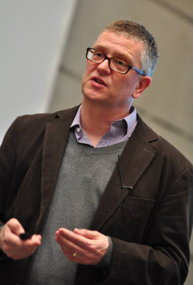 Paul Frankland合作论文数Department of Psychology, Faculty of Arts & Science, University of Toronto;Hospital for Sick Children7
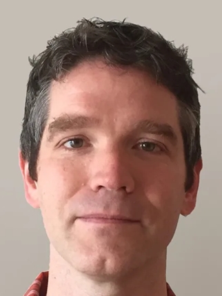 Jason Lerch合作论文数Preclinical Imaging Group, Nuffield Department of Clinical Neurosciences, Medical Sciences Division, University of Oxford;Wellcome Centre for Integrative Neuroimaging, University of Oxford5
 Sheena Josselyn合作论文数Department of Psychology, Faculty of Arts and Science, University of Toronto4
 Kuzyk Alexandra合作论文数4
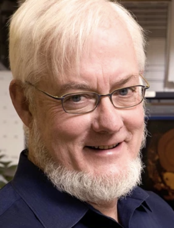 Mark Henkelman合作论文数Departments of Medical Biophysics and Medical Imaging, University of Toronto;Mouse Imaging Centre, The Hospital for Sick Children4
 Kirill Zaslavsky合作论文数Program in Neurosciences and Mental Health;Hospital for Sick Children;Program in Neurosciences and Mental Health, Hospital for Sick Children4Sara Mirali合作论文数3
 Xin Wang合作论文数Hosp Sick Children, Arthur & Sonia Labatt Brain Tumour Res Ctr, Div Neurosurg, Toronto, ON M5G 1X8, Canada3
 Kathy Boutis合作论文数Department of Paediatrics, Temerty Faculty of Medicine, University of Toronto;Division of Emergency Medicine, The Hospital for Sick Children3Crudden Johanna合作论文数3
 Tran Lina合作论文数3
 Binesh Marvasti Tina合作论文数3
 Ayan Dey合作论文数Department of Laboratory Medicine, All India Institute of Medical Sciences3
 Justin W Kenney合作论文数Department of Psychology, Temple University3
 Jacob Ellegood合作论文数Holland Bloorview Kids Rehabilitation Hospital;University of Toronto3S Markx合作论文数2Taline Naranian合作论文数2Elina K. Cook合作论文数2
 Jennifer Ji合作论文数2
 Abdullah Ishaque合作论文数Neurosci & Mental Hlth Inst, Univ Alberta2Frances Xia合作论文数Hosp Sick Children, Program Neurosci & Mental Hlth, Toronto, ON, Canada2
 Alexander Levit合作论文数Department of Psychiatry, University of British Columbia2
 Norman Rosenblum合作论文数Department of Laboratory Medicine and Pathobiology, Temerty Faculty of Medicine, University of Toronto2
 Joseph A. Gogos合作论文数Vagelos College of Physicians and Surgeons, Columbia University;Mortimer B. Zuckerman Mind Brain Behavior Institute, Columbia University2
 John Paul Oliveria合作论文数Department of Medicine, McMaster University2
 David Twa合作论文数Department of Lymphoid Cancer Research, BC Cancer Agency2
 Jordan Squair合作论文数NeuroRestore Center, École Polytechnique Fédérale de Lausanne;Brain Mind Institute, School of Life Sciences, École Polytechnique Fédérale de Lausanne;Department of Clinical Neuroscience, Lausanne University Hospital2
 Skinnider Michael A合作论文数M. G. DeGroote Institute for Infectious Disease Research, McMaster University2
 Zhou Tianwei合作论文数2Raymond Lynn A合作论文数2Genç C合作论文数2
 Barton Kristen I合作论文数Faculty of Kinesiology, University of Calgary2Maria Karayiorgou合作论文数2
 Mark Eisenberg合作论文数Department of Epidemiology, Biostatistics and Occupational Health, School of Population and Global Health, McGill University;Division of Clinical Epidemiology, Jewish General Hospital;Center for Clinical Epidemiology and Community Studies2
 D. Alan Underhill合作论文数Department of Biochemistry, McGill University2
 Gisella Vetere合作论文数Hosp Sick Children, Program Neurosci & Mental Hlth, Toronto, ON M5G 1X8, Canada2
 Steven Kushner (S.A.)合作论文数Department of Neuroscience, Erasmus MC;Department of Psychiatry, Erasmus MC;Population Neuroscience Academic Center for Excellence, Erasmus MC2
 Frank A. Provenzano合作论文数Department of Neurology, Columbia University;The Taub Institute, Columbia University2
 Matthew G.K. Benesch合作论文数School of Molecular and Systems Medicine, University of Alberta2
 Peter J Margetts合作论文数Centre for Gene Therapeutics, McMaster University2
 Lindsay S. Cahill合作论文数Department of Physics, University of Warwick2James F. Lewis合作论文数2Ju Hee Lee合作论文数1J. Dazai合作论文数1C. Laliberte合作论文数1Tianwei (Ellen) Zhou合作论文数1Andrew J Mocle合作论文数1Amirah Momen合作论文数1Ege Babadagli合作论文数1




