This paper describes the design and assembling process of two experimental structures located in the University of Minho’s School of Architecture, Art and Design, carried out between 2020 and 2022. Both were designed using the concept of active bending, assured by poles made of Glass Fibre Reinforced Polymer (GFRP) as supporting elements to membrane mesh made in PVC coated polyester. The assembling process allowed participants to experiment all the steps related with the design and construction of membrane structures with active bending elements, considering their advantages and disadvantages. The ability of membrane structures for the functional conditioning of the built environment was explored in two different scenarios, showing that they can be adequate both in outdoor as in indoor contexts. The idea of learning by doing served as the foundation for the pedagogical practices used. Each practice was discussed and tailored to the specific situation, regarding the conditions of the site, the participants involved and the functional conditioning requirements of each space.
This paper presents a house design that uses adobe in the walls and wood in the roof, a mixed building system that vernacular houses in the region where it is located already use, however it presents a contemporary strategy based on less, more natural, reused, and local materials, implying also less transport. The environmental impact analysis of the Case Study was made considering the Embodied Carbon of the construction materials used and compared with conventional building systems. In spite of presenting a much lower embodied carbon than all the conventional solutions in comparison, it turns to be more expensive.
Energy retrofitting is one of the critical elements in national energy efficiency action plans in Egypt and Portugal to achieve energy efficiency in the buildings' sector. Therefore, this study explores the techno-economic feasibility of proposed simple passive retrofitting solutions using nature-based and conventional thermal insulation materials to the existing dwellings in the traditional coastal settlements under the Mediterranean climate. Two case studies, one representing a typical coastal, traditional wooden house in the west center of Portugal and another, a conventional brick house in the Delta region in Egypt, were selected. Each case study house was simulated using energy performance assessment tools (Energy+) before and after retrofitting in both locations using similar insulation properties. The results showed that energy consumption decreased by (29% and 43%) for cooling and (23% and 45%) for heating under different circumstances. Furthermore, the shortest simple payback model was projected by 16 and 25 years, with profitability achieved in 17 and 27 years for a proposed retrofitting solution. Also, a new propinquity between Portuguese and Egyptian Architecture was brought out. (C) 2022 American Society of Civil Engineers.
The development strategy based on unlimited economic growth has been increasing extraction and consumption of natural resources and is responsible for pollutant emissions and wastes, on such a way that we risk of rapidly depleting planet earth. And although there is an awareness on optimizing available economic resources and an effort to reduce the environmental impact of production, what is certain is that pollutant emissions have not been reduced at a global level, quite the opposite. The construction industry is one of the sectors with great responsibility for greenhouse effect gas emissions. This work intends to demonstrate that, by avoiding the use of new and industrialized materials, replacing them as much as possible with reused, natural and locally available ones, such as adobe and wood is possible to reduce the environmental impact, but still being economically feasible. As a case study, a single-family housing project developed according to these principles is presented.
State-of-the-art methods for 3D reconstruction of faces from a single image require 2D-3D pairs of ground-truth data for supervision. Such data is costly to acquire, and most datasets available in the literature are restricted to pairs for which the input 2D images depict faces in a near fronto-parallel pose. Therefore, many data-driven methods for single-image 3D facial reconstruction perform poorly on profile and near-profile faces. We propose a method to improve the performance of single-image 3D facial reconstruction networks by utilizing the network to synthesize its own training data for fine-tuning, comprising: (i) single-image 3D reconstruction of faces in near-frontal images without ground-truth 3D shape; (ii) application of a rigid-body transformation to the reconstructed face model; (iii) rendering of the face model from new viewpoints; and (iv) use of the rendered image and corresponding 3D reconstruction as additional data for supervised fine-tuning. The new 2D-3D pairs thus produced have the same high-quality observed for near fronto-parallel reconstructions, thereby nudging the network towards more uniform performance as a function of the viewing angle of input faces. Application of the proposed technique to the fine-tuning of a state-of-the-art single-image 3D-reconstruction network for faces demonstrates the usefulness of the method, with particularly significant gains for profile or near-profile views.
Assessing the severity of liver fibrosis has direct clinical implications for patient diagnosis and treatment. Liver biopsy, typically considered the gold standard, has limited clinical utility due to its invasiveness. Therefore, several imaging-based techniques for staging liver fibrosis have emerged, such as magnetic resonance elastography (MRE) and ultrasound elastography (USE), but they face challenges that include limited availability, high cost, poor patient compliance, low repeatability, and inaccuracy. Computed tomography (CT) can address many of these limitations, but is still hampered by inaccuracy in the presence of confounding factors, such as liver fat. Dual-energy CT (DECT), with its ability to discriminate between different tissue types, may offer a viable alternative to these methods. By combining the "multi-material decomposition" (MMD) algorithm with a biologically driven hypothesis we developed a method for assessing liver fibrosis from DECT images. On a twelve-patient cohort the method produced quantitative maps showing the spatial distribution of liver fibrosis, as well as a fibrosis score for each patient with statistically significant correlation with the severity of fibrosis across a wide range of disease severities. A preliminary comparison of the proposed algorithm against MRE showed good agreement between the two methods. Finally, the application of the algorithm to longitudinal DECT scans of the cohort produced highly repeatable results. We conclude that our algorithm can successfully stratify patients with liver fibrosis and can serve to supplement and augment current clinical practice and the role of DECT imaging in staging liver fibrosis.
Texture analysis plays an important role in many image processing tasks. In this work, we present a texture descriptor based on the topology of excursion sets, derived from the concept of Minkowski functionals, and evaluate their usefulness in the detection of breast masses in 2D breast ultrasound images. The application includes three major stages: preprocessing, including candidate generation through computation of gradient concentration under a Fisher-Tippet noise model (in itself another contribution of the paper); texture feature extraction; and region classification using a Random Forests classifier. Performance of the proposed method is evaluated on 135 2D BUS images with 139 masses. Our method reaches 91% sensitivity with an averaged 1.19 false detections, and the proposed texture feature compares favorably against the often-used grey level co-occurrence matrices on the exact the same task.
Digital breast tomosynthesis (DBT) is a new modality that has strong potential in improving the sensitivity and specificity of breast mass detection. However, the detection of microcalcifications (MCs) in DBT is challenging because radiologists have to search for the often subtle signals in many slices. We are developing a computer-aided detection (CAD) system to assist radiologists in reading DBT. The system consists of four major steps, namely: image enhancement; pre-screening of MC candidates; false-positive (FP) reduction, and detection of MC cluster candidates of clinical interest. We propose an algorithm for reducing FPs by using 3D characteristics of MC clusters in DBT. The proposed method takes the MC candidates from the pre-screening step described in [14] as input, which are then iteratively clustered to provide training samples to a random-forest classifier and a rule-based classifier. The random-forest classifier is used to learn a discriminative model of MC clusters using 3D texture features, whereas the rule-based classifier revisits the initial training samples and enhances them by combining median filtering and graph-cut-based segmentation followed by thresholding on the final number of MCs belonging to the candidate cluster. The outputs of these two classifiers are combined according to the prediction confidence of the random-forest classifier. We evaluate the proposed FP-reduction algorithm on a data set of two-view DBT from 40 breasts with biopsy-proven MC clusters. The experimental results demonstrate a significant reduction in FP detections, with a final sensitivity of 92.2% for an FP rate of 50%.
The ability of dual-energy computed-tomographic (CT) systems to determine the concentration of constituent materials in a mixture, known as material decomposition, is the basis for many of dual-energy CT's clinical applications. However, the complex composition of tissues and organs in the human body poses a challenge for many material decomposition methods, which assume the presence of only two, or at most three, materials in the mixture. We developed a flexible, model-based method that extends dual-energy CT's core material decomposition capability to handle more complex situations, in which it is necessary to disambiguate among and quantify the concentration of a larger number of materials. The proposed method, named multi-material decomposition (MMD), was used to develop two image analysis algorithms. The first was virtual unenhancement (VUE), which digitally removes the effect of contrast agents from contrast-enhanced dual-energy CT exams. VUE has the ability to reduce patient dose and improve clinical workflow, and can be used in a number of clinical applications such as CT urography and CT angiography. The second algorithm developed was liver-fat quantification (LFQ), which accurately quantifies the fat concentration in the liver from dual-energy CT exams. LFQ can form the basis of a clinical application targeting the diagnosis and treatment of fatty liver disease. Using image data collected from a cohort consisting of 50 patients and from phantoms, the application of MMD to VUE and LFQ yielded quantitatively accurate results when compared against gold standards. Furthermore, consistent results were obtained across all phases of imaging (contrast-free and contrast-enhanced). This is of particular importance since most clinical protocols for abdominal imaging with CT call for multi-phase imaging. We conclude that MMD can successfully form the basis of a number of dual-energy CT image analysis algorithms, and has the potential to improve the clinical utility of dual-energy CT in disease management.
We develop a novel deformable atlas method for multistructure segmentation that seamlessly combines the advantages of image-based and atlas-based methods. The method formulates a probabilistic framework that combines prior anatomical knowledge with image-based cues that are specific to the subject's anatomy, and solves it using expectation-maximization method. It improves the segmentation over conventional label fusion methods especially around the structure boundaries, and is robust to large anatomical variation. The proposed method was applied to segment multiple structures in both normal and diseased brains and was shown to significantly improve results especially in diseased brains.
Nowadays, there is an increasing need for alternative construction technologies that allow, among others, reducing construction wastes and energy consumption during the buildings' life-cycle. In this context, this paper presents results of a research project which goal is to develop an innovative solution for partition walls. This solution is based on a masonry block made of an eco-efficient new composite material. The composite material used in the production of the blocks results from the combination of three industrial by-products, namely: flue-gas desulfurization gypsum; granulated cork; and textile fibers resulting from the tyre recycling process. Besides the raw materials, the innovation of the solution results also from the new design of the block, whose shape enables the positioning of the infra-structures during the assembling of the indoor wall.In this paper, details of the design process of the block and both the optimization of the composition of the material and construction technology are provided. The validation of the partition wall solution in the point of view of mechanical, thermal and acoustic performance was also carried out.From the results obtained, it is possible to conclude that the solution fulfils all the requirements of structural stability adequate for this type of wall. In terms of thermal performance, the proposed solution presents very good behaviour, being the acoustic performance slightly lower than the traditional solution. (C) 2013 Elsevier Ltd. All rights reserved.
The diagnosis and treatment of fatty liver disease requires accurate quantification of the amount of fat in the liver. Image-based methods for quantification of liver fat are of increasing interest due to the high sampling error and invasiveness associated with liver biopsy, which despite these difficulties remains the gold standard. Current computed tomography (CT) methods for liver-fat quantification are only semi-quantitative and infer the concentration of liver fat heuristically. Furthermore, these techniques are only applicable to images acquired without the use of contrast agent, even though contrast-enhanced CT imaging is more prevalent in clinical practice. In this paper, we introduce a method that allows for direct quantification of liver fat for both contrast-free and contrast- enhanced CT images. Phantom and patient data are used for validation, and we conclude that our algorithm allows for highly accurate and repeatable quantification of liver fat for spectral CT.
The use of image-based automatic defect recognition (ADR) systems in a production line often requires strict processing-time specifications. On the other hand, the typical high-performance requirement of such system calls for the use of sophisticated, computationally-complex algorithms. Addressing the conflicting requirements of fast throughput and high detection performance is a significant challenge. In this paper we present a 3D learning-based ADR approach for industrial parts. The proposed method first extracts defect candidate regions using morphological closing and template matching. Then a local registration-based approach is utilized to produce accurate defect segmentation mask. Finally, 29 features including geometric features and texture features derived from grey level co-occurrence matrix are calculated for each candidate region, and a fast random forests classifier is used to classify the candidate regions as defect or defect-free. This approach was developed into a fully automated system for detecting casting defects in aluminum industrial parts depicted in 3D Computed Tomographic (CT) images. The system was tested on 31 images with 49 cavities and porosities defects, achieving a sensitivity of 94% with an average 3.5 false detections per part.
We introduce an automated method for the 3D tracking of carotids acquired as a sequence of 2D ultrasound images. The method includes an image stabilization step that compensates for the cardiac and respiratory motion of the carotid, and tracks the carotid wall via a shape and appearance model trained from representative images. Envisaging an application in automatic detection of plaques, the algorithm was tested on ultrasound volumes from 4,000 patients and its accuracy was evaluated by measuring the distance between the location of more than 4,000 carotid plaques and the location of the carotid wall as estimated by the proposed algorithm. Results show that the centroids of over 95% of the carotid plaques in the dataset were located within 3 mm of the estimated carotid wall, indicating the accuracy of the tracking algorithm.
 Rahul Bhotika合作论文数lululemon25
 James G. Miller合作论文数Laboratory of Ultrasonics, Department of Physics, Arts & Sciences, Washington University in St.Louis11
 Roberto Cipolla合作论文数Department of Engineering, University of Cambridge;Cambridge Research Laboratory, Toshiba Europe Ltd9Fei Zhao合作论文数One Research Circle, GE Global Research8
 Dirk R. Padfield合作论文数GE Research Center8Peter Lamb合作论文数7
 Robert Kaucic合作论文数Amazon6Ali Can合作论文数Amazon Global Robotics6Saad Sirohey合作论文数GE Healthcare5
 Xiaodong Tao合作论文数IFlyHealth5thomas baby sebastian合作论文数4matthew william turek合作论文数4robert august kaucic合作论文数4
 Shubao Liu (刘书宝)合作论文数Chongqing Key Lab Neurobiol, Third Mil Med Univ4Peter H. Tu合作论文数Computer Science & Engineering department at the University of Washington4
 Dushyant Sahani合作论文数Department of Radiology, University of Washington4
 Wesley D. Turner合作论文数One Research Circle, GE Global Research4matthieu denis ferrant合作论文数3
 Aires Camoes合作论文数3xiaoye wu合作论文数3Brian Thomsen合作论文数3
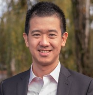 Tony Pan合作论文数Modern Hydrogen;Unreasonable;YPO3Graça Vasconcelos合作论文数Department of Civil Engineering, University of Minho3
 Harvey E. Cline合作论文数General Electric Company3
 András Kriston合作论文数3
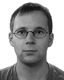 Nils Krahnstoever合作论文数3jerome knoplioch合作论文数2Clara Vieira合作论文数2glenn robert shaffer合作论文数2joseph manuel portaz合作论文数2arunabha roy合作论文数2Masayuki Kudo合作论文数2Mahnaz Maddah合作论文数2
 Ricardo Mateus合作论文数2sandeep dutta合作论文数2Kosuke Sasaki合作论文数2
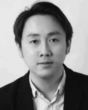 Qi Song合作论文数CuraCloud Corp, Seattle, WA 98104 USA2
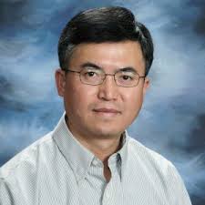 Zhaohui Sun合作论文数Kitware, Inc.2Li Xiaoxing合作论文数2
 Srikanth Suryanarayanan合作论文数2
 Laszlo Rusko合作论文数2
 Paulo B. Lourenco合作论文数Department of Civil Engineering, University of Minho;Institute for Sustainability and Innovation in Structural Engineering2
 Glen Brooksby合作论文数GE Global Research2
 Ajay Gopinath合作论文数Department of Computer Sciences and Institute of Computational;Engineering & Sciences;University of Texas at Austin;Department of Computer Sciences and Institute of Computational, University of Texas at Austin2Rakesh Mullick合作论文数John F. Welch Technology Center2Xiaofeng Liu合作论文数Departments of Computer Science,2
 Ricardo Avila合作论文数GE Corporate Research & Development2
 Jens Rittscher合作论文数Nuffield Department of Medicine, Harris Manchester College, University of Oxford2




