Recent studies have described acquired ring chromosomes (aRCs), marker chromosomes, and extrachromosomal DNA (ecDNA) in various tumors. However, the genomic copy number aberrations (CNAs) and gene rearrangements within these aRCs require further genomic analysis, and the knowledge base to interpret their clinical implications remains largely unformed. A working group was organized by the International Consortium of Human Ring Chromosomes (ICHRC) to conduct a systematic evidence review of the role of aRCs in hematologic malignancies. This retrospective review summarizes the current molecular cytogenetic and genomic technologies for aRC analysis, presents an overview of the types of aRCs in various hematological malignancies, and provides evidence to support cytogenomic analysis and diagnostic interpretation. The findings from this study recommend an integrated cytogenomic analysis for aRCs and prompt further functional analyses to elucidate the molecular mechanisms for disease causation and targeted treatment.
Molecular characterization of balanced complex chromosomal rearrangements (CCR) aids in understanding the pathophysiological mechanism and corresponding genotype-phenotype correlations. The present case describes a male child with intellectual disability, developmental delay, and dysmorphism. A thorough and sequential genetic evaluation using karyotyping, fluorescence in situ hybridization (FISH), chromosomal microarray (CMA), and long read sequencing (LRS) identified a genomically balanced CCR. The CCR involved eight chromosomes, the largest to be documented till date for chromoanagenesis and being balanced despite the high level of complex chromosomal involvement. Translocations accounted for the majority of the rearrangements along with an insertion, inversion, and a small deletion likely driven by chromoplexy. Although the CCR was genomically balanced, it may still result in functionally significant genomic consequences including gene disruptions, gene fusions, and position effects. Long read whole genome sequencing using PacBio was used for breakpoint characterization that revealed three protein-coding genes to be disrupted, namely, NLGN4X, LAMA4, and ALG6. Of these, a candidate association was observed for the NLGN4X gene with the intellectual disability phenotype reported in the proband, which is likely due to disruption of transcription and nonsense mediated decay. We show combinatorial application of advanced genomic technologies with orthogonal cytogenetic techniques in delineating balanced CCRs and understanding the biological and potential clinical implications of balanced yet functionally disruptive CCRs.
This case demonstrates the value of cell-free DNA (cfDNA) screening for detecting subchromosomal microdeletions in fetuses with non-specific prenatal screening abnormalities and no overt structural malformations on ultrasound; CMA and karyotyping confirmation and integrated genetic counseling are essential for diagnosing 5q14.3q15 deletion-related BBSOAS and guiding parental decision-making.
Background Artificial intelligence (AI)-assisted image capture and signal pattern classification for digital fluorescence in situ hybridization (FISH) are important progression toward analytical automation in clinical cytogenetics laboratories. We provide a systematic approach for the validation and integration of digital FISH using the Applied Spectral Imaging (ASI) on FISH panels of myelodysplastic neoplasms (MDS), acute myeloid leukemia (AML), chronic lymphocytic leukemia (CLL), and B-cell lymphoma. Methods Analytical validity was evaluated using normal cutoff values by beta-inverse function on FISH scoring from 20 normal cases and reproducibility by coefficient of variation (CV) from three normal cases. Analytical accuracy was assessed by linear regression to correlate the percentages of abnormal cells between digital and manual FISH scoring and by concordance of FISH with karyotyping results in abnormal cases. Results Normal cutoff values showed comparable and acceptable ranges of 3–11%, 6–13%, and 4–8% from manual, digital, and combined manual/digital scoring, respectively. Intra-assay and inter-assay reproducibility showed CV in the range of 0-3.6%. Abnormal-cell percentages between manual and digital scores showed strong correlation with R 2 values of 0.76–0.93. For FISH tests with five or more abnormal cases, concordant abnormal patterns between FISH and chromosome results showed a range of 76%-100% with an overall concordance of 87%. Discordant cases were primarily attributable to cryptic deletions that were undetectable by conventional karyotyping. Conclusion This study demonstrated that validated digital FISH image capture and re-classification by a technologist integrated with independent manual scoring could ensure accuracy and improve efficacy for the detection of recurrent abnormalities in various leukemias.
Trisomy 8 mosaicism (T8M) syndrome is a rare aneuploidy condition affecting 1/25,000-50,000 live births. Affected individuals have highly variable phenotypes from very mild dysmorphism to severe structural anomalies caused by chromosomal mosaicism and possibly undetected molecular aberrations. The utilization of chromosome microarray analysis (CMA) and exome sequencing (ES) in clinical laboratories enable the identification of genomic copy number imbalances and pathogenic gene variants. We presented one patient with a double aneuploid mosaic pattern of Monosomy X and Trisomy 8 for a compound phenotype of Turner syndrome (TS) and T8M syndrome, the second patient with T8M and a mosaic pathogenic variant in the PTEN gene detected by ES, and the third patient with typical phenotypic constellation of malformations with no other genetic aberrations detected by CMA and ES. Classification of mosaic findings was provided using a recommended six-attribute scheme. Review of the literature summarized cases of T8M with concomitant molecular defects of a deletion at 22q11.2 and pathogenic variants in the SALL1, RECQL4, NF1, CASK, and PAH genes. These observations indicated that integrated cytogenetic and genomic analyses should be offered to patients with phenotypic abnormalities outside the spectrum of the T8M syndrome for comprehensive laboratory diagnosis and clinical management.
Modern genetics began with the rediscovery of Mendelian inheritance in 1900. The discovery of the DNA double helix in 1953 laid the foundation of molecular genetics. The Human Genome Project from 1990 to 2003 provided a fundamental genetic blueprint of human biology, with a lasting and expanding impact on the development of numerous genomic, multi-omics, and gene-editing technologies for precision medicine. To highlight the contributions of Chinese geneticists with work and training experience across the Pacific, this historical review presents the breakthrough discoveries by prominent Chinese geneticists during the early pioneering generation (1920-1950), the molecular genetics era (1951-1990), the transition to human and medical genetics, and current progress in genetic and genomic medicine (1991-present). These world-renowned geneticists have made significant contributions to understanding the molecular mechanisms that regulate biological processes, the etiology and pathogenesis of genetic diseases, and the development of novel genetic engineering and therapeutic approaches. They also played leading roles in various professional organizations, academic centers, and industry. Standing on the shoulders of these prominent Chinese geneticists, current and future generations of geneticists and researchers could have a broader vision and more deeply explore the frontiers of genetics.
Induced pluripotent stem cells (iPSCs) hold great promise for the treatment of cardiovascular diseases through cell-based therapies, but these therapies require extensive preclinical testing that is best done in species-in-species experiments. Pigs are a good large animal model for these tests due to the similarity of their cardiovascular system to humans. However, a lack of adequate pig iPSCs (piPSCs) that are analogous to human iPSCs has greatly limited the potential usefulness of this model system. Herein, transgene-free piPSCs with true pluripotency were generated by using reprogramming factors in an optimized pig pluripotency medium. Using an effective differentiation protocol, piPSCs were used to derive endothelial cells (ECs) which displayed EC markers and functionality comparable to native pig ECs. Further, piPSC-ECs demonstrated suitability for vascular tissue engineering, producing a tissue engineered vascular conduit (TEVC) that displayed the upregulation of flow responding markers. In an in vivo functional study, these piPSC-EC-TEVCs maintained the expression of endothelial markers and prevented thrombosis as interposition inferior vena cava grafts in immunodeficient rats. The piPSCs described in this study open up the possibility of unique preclinical species-in-species large animal modeling for the furtherance of modeling of cell-based cardiovascular tissue engineering therapies. Statement of Significance While there has been significant progress in the development of cellularized cardiovascular tissue engineered therapeutics using stem cells, few of them have moved into clinical trials. This is due to the lack of a robust preclinical large animal model to address the high safety and efficacy standards for transplanted therapeutics. In this study, pig stem cells that are analagous to human's were created to address this bottleneck. They demonstrated the ability to differentiate into functional endothelial cells and were able to create a tissue engineered therapeutic that is analogous to a human therapy. With these cells, future experiments testing the safety and efficacy of tissue engineered constructs are possible, bringing these crucial therapeutics closer to the patients that need them.
We report a case of a 61-year-old male patient with a complex hematologic history including pre-B acute lymphocytic leukemia, allogeneic stem cell transplant, and newly treated colon cancer followed by diagnosis of high-risk myelodysplastic syndrome (MDS), who exhibited abolishment of D antigen production. The patient's red blood cells (RBCs), which originally typed as group A, D+, again typed as group A, D+, after receiving stem cells from a female donor with the same blood type. The reactivity of the patient's RBCs was strong when tested with anti-D reagent until he was treated for colon cancer. Within 6 months of diagnosis of cancer, he developed a mixed-field D typing result followed by a complete D- phenotype over the course of a few weeks, coinciding with the new diagnosis of MDS and initiation of immunosuppressive therapy. Polymerase chain reaction (PCR) testing revealed no weak or partial D variants, and subsequent Sanger sequencing confirmed the presence of a conventional RHD gene. A more focused analysis by chromosomal microarray identified deletions involving the RHD locus, supporting the hypothesis that active MDS disrupted gene expression. The patient received another stem cell transplant, this time from a group AB, D+ male donor. In a short period of time, MDS re-emerged along with re-identification of microarray mutations encompassing RHD in female cells (from the first donor) even after the second stem cell transplant. However, D expression remained strong, underscoring the presence of enough male donor RHD expression to maintain the D+ phenotype.
List of somatic SNV/Indel present in the concordance study samples and reported by at least one assay.
Introduction B-cell acute lymphoblastic leukemia (B-ALL) is a hematologic malignancy characterized by immature B-lymphoid cell proliferation. Conventional cytogenetic analyses, including fluorescence in situ hybridization (FISH) and chromosome analysis, play critical roles in diagnosis, but these methods may miss cryptic structural variants. Microarray technology offers higher-resolution genome-wide analysis, however, it has limitations in identifying balanced structural variants. Recent advancements in long-read sequencing enable detection of complex genomic rearrangements. Here, we present a pediatric B-ALL case with complex rearrangements and gene fusions detected using multiple genomic techniques. Case Presentation A 15-year-old boy with no previous medical history presented at the emergency department with fatigue, pale skin, weakness, fever, and nausea. Blood tests revealed anemia and leukocytosis with 93% of blast cells. Flow cytometry confirmed the diagnosis of B-lymphoblastic leukemia, showing that 96% of the analyzed blood cells were lymphoblasts with the following immunophenotype: CD45(dim)+, CD34(var), CD19(dim)+, CD10+, CD20(var), CD79a+, CD38+, CD123+, CD58(dim)+, cytoCD3-, CD5-, TDT(dim)+, MPO-, CD117-, CD13-, CD33-, glyco-, CD41a-, CD4-, CD8-, and CD56-. Approximately 60% of the lymphoblasts expressed CD22+. As a newly diagnosed high-risk B-ALL patient, he was enrolled in the Children's Oncology Group (COG). Diagnostic Workup and Management G-banding revealed an abnormal clone with the majority of cells exhibiting a rearrangement between chromosomes 7 and 20 46,XY,t(7;20)(q34;q13.3)[18]/46,XY[2]. B-ALL FISH probes did not identify any remarkable findings. Microarray analysis identified a deletion at 9p21.3 encompassing the CDKN2A and CDKN2B genes, and revealed contiguous deletions and a suspected duplication at the 9q34.12q34.2 region, affecting the ABL1, NUP214, and TSC1 genes. The microarray result suggested the presence of a NUP214-ABL1 gene fusion. Long-read sequencing revealed cryptic rearrangements involving NUP214-ABL1 and TSC1-ABL1 at the 9q34.12q34.2 region. In addition, the VAPB::TRBV30 fusion was found to correspond with the observed karyotypic abnormality. These findings were validated through RNA transcript evidence. Importantly, the identification of the NUP214-ABL1 fusion highlighted a clinically actionable target for tyrosine kinase inhibitor therapy, specifically imatinib. Discussion and Conclusion Microarray and long-read sequencing identified deletions in CDKN2A, CDKN2B, and IKZF1, all recognized as poor prognostic markers in ALL. The identification of NUP214-ABL1 and ABL1-TSC1 fusions highlights the complex genomic landscape with multiple rearrangements and underscores the importance of advanced techniques in uncovering hidden genetic alterations that may serve as therapeutic targets. This study emphasizes the need for integrated sequencing approaches to better characterize major clones and their roles, ultimately advancing precision oncology in B-ALL.
Purpose:Ring chromosomes (RCs) are rare cytogenetic abnormalities involving copy-number variants and chromosome instability. Identifying the breakage-fusion sequences of RCs at nucleotide-level resolution can elucidate the cytogenomic rearrangements and ring formation mechanisms. Methods:This study used short-read genomic sequencing (srGS) and long-read genomic sequencing (lrGS) alongside the telomere-to-telomere reference genome to characterize the breakage-fusion events of 17 RC cases. Results:Complete RCs without loss of euchromatin by a fusion of subtelomeric or telomeric regions were noted in a RC14 and a RC20. Incomplete RCs with intrachromosomal copy-number variants were noted in 15 cases, including a RC3, a RC4, 4 RC13s, a RC14, 3 RC18s, a RC21, 3 RC22s, and an RCY. srGS defined breakage-fusion sequences in single-copy sequences, and lrGS mapped subtelomeric and pericentric repetitive sequences using the telomere-to-telomere reference genome. The breakage-fusion sequences revealed ring formation mechanisms by intrastrand nonhomology end joining in 5 RCs, microhomology-mediated end joining in 8 RCs, and microhomology-mediated break-induced replication in 4 RCs. Conclusion:This study demonstrated the analytic validity and diagnostic utility of srGS and lrGS in delineating the genomic rearrangements in RCs for better interpreting clinico-cytogenomic correlations and further analysis of RC behavior in cell cycles.
Summary and list of detected and reported alterations by alteration type (CNV, SNV/Indel) and assay (both assays, Central Lab (CL) or Assay H only, Pilot Lab only). For both detected and reported variants, positive percent agreement (PPA) with Pilot Lab and then CL as reference along with average percent agreement (APA), which is a weighted average of each PPA, were calculated.
Summary of detected and reported CNV alterations by assay (both assays, Central Lab (CL) or assay H only, Designated Lab (DL) assay only). For both detected and reported variants, positive percent agreement (PPA) with DL and then CL as reference along with average percent agreement (APA), which is a weighted average of each PPA, were calculated.
Scatter plots of continuous CN results obtained by NGS Central Lab (CL) or assay H, or NGS assay U and non-NGS techniques: A) ddPCR, B) microarray, C) FISH (Raw values) and D) FISH (Ratio). The solid line demonstrates a Deming regression line, while the dashed line demonstrates the identity line.
Chromosomal microarray analysis (CMA) detects pathogenic copy-number variants (pCNVs) and regions of homozygosity (ROHs) in prenatal genetic analysis. This study evaluates the clinical significance of ROH detection in prenatal settings. We reviewed 178 fetuses with ROH detected by CMA among 20,546 fetuses from 2015 to 2023. Clinical and laboratory results, including ultrasound anomalies, cell-free DNA (cfDNA) screening, karyotyping, exome sequencing (ES), and methylation-specific multiplex ligation-dependent probe amplification (MS-MLPA), were analyzed. These 178 fetuses with ROH accounted for 0.87% of prenatal cases. Among them, 24.2% had positive cfDNA screening results, and 52.8% underwent follow-up ES, trio CMA, and MS-MLPA. Follow-up studies detected pathogenic homozygous variants within ROH in two fetuses and uniparental disomy (UPD)-related diseases in five fetuses. Our results and findings from the other five large prenatal case series from literature indicated that ROH detection in prenatal CMA has a baseline positive predictive value of 2.7% for autosomal-recessive disorders, 9.6% for UPD-related diseases, and 0.04% overall additive diagnostic yield. These findings support the use of ES and MS-MLPA for follow-up testing and provide guidance for genetic counseling in fetuses with ROH.
B-cell acute lymphoblastic leukemia (B-ALL) is a heterogeneous hematologic malignancy caused by diverse genetic alterations. While cytogenetic methods such as karyotyping and FISH are routinely used in diagnostics, cryptic and novel oncogenic gene fusions often go undetected. We report a 15-year-old male diagnosed with high-risk B-ALL, presenting with anemia, leukocytosis, and significant lymphoblast burden. Initial karyotyping identified an abnormal clone with a t(7;20)(q34;q13.3) and FISH detected a partial deletion of the ABL1 gene. Chromosome microarray analysis revealed a deletion at 9p21.3 encompassing CDKN2A/CDKN2B and several contiguous copy number aberrations at 9q34.12q34.2. Further long-read genomic sequencing uncovered cryptic NUP214::ABL1 and ABL1::TSC1 fusions, as well as a novel VAPB::TRBV30 rearrangement from the t(7;20). RNA sequencing confirmed the transcripts for both ABL1 fusions and a noncoding rearrangement involving the TCRB locus. Notably, the presence of NUP214::ABL1 identified a clinically actionable target, supporting the use of tyrosine kinase inhibitors (TKIs) such as imatinib. This case underscores the critical role of integrated sequencing approaches in identifying cryptic genetic alterations in B-ALL for precise classification of oncogenic drivers and for targeted therapeutic strategies to improve patient outcomes.
Introduction Mature T-cell lymphomas, including aggressive nodal PTCL, advanced stage CTCL, and extranodal cytotoxic lymphomas, are associated with poor outcomes. While karyotype (KT) abnormalities are frequently detected in these diseases, limited data exist on their prognostic significance. We evaluated the impact of KT abnormalities, including complex KT and specific chromosomal lesions, along with clinical and demographic factors, on overall survival (OS) in a single-center cohort. Methods We retrospectively reviewed 189 patients (pts) diagnosed with T-cell lymphoma who had successful metaphase KT at diagnosis from peripheral blood (PB), bone marrow (BM), or tumor tissue. T-cell lymphomas were classified according to WHO 2022 into nodal, extranodal/cytotoxic, and cutaneous subtypes. CTCLs were stratified into indolent (Early-stage CTCL, IA–IB) and aggressive (Sézary syndrome, transformed mycosis fungoides). KTs classified as normal, abnormal, and complex (≥3 abnormalities), and recurrent abnormalities were recorded. Baseline clinical and lab data were collected. OS was estimated using the Kaplan-Meier (KM) method with 95% confidence intervals (CI). Univariate Cox proportional hazards (CPH) model identified factors associated with OS, and significant variables were entered into multivariate CPH models. Results The median age was 66 years (IQR 55-74); 65% were male and 35% were female. Overall, 58% of pts were alive at analysis. Nodal PTCL was the most common subtype (113 pts, 60%), followed by early CTCL (30, 16%), aggressive CTCL (24, 13%), and extranodal lymphomas (22, 12%). KT was obtained from 166 BM (86%), 26 PB (13%), and 2 tissue (1%) specimens. Among abnormal KTs, 63% were from BM, 35% from PB, and 2% from tissue. Specific abnormalities included gain of chr1 (5%), gain of chr3 (5%), gain of chr7 (5%), gain of chr8 (5%), monosomy 5 or del(5q) (4%), monosomy 9 or del(9q13q22) (4%), Monosomy 10 (4%), loss of chrY (5%), and presence of marker chromosomes (5%).Of the 189 pts, 48 (25%) had an abnormal KT, while 141 (75%) had a normal KT. Complex KT was observed in 31 (16%) pts. Abnormal non-complex and complex KTs were seen respectively in: aggressive CTCL (8%, 42%), nodal PTCL (4%, 13%), extranodal (9%, 14%), and early CTCL (27%, 10%). KM survival analysis showed a median OS for the entire cohort of 95 months (95% CI: 65-245). Pts with a normal KT had a median OS of 143 months (95% CI: 81-inf) compared with 34 months (95% CI: 18-83) for those with an abnormal KT. Pts without a complex KT had a median OS of 144 months (95% CI: 85-inf), whereas those with a complex KT had 14 months (95% CI: 4-25). For specific abnormalities, pts with gain of chr7 had OS of 3 months, gain of chr8 had 19 months, monosomy 5 or del(5q) had 4 months, loss of chrY had 30 months, gain of chr1 had 4.5 months, gain of chr3 had 7.5 months, monosomy 9 or del(9q13q22) had 2 months, monosomy 10 had 17 months, and marker chromosome presence had 5.5 months. Univariate CPH identified complex KT (HR = 3.9, p < 0.001), gain of chr1 (HR = 5.7, p < 0.001), gain of chr3 (HR = 4.6, p < 0.001), monosomy 5 or del(5q) (HR = 4.6, p < 0.001), gain of chr7 (HR = 4.2, p < 0.001), gain of chr8 (HR = 3.3, p < 0.001), monosomy 9 or del(9q13q22) (HR = 4.9, p < 0.001), monosomy 10 (HR = 2.8, p = 0.008), and marker chromosomes (HR = 4.6, p < 0.001) as adverse factors. Higher platelet count was protective (HR = 0.9, p < 0.001), while greater number of abnormalities increased hazard (HR = 1.1, p < 0.001). In multivariate CPH model, complex KT (HR = 3.1, 95% CI: 1.2-8.2, p = 0.019), monosomy 9 or del(9q13q22) (HR = 4.4, 95% CI: 1.2-15.4, p = 0.022), gain of chr1 (HR = 3.5, 95% CI: 1.1-11.8, p = 0.042), and low platelet count (HR = 0.98, 95% CI: 0.97-0.99, p = 0.006) remained independent predictors of poor OS. Conclusion: We demonstrate that cytogenetic abnormalities detected in PB or BM, most notably complex KTs, confer a markedly poor prognosis in aggressive T-cell lymphomas. The integration of KT data with clinical parameters, such as platelet count, offers a powerful approach to refine risk stratification, identify biologically high-risk subgroups, and potentially guide personalized therapeutic strategies in this challenging disease spectrum. To our knowledge, this is one of the largest single-center studies assessing the prognostic significance of KT in mature T-cell lymphomas, providing novel insights into the role of specific chromosomal lesions in patient outcomes.
Background: Copy number variants of uncertain significance (CNVus) from chromosome microarray analysis (CMA) presents unresolved challenges for clinical geneticists, genetic counselors, and patients. We performed a systematic reevaluation of reported CNVus and reanalysis of selected CNVus by whole genome sequencing (WGS) to assess the diagnostic value and clinical impact on CNVus reclassification. Methods: We retrospectively reviewed 5277 consecutive pediatric cases by CMA from the Yale Clinical Cytogenetics Laboratory over a 13-year period. Reevaluation was performed on all reported CNVus following current ACMG/ClinGen guidelines. Reanalysis by WGS was applied to selected cases for reclassification of CNVus. Results: A total of 567 CNVus from 480 cases were reported, which accounted for 9.1% of pediatric cases. A total of 4 CNVus in 4 cases (0.8%, 4/480) were reclassified to pathogenic/likely pathogenic CNVs (pCNVs/lpCNVs); while 23 CNVus in 23 cases (4.8%, 23/480) were reclassified to benign/likely benign CNVs (bCNVs/lbCNVs). The overall rate of reclassification was 5.6%. WGS performed on selected cases further defined breakpoints and ruled out additional causative genetic variants. Conclusions: The results from this study demonstrated the diagnostic value of periodic reevaluation of CNVus and reanalysis by WGS in an interval of 3–5 years and provided evidence to support standardized laboratory reevaluation and reanalysis.
Scatter plots of continuous copy number results obtained from Central Lab (CL) or assay H and Designated Lab (DL) assay. The solid line demonstrates a Deming regression line, while the dashed line demonstrates the identity line. Black points denote variants which were reported by both assays, light green is for variants detected by both assays but not reported by at least one assay, and orange points denote variants that were only detected by CL. Circle is for variants detected by both assays. Triangle is for a variant detected by both assays but reported only by DL. Diamond is for a variant reported only by CL.
 Hongyan Chai (柴红燕)合作论文数Dept Genet, Yale Univ33Autumn J Diadamo合作论文数24Wen Jiadi合作论文数Department of Pathology and Laboratory Medicine, University of British Columbia;Child &Family Research Institute21Fang Xu合作论文数18
 Hui ZHANG合作论文数16Brittany Grommisch合作论文数13
 Bixia Xiang合作论文数11Weiqing Wu合作论文数10
 Jiansheng Xie (谢建生)合作论文数深圳市妇幼保健院10Jn Thompson合作论文数9
 Qinghua Zhou (周庆华)合作论文数College of Life Science and Technology, Jinan University9
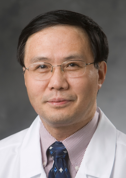 Yong-Hui Jiang合作论文数School of Medicine, Yale University9Qian Geng合作论文数8QiPing Hu (胡启平)合作论文数8
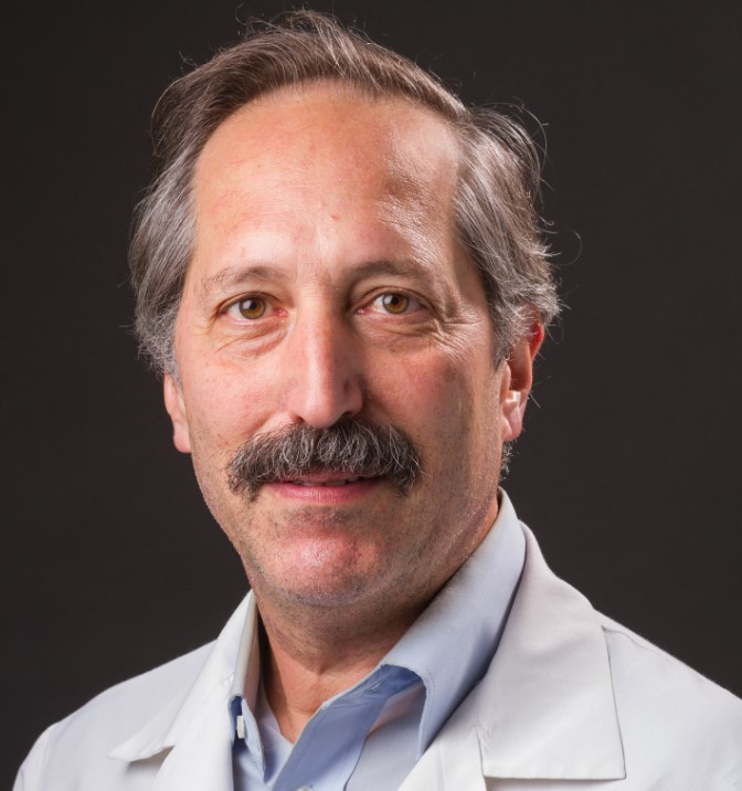 Allen Bale合作论文数NA Diagnostics Laboratory, Department of Genetics, Yale School of Medicine, Yale University;Yale Cancer Center, Yale School of Medicine, Yale University;Smilow Cancer Hospital, Yale New Haven Children’s Hospital8Zhiyong Xu合作论文数7
 Pei Hui合作论文数Pathology Faculty, Yale School of Medicine, Yale University6
 Liehr Thomas合作论文数Institut fur Humangenetik, Universitätsklinikum Jena6Michele Spencer-Manzon合作论文数5FuWei Luo (罗福薇)合作论文数5
 Gang Peng合作论文数Department of Medical and Molecular Genetics, School of Medicine, Indiana University5Amato Katherine合作论文数5Youwen Liu (刘友文)合作论文数School of Materials Science and Engineering, Huazhong University of Science and Technology5
 Hongyu Zhao (赵宏宇)合作论文数School of Public Health, Yale University5
 Yunji Meng合作论文数Department of Applied Physics, Nanjing University of Aeronautics and Astronautics5Mei Ling Chong合作论文数4
 Yang Liu合作论文数4
 Xiaolei Xie (谢小雷)合作论文数Guangzhou Medical University4
 Weizhen Ji合作论文数Department of Pediatrics, Yale School of Medicine, Yale University4
 Yiping Shen (沈亦平)合作论文数Boston Children's Hospital;Department of Neuroscience, Harvard Medical School, Harvard University4
 Marilyn M. Li合作论文数Perelman School of Medicine, University of Pennsylvania;The Children's Hospital of Philadelphia4
 Maurice Mahoney合作论文数4Deqiong Ma合作论文数4Katherine Wilcox合作论文数3Bs Brittany Grommisch合作论文数3
 Michael R Rossi合作论文数3Daniel Dykas合作论文数3Renu Bajaj合作论文数3Kangmo Lu合作论文数3Cheng-Hua Cui合作论文数3Bs Autumn J. DiAdamo合作论文数3Shengming Wu合作论文数3
 Anna Szekely合作论文数Department of Neurology, Yale School of Medicine, Yale University3
 Guilin Wang合作论文数School of Medicine, Yale University3
 Francine M. Foss合作论文数Department of Internal Medicine, Yale School of Medicine, Yale University3Natalia Buza合作论文数Pathology Faculty, Yale School of Medicine, Yale University3虞京葳合作论文数加州大学旧金山分校3
 Patrick Gallagher合作论文数School of Medicine, Yale University3Michael Girardi合作论文数3




