Almost fifty years after the identification of ataxia telangiectasia (A-T) as a radiosensitive disorder and thirty years following the identification of ataxia telangiectasia mutated (ATM) as the defective gene, clinicians and scientists gathered at Loughborough University, UK from June 25th -27th 2025 for an Ataxia Telangiectasia Clinical Research Conference. The mix of expertise of clinicians and scientists with basic and translational expertise ensured that a focus was on how to exploit our knowledge of ATM’s function to clinical benefit. Considerable emphasis was placed on the role of ATM in the DNA damage response and the consequences in this multisystem disease, including the neurodegenerative phenotype. The increasingly recognized role of ATM in oxidative stress was also considered and how it was pertinent to mitochondrial dysfunction, metabolic abnormalities and energy metabolism in A-T. The implications of these roles of ATM in protecting the genome/cell and the development of new technology, such as organoids, were widely discussed in the clinical setting of patients with A-T. An important contribution to the meeting was the description of pathways /mechanisms that have led to the development of therapeutic approaches for A-T including the use of specific antisense oligonucleotides to restore ATM function in patients; delivery of full-length ATM cDNA to A-T cells; eDSP (formerly EryDex) that encapsulates dexamethasone sodium phosphate in a patient’s own red blood cells and the use of small molecules (triheptanoin, nicotinamide riboside and N-acetyl leucine) to correct mitochondrial and metabolic function.
Inborn errors of T cell development present a pediatric emergency in which timely curative therapy is informed by molecular diagnosis. In 11 affected patients across four consanguineous kindreds, we detected homozygosity for a single deleterious missense variant in the gene NudC domain–containing 3 ( NUDCD3 ) . Two infants had severe combined immunodeficiency with the complete absence of T and B cells (T - B - SCID), whereas nine showed classical features of Omenn syndrome (OS). Restricted antigen receptor gene usage by residual T lymphocytes suggested impaired V(D)J recombination. Patient cells showed reduced expression of NUDCD3 protein and diminished ability to support RAG-mediated recombination in vitro, which was associated with pathologic sequestration of RAG1 in the nucleoli. Although impaired V(D)J recombination in a mouse model bearing the homologous variant led to milder immunologic abnormalities, NUDCD3 is absolutely required for healthy T and B cell development in humans.
New development and optimization of oncologic strategies are steadily increasing the number of long-term cancer survivors being at risk of developing second primary neoplasms (SPNs) as a late consequence of genotoxic cancer therapies with the highest risk among former childhood cancer patients. Since risk factors and predictive biomarkers for therapy-associated SPN remain unknown, we examined the sensitivity to mild replication stress as a driver of genomic instability and carcinogenesis in fibroblasts from 23 long-term survivors of a pediatric first primary neoplasm (FPN), 22 patients with the same FPN and a subsequent SPN, and 22 controls with no neoplasm (NN) using the cytokinesis-block micronucleus (CBMN) assay. Mild replication stress was induced with the DNA-polymerase inhibitor aphidicolin (APH). Fibroblasts from patients with the DNA repair deficiency syndromes Bloom, Seckel, and Fanconi anemia served as positive controls and for validation of the CBMN assay supplemented by analysis of chromosomal aberrations, DNA repair foci (γH2AX/53BP1), and cell cycle regulation. APH treatment resulted in G2/M arrest and underestimation of cytogenetic damage beyond G2, which could be overcome by inhibition of Chk1. Basal micronuclei were significantly increased in DNA repair deficiency syndromes but comparable between NN, FPN, and SPN donors. After APH-induced replication stress, the average yield of micronuclei was significantly elevated in SPN donors compared to FPN (p = 0.013) as well as NN (p = 0.03) donors but substantially lower than for DNA repair deficiency syndromes. Our findings suggest that mild impairment of the response to replication stress induced by genotoxic impacts of DNA-damaging cancer therapies promotes genomic instability in a subset of long-term cancer survivors and may drive the development of an SPN. Our study provides a basis for detailed mechanistic studies as well as predictive bioassays for clinical surveillance, to identify cancer patients at high risk for SPNs at first diagnosis.
Supplementary Methods, Figure Legends 1-4 from The Limitations of the G<sub>1</sub>-S Checkpoint
Abstract It has been proposed that the G1-S checkpoint is the critical regulator of genomic stability, preventing the cell cycle progression of cells with a single DNA double-strand break. Using fluorescence-activated cell sorting analysis of asynchronous cells and microscopic analysis of asynchronous and synchronized cells, we show that full blockage of S-phase entry is only observed >4 hours after irradiation. The process is ataxia-telangiectasia mutated (ATM) dependent and Chk1/2 independent and can be activated throughout G1 phase. By monitoring S-phase entry of irradiated synchronized cells, we show that the duration of arrest is dose dependent, with S-phase entry recommencing after arrest with kinetics similar to that observed in unirradiated cells. Thus, G1-S checkpoint arrest is not always permanent. Following exposure to higher doses (≥2 Gy), G1-S arrest is inefficiently maintained, allowing progression of G1-phase cells into G2 with elevated γH2AX foci and chromosome breaks. At early times after irradiation (≤4 h), G1-S checkpoint arrest is not established but cells enter S phase at a reduced rate. This early slowing in S-phase entry is ATM and Chk2 dependent and detectable after 100 mGy, showing a novel and sensitive damage response. However, the time needed to establish G1-S checkpoint arrest provides a window when cells can progress to G2 and form chromosome breaks. Our findings detail the efficacy of the G1-S checkpoint and define two significant limitations: At early times after IR, the activated checkpoint fails to efficiently prevent S-phase entry, and at later times, the checkpoint is inefficiently maintained. Cancer Res; 70(11); 4412–21. ©2010 AACR.
Supplementary Data from Sensitization to Radiation and Alkylating Agents by Inhibitors of Poly(ADP-ribose) Polymerase Is Enhanced in Cells Deficient in DNA Double-Strand Break Repair
Supplementary Figure 1 from ATM and DNA-PK Function Redundantly to Phosphorylate H2AX after Exposure to Ionizing Radiation
Supplementary Methods, Figure Legends 1-4 from The Limitations of the G1-S Checkpoint
The PBRM1 subunit of the PBAF (SWI/SNF) chromatin remodeling complex is mutated in ∼40% of clear cell renal cancers. PBRM1 loss has been implicated in responses to immunotherapy in renal cancer, but the mechanism is unclear. DNA damage-induced inflammatory signaling is an important factor determining immunotherapy response. This response is kept in check by the G2/M checkpoint, which prevents progression through mitosis with unrepaired damage. We found that in the absence of PBRM1, p53-dependent p21 up-regulation is delayed after DNA damage, leading to defective transcriptional repression by the DREAM complex and premature entry into mitosis. Consequently, DNA damage-induced inflammatory signaling pathways are activated by cytosolic DNA. Notably, p53 is infrequently mutated in renal cancer, so PBRM1 mutational status is critical to G2/M checkpoint maintenance. Moreover, we found that the ability of PBRM1 deficiency to predict response to immunotherapy correlates with expression of the cytosolic DNA-sensing pathway in clinical samples. These findings have implications for therapeutic responses in renal cancer.
Cancer risk after ionizing radiation (IR) is assumed to be linear with the dose; however, for low doses, definite evidence is lacking. Here, using temporal multi-omic systems analyses after a low (LD; 0.1 Gy) or a high (HD; 1 Gy) dose of X-rays, we show that, although the DNA damage response (DDR) displayed dose proportionality, many other molecular and cellular responses did not. Phosphoproteomics uncovered a novel mode of phospho-signaling via S12-PPP1R7, and large-scale dephosphorylation events that regulate mitotic exit control in undamaged cells and the G2/M checkpoint upon IR in a dose-dependent manner. The phosphoproteomics of irradiated DNA double-strand breaks (DSBs) repair-deficient cells unveiled extended phospho-signaling duration in either a dose-dependent (DDR signaling) or independent (mTOR-ERK-MAPK signaling) manner without affecting signal magnitude. Nascent transcriptomics revealed the transcriptional activation of genes involved in NRF2-regulated antioxidant defense, redox-sensitive ERK-MAPK signaling, glycolysis and mitochondrial function after LD, suggesting a prominent role for reactive oxygen species (ROS) in molecular and cellular responses to LD exposure, whereas DDR genes were prominently activated after HD. However, how and to what extent the observed dose-dependent differences in molecular and cellular responses may impact cancer development remain unclear, as the induction of chromosomal damage was found to be dose-proportional (10–200 mGy).
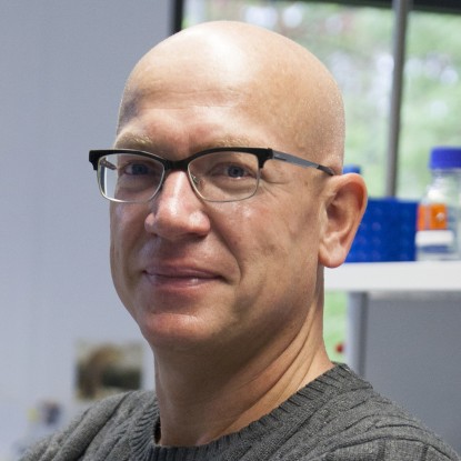 Markus Löbrich合作论文数Workgroup for Radiation Biology and DNA Repair, Department of Biology, Technische Universität Darmstadt45
 Goodarzi Aaron A合作论文数Genome Damage & Stabil Ctr, Univ Sussex40
 O'Driscoll Mark合作论文数30
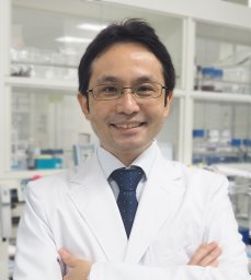 Atsushi Shibata合作论文数30
 Lisa Woodbine合作论文数18Boris Kysela合作论文数17
 Steve Jackson合作论文数Cancer Research UK Gurdon Institute, University of Cambridge;Department of Biochemistry, University of Cambrige15Dorothee Deckbar合作论文数15A Priestley合作论文数13
 Andrew Gennery合作论文数Faculty of Medical Sciences, Newcastle University;Institute of Translational and Clinical Research, Paediatric Immunology Department, Great North Children's Hospital13G E Taccioli合作论文数12Thomas Stiff合作论文数12Caroline Reis合作论文数11E Riballo合作论文数10Tom Stiff合作论文数10Aidan J. Doherty合作论文数10Angela T Noon合作论文数9Barbara Koch合作论文数9
 Andreas Kakarougkas合作论文数9
 Tracy Blunt合作论文数9Jessica A Downs合作论文数8Olivia Barton合作论文数8R Holliday合作论文数7B K Singleton合作论文数7Tomoo Ogi合作论文数Genome Damage and Stability Centre;University of Sussex;Genome Damage and Stability Centre, University of Sussex7Enriqueta Riballo合作论文数7
 Nicolas Foray合作论文数6
 Frederick W. Alt合作论文数Department of Genetics, Blavatnik Institute, Harvard Medical School;Department of Pediatrics, Boston Children's Hospital6
 Majid Hafezparast合作论文数Department of Neurodegenerative Disease, the National Hospital for Neurology and Neurosurgery6T. Stiff合作论文数5
 Patrick Concannon合作论文数Department of Pathology, Immunology, and Laboratory Medicine, College of Medicine, University of Florida5
 Holly Brunton合作论文数Faculty of Life Sciences, The University of Manchester5
 Pierre-Marie Girard合作论文数Institut Curie, Centre de Recherche, Orsay, France;CNRS UMR 3348, Centre Universitaire;Institut Curie, Centre Universitaire5
 Keith Caldecott合作论文数Caldecott Lab, School of Life Sciences, University of Sussex5
 Tony Carr合作论文数School of Life Sciences, University of Sussex;Genome Damage and Stability Centre, University of Sussex5
 Kai Rothkamm合作论文数Health Protection Agency5Sandro Conrad合作论文数5
 Amani Ismail合作论文数Urology and Nephrology Center, Mansoura University5David a Gell合作论文数5Graham Davies合作论文数Great Ormond Street Hospital for Children5Raghbir S. Athwal合作论文数5
 Lara Barazzuol合作论文数5
 Andrew Jackson合作论文数Institute of Genetics and Cancer, University of Edinburgh5Amruta Shrikhande合作论文数4Andrea Beucher合作论文数4Andrea Krempler合作论文数4GP KAUR合作论文数4Katsuki Yoko合作论文数4Julie Birraux合作论文数4




