Ataxia telangiectasia (AT) is a genetic condition caused by mutations involving ATM (Ataxia Telangiectasia Mutated). This gene is responsible for the expression of a DNA double stranded break repair kinase, the ATM protein kinase. The syndrome encompasses combined immunodeficiency and various degrees of neurological abnormalities and increased risk of malignancy. Typically, patients present early in life with delay in neurological milestones, but very infrequently, with life threatening infections typical of a profound T cell deficiency. It would therefore be unexpected to identify this condition immediately after birth using T cell receptor excision circle (TREC)-based newborn screening (NBS) for SCID. We sought to evaluate the frequency of AT detected by NBS, and to assess immunity as well as the genetic aberrations associated with this early presentation. Here, we describe the clinical, laboratory, and genetic features of patients diagnosed with AT through the Ontario NBS program for SCID, and followed in our center since its inception in 2013. Four patients were diagnosed with AT as a result of low TRECs on NBS. In each case, whole exome sequencing was diagnostic. All of our patients had compound heterozygous mutations involving the FRAP-ATM-TRRAP (FAT) domain of the ATM gene, which appears critical for kinase activity and is highly sensitive to mutagenesis. Our patients presented with profound lymphopenia involving both B and T cells. The ratio of naïve/memory CD45+RA/RO T cells population was variable. T cell repertoire showed decreased T cell diversity. Two out of four patients had decreased specific antibody response to vaccination and hypogammaglobulinemia requiring IVIG replacement. In two patients, profound decreased responses to phytohemagglutinin stimulation was observed. In the other two patients, the initial robust response declined with time. In summary, the rate of detection of AT through NBS had been surprisingly high at our center. One case was identified per year, while the total rate for SCID has been five new cases per year. This early detection may allow for better prospective evaluation of AT shortly after birth, and may assist in formulating early and more effective interventions both for the neurological as well as the immune abnormalities in this syndrome.
The use of immunoglobulin therapy has grown steadily over the past 3 decades, mainly due to the increased awareness and expansion of indications. This limited resource is now shared between patients with immunodeficiency disorders who need it as replacement therapy, and individuals who suffer a variety of autoimmune or inflammatory disorders. While alternative therapies exist for the latter diseases, immunodeficient patients are dependent upon this treatment for life. Due to the long-term cost burden of this treatment on healthcare systems, healthcare providers have attempted to moderate its use but are frequently seeking evidence from experts in the field. This raised the critical need for recommendations from experts. To this end, a group a Canadian immunologists representing all regions of the country have formed a panel, the National Immunoglobulin replacement Expert Committee (NIGEC), and formulated a set of unanimously agreed upon recommendations for the use of immunoglobulin replacement in primary immunodeficiency.
Intra-articular injection of long-acting insoluble corticosteroids produces rapid resolution of active arthritis in nearly all injected joints. Almost all of our information on the use of intra-articular corticosteroids in children comes from observational or retrospective analyses or, by inference, from studies in adult patients with arthritis. The duration of response has been found to vary according to the subtype of arthritis, the dose of injected steroids, the accuracy of injection, the duration of disease prior to injection, and possibly the age of the patient. Although the duration of follow-up in most studies has been short, intra-articular steroid therapy seems to be remarkably free of clinically important detrimental effects. Side effects are relatively uncommon and include subcutaneous atrophy and radiologically detectable structural changes or calcification. There is transient suppression of endogenous cortisol production, which may not be clinically important. Although intra-articular steroid therapy is most effective in pauciarticular juvenile rheumatoid arthritis, there are still no solid data to indicate whether it should be used earlier in the course of the disease instead of or along with systemic anti-inflammatory therapy. It has been suggested that repeated injection of the same joint decreases the likelihood of a favorable response. There are still many unanswered questions about how steroids exert their beneficial effects. Newer imaging techniques promise to provide insight into the mechanism of action and possibly to a more informed basis for the use of intra-articular steroids.
140.240, an IgG2a mouse monoclonal antibody raised against a cultured human melanoma cell line, was highly specific for melanoma cells as determined by direct and absorption analyses in a mixed hemadsorption assay. Supernatants of doubly cloned hybridomas producing antibody 140.240 reacted with all cultured and fresh melanomas tested but failed to react with a variety of carcinomas, sarcomas, lymphomas, leukemias and other tumors of neuroectodermal origin. This antibody did not react with B-lymphoid cell lines, ruling out HLA-DR specificity. Non-reactivity of antibody 140.240 with peripheral blood lymphocytes obtained from the donor of the immunizing melanoma line excluded the possibility of detecting histocompatibility antigens. Nevus cells were also non-reactive. However, antibody 140.240 did identify an antigenic determinant on tissue homogenates prepared from fetuses of 10-14 weeks' gestation. The antigen involved was shed by cultured melanoma lines and, by immunoprecipitation analysis of radiolabelled lysates, had a molecular weight of approximately 87kdal. Thus, the structure identified by monoclonal antibody 140.240 is a melanoma-specific oncofetal antigen.
The effect of human leukocyte Interferon (IFN) on the in vitro growth and expression of melanoma-associated antigens (MAA), β2-microglobulin (β2m) and HLA-DR antigen on cultured human melanoma cells was studied. Exposure of melanoma cells to IFN for 64 hours resulted in a dose-dependent inhibition of growth with 46% reduction in cell number at 103 U IFN/ml and 74% reduction at 105 U/ml. Quantitative absorption experiments in the mixed hemadsorption assay determined that the expression of MAA and β2m on treated cells was enhanced at 102–105 U IFN/ml, twofold to fivefold for MAA and fivefold to twelvefold for β2m. No change was seen in HLA-DR antigen expression. The IFN-induced enhancement of MAA and β2m could be detected as early as after 16 hours and a maximum expression was reached at 96 hours after IFN exposure. The IFN-induced enhancement of MAA and β2m on melanoma cells was reversible. Studies with melanoma cells grown in stationary phase and serum-deprived conditions indicated that IFN-induced augmentation of MAA and β2m did not require cell proliferation. The data suggest that the effect of IFN on antigen expression is independent of its effect on cell growth. Further studies are needed to fully elucidate the mechanism.
A patient with malignantmelanoma(RL) received a courseof 28 immunizations with autologous,irradiated cuituredmelanomacells plusBacillusCalmette-Guérin. The postimmune serumandthe preimmuneplasmawere tested for their reactivityin mixed hemadsorption mi croassaysagainst five allogeneicmelanomacell lines andfive non-melanoma lines.No reactivitywasdetected in the preimmuneplasmaagainstany of the cell lines tested, while strongreactivityagainst5 of 5 melanoma lines(1:2,560to 1:40,960)and2 of 5 non-melanoma lines (1:320 and 1:1,280) were found in the unabsorbed postim mune serum.After absorptionwith humanAB erythro cytes, pooled peripheral blood leukocytes, and spleen homogenate of normaladuits,reactivityagainstthesecell lines did not alter. Furtherabsorptionwith cells of a humanlaryngealcarcinoma(KB)line resultedin 4- to 16- foldreductionin antibodytiter andyet retainedreactivity (1:160to I :5,120)dIrectedagainstallfivemelanomalines. The sameabsorbedserumno longerreactedwithany of the five non-melanomalines. Removal of reactivity againstfetal calf serumby absorptionwith Insolubilized fetal calf serumreducedthe reactivityagainstmelanoma cell linesto the same extentas did absorptionwith KB cells, but failed to removereactivityagainstnon-mela noma cell lines completely.Furtherabsorptionof the postimmune serumwith humanfetal homogenate or Ba cillus Calmette-Guèrin had no effect on the reactivity againstmelanomalines. Bloodgroupisoantigens,HLA antigens,andfetalcalfserumwereruledoutascausesof the observedseropositivity againstmelanomacell lines in the final two stages of sequential absorptions. These data providefurtherevidencefor the existenceof com monmelanoma-associated antigens.
 BJ CLARKE合作论文数4Mohammed J. Khosravi合作论文数4PC KWONG合作论文数3SK LIAO合作论文数2susan liao合作论文数2
 Chaim Roifman合作论文数Department of Immunology, Temerty Faculty of Medicine, University of Toronto;Immunology Service Laboratory, Division of Immunology/Allergy, The Hospital for Sick Children;Research Institute,The Hospital for Sick Children2
 Shuen-Kuei Liao合作论文数2
 Brenda Reid合作论文数2Bob Schellenberg合作论文数1Alison Haynes合作论文数1Rae Brager合作论文数1Stanley P. L. Leong合作论文数1
 Stephen Betschel合作论文数Department of Medicine, Temerty Faculty of Medicine, University of Toronto1
 Bruce Mazer合作论文数Division of Experimental Medicine, Department of Medicine, Faculty of Medicine and Health Sciences, McGill University1
 Stuart Turvey合作论文数Division of Allergy and Immunology, Faculty of Medicine, University of British Columbia;Department of Pediatrics, Faculty of Medicine, University of British Columbia\;BC Children's Hospital1
 Amarilla Mandola合作论文数1
 Nancy T Walker合作论文数Department of Pediatrics, State University of New York–Health Science Center1
 Dennis Bulman合作论文数Centre for Neuromuscular Disease, University of Ottawa Ottawa Health Research Institute1
 Elie Haddad合作论文数Department of Pediatrics, Faculty of Medicine, University of Montreal;Department of Microbiology, Immunology et Infectiology, Faculty of Medicine, University of Montreal;Immuno-Allergy and Rheumatology Division, CHU Sainte-Justine1Carl M. Sutherland合作论文数1
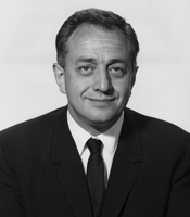 Robert A. Good合作论文数University of South Florida;All Children’s Hospital1
 Brenda Gallie合作论文数Department of Medical Biophysics, Temerty Faculty of Medicine, University of Toronto1
 Gordon Sussman合作论文数Department of Medicine, Temerty Faculty of Medicine, University of Toronto1Susan Waserman合作论文数1




