Magnetic resonance guided focused ultrasound (MRgFUS) thalamotomy has been an effective treatment option in the management of various tremor etiologies. The process of targeting the VIM thalamic location relies on indirect atlas/coordinate-based systems using anatomic reference points. With advancing imaging modalities, thalamic structures can be better visualized resulting in additional targeting techniques and improved accuracy. To identify and evaluate MRgFUS thalamotomy targeting techniques for tremor beyond atlas/coordinate-based systems. A systematic review was conducted in Embase, Cochrane, and PubMed. Studies including MRgFUS for tremor, targeting methods, and tremor outcomes were included. After review of 297 studies, 13 met inclusion criteria. All 13 studies used tractography to visualize various combinations of white matter tracts to guide target acquisition. These tracts include the medial lemniscus (ML), corticospinal tract (CST), and the dentatorubrothalamic tracts (DRTT) which was further delineated into the non-decussating (ndDRTT) and a decussating components (dDRTT) by some studies. At least 50
BACKGROUND:Parkinson's disease (PD) presents with tremor, rigidity, bradykinesia, and postural instability. Commonly, PD is diagnosed around age 60 with the rate of diagnosis increasing with age. Deep brain stimulation (DBS) is an invasive surgical treatment for PD. Older patients, who are more likely to be frail, may experience higher risk of adverse outcomes following DBS. METHODS:This retrospective cohort study analyzed NIS data from 2016 to 2020, identifying PD patients who received DBS. Frailty was determined using the 11-item Modified Frailty Index (mFI-11), with patients classified into frail and non-frail groups. Primary outcomes included prolonged length of stay (LOS) beyond the 75th percentile, non-routine discharge, and postoperative complications. Secondary outcomes involved total hospital costs. Univariate and multivariate analyses were performed, adjusting for demographics, comorbidities (CCI), and hospital characteristics. RESULTS:A total of 3,472 patients met the inclusion criteria. In univariate analysis, frailty was significantly associated with increased odds of unfavorable discharge (OR: 1.76, 95 % CI: 1.44-2.16), prolonged length of stay (OR: 1.99, 95 % CI: 1.57-2.54), postoperative complications (OR: 1.89, 95 % CI: 1.22-3.01), and higher hospital costs (mean difference: $6,780; p = 0.001). After adjustment for demographic, clinical, and hospital factors, frailty remained independently associated with unfavorable discharge (aOR: 1.27, 95 % CI: 1.01-1.60), but not with prolonged length of stay, complications, or cost. In stratified analysis, frailty was significantly associated with adverse outcomes in patients with lower comorbidity burden (CCI < 2), but not in those with higher CCI scores. CONCLUSIONS:Frailty is a strong predictor of adverse outcomes in PD patients undergoing DBS, particularly non-routine discharge. These findings suggest that incorporating frailty assessments into preoperative planning may improve outcomes and reduce healthcare costs.
Subcortical brain structures such as the subthalamic nucleus or the thalamus are involved in regulating motor and cognitive behavior. However, their contribution to perceptual consciousness remains unclear, due to the inherent difficulties of recording subcortical neuronal activity in humans. Here, we asked neurological patients undergoing surgery for deep brain stimulation to detect weak vibrotactile stimuli applied on their hand while recording single neuron activity from the tip of a microelectrode. We isolated putative single neurons in the subthalamic nucleus and thalamus. A significant proportion of neurons modulated their activity while participants were expecting a stimulus. We found that the firing rate of 23% of these neurons differed between detected and undetected stimuli. Our results provide direct neurophysiological evidence of the involvement of the subthalamic nucleus and the thalamus for the detection of vibrotactile stimuli, thereby calling for a less cortico-centric view of the neural correlates of consciousness.
INTRODUCTION: Electrical stimulation of the vagus nerve activates the inflammatory reflex to inhibit cytokines and decrease clinical signs and symptoms of chronic inflammatory disease such as rheumatoid arthritis (RA) (Genovese et al. Lancet Rheum 2020). METHODS: The device system consists of two implanted components: a miniature integrated pulse generator and a silicon sleeve positioning device that holds the generator in apposition to the nerve. There are two external components: a wireless charger and an iPad application for programming the pulse generator. Subjects were randomly assigned (1:1) after device implantation on the left vagus nerve to receive active or sham stimulation. The risks of the surgical procedure, device, and stimulation were blindly assessed after 12 weeks of stimulation therapy in the first 60 subjects enrolled in the study. RESULTS: All implant procedures were completed without intraoperative complications, infections, or surgical revisions. No unanticipated adverse events (AEs) were reported during the perioperative period and at the end of 12 weeks of follow-up. No serious AEs related to the device, stimulation, or explant procedures were reported. Vocal cord paresis and prolonged hoarseness were reported in two subjects. The former resolved following vocal cord augmentation with injectable filler; the latter is ongoing and improving with speech therapy. CONCLUSIONS: Initial results demonstrated that implantation and programming of the novel neuroimmune modulation device were safe, and the surgical procedure and device were well tolerated.
OBJECTIVE:Subthalamic nucleus (STN) and globus pallidus internus (GPI) deep brain stimulation (DBS) effectively treat motor symptoms in Parkinson's disease (PD) but may be associated with cognitive and psychiatric changes in some patients. Evaluation of changes in cognitive and psychiatric symptoms following DBS is complicated by changes in these symptoms that occur as part of the natural disease course. The aim of this study was to evaluate whether electrode position was associated with changes in neurocognitive symptoms in patients who underwent STN and GPI DBS. METHODS:A single-institution retrospective cohort study was conducted on patients with PD who underwent DBS from 2008 to 2019. Cognitive and psychiatric outcomes included Beck Depression Inventory II (BDI-II) score, presence of impulsive-compulsive behavior (ICB), Mini-Mental State Examination (MMSE) score, and overall cognitive status grade determined by comprehensive neuropsychology testing (normal, mild impairment, moderate impairment, and dementia). Pre- and postoperative comparisons were performed using a Wilcoxon signed-rank test or paired t-test. Patients with and without cognitive decline were compared using a Mann-Whitney U-test or unpaired t-test. A chi-square test was used for categorical comparisons. RESULTS:One hundred thirty patients were included (mean age 62.5 ± 7.9 years). At a mean postoperative follow-up from DBS of 13.0 ± 12.7 (range 6-66) months, there was an improvement in ICB (26.3% preoperatively vs 15.0% postoperatively, p = 0.017), but a decline in MMSE score (28.6 ± 1.6 vs 27.6 ± 2.0, p < 0.001) and overall cognitive status (normal: 66.2% vs 39.2%; mild: 12.3% vs 17.7%; moderate: 21.5% vs 33.1%; dementia: 0.0% vs 10.0%; p < 0.001). Patients undergoing STN DBS had a worse decline in overall cognitive status than patients who underwent GPI DBS (p = 0.006). Postoperative cognitive decline was associated with a more medial electrode position only for patients who underwent STN DBS. CONCLUSIONS:Cognitive change was observed in some patients with PD who underwent both GPI and STN DBS, likely due partly to underlying disease progression. Compared with GPI DBS, STN DBS was associated with a greater likelihood of cognitive decline. In STN but not GPI DBS, cognitive decline was associated with medialized electrode position, suggesting modulation of nonmotor STN divisions may contribute to cognitive changes following STN DBS.
Background Rheumatoid arthritis (RA) is a chronic inflammatory autoimmune disease that causes persistent synovitis, bone damage, and progressive joint destruction. Neuroimmune modulation through electrical stimulation of the vagus nerve activates the inflammatory reflex and has been shown to inhibit the production and release of inflammatory cytokines and decrease clinical signs and symptoms in RA. The RESET-RA study was designed to determine the safety and efficacy of an active implantable device for treating RA. Methods The RESET-RA study is a randomized, double-blind, sham-controlled, multi-center, two-stage pivotal trial that enrolled patients with moderate-to-severe RA who were incomplete responders or intolerant to at least one biologic or targeted synthetic disease-modifying anti-rheumatic drug. A neuroimmune modulation device (SetPoint Medical, Valencia, CA) was implanted on the left cervical vagus nerve within the carotid sheath in all patients. Following post-surgical clearance, patients were randomly assigned (1:1) to active stimulation or non-active (control) stimulation for 1 min once per day. A predefined blinded interim analysis was performed in patients enrolled in the study’s initial stage (Stage 1) that included demographics, enrollment rates, device implantation rates, and safety of the surgical procedure, device, and stimulation over 12 weeks of treatment. Results Sixty patients were implanted during Stage 1 of the study. All device implant procedures were completed without intraoperative complications, infections, or surgical revisions. No unanticipated adverse events were reported during the perioperative period and at the end of 12 weeks of follow-up. No study discontinuations were due to adverse events, and no serious adverse events were related to the device or stimulation. Two serious adverse events were related to the implantation procedure: vocal cord paresis and prolonged hoarseness. These were reported in two patients and are known complications of surgical implantation procedures with vagus nerve stimulation devices. The adverse event of vocal cord paresis resolved after vocal cord augmentation injections with filler and speech therapy. The prolonged hoarseness had improved with speech therapy, but mild hoarseness persists. Conclusions The surgical procedures for implantation of the novel neuroimmune modulation device for the treatment of RA were safe, and the device and its use were well tolerated. Trial registration NCT04539964; August 31, 2020.
Drug-induced tremor is a common side effect of lithium with an occurrence of approximately 25% of patients. Cessation of the offending drug can be difficult, and many medical treatments for drug-induced tremor are ineffective. Deep brain stimulation (DBS) has been shown in a limited number of case reports to effectively reduce drug-induced tremor, however, which remains an invasive therapeutic option. MR-guided focused ultrasound (MRgFUS) thalamotomy is an FDA-approved non-invasive treatment for essential tremor (ET). To the best of our knowledge, MRgFUS thalamotomy has never been reported to treat drug-induced tremor. Here, we present a case of a left-handed 55-year-old man with a progressive, medically refractory lithium-induced tremor of the bilateral upper extremities. The patient underwent MRgFUS thalamotomy targeting the right ventral intermediate nucleus (VIM) of the thalamus to treat the left hand. There was almost complete resolution of his left-hand tremor immediately following MRgFUS. There were no side effects. The patient continues to show excellent tremor control at 90-day follow-up and remains free from side effects. This case demonstrates MRgFUS thalamotomy as a possible novel treatment option to treat drug-induced tremor.
We describe a novel system for electrophysiologic mapping and brain-computer interface technology, comprising conformable thin-film electrode arrays and a minimally invasive surgical delivery system that together facilitate bidirectional data exchange from large portions of the cortical surface.
Magnetic resonance-guided, focused ultrasound thalamotomy is a neurosurgical treatment for refractory essential tremor. This study examined cognitive outcomes following unilateral magnetic resonance-guided, focused ultrasound thalamotomy, targeting the ventral intermediate nucleus of the thalamus for essential tremor. The research was conducted at two sites: Sunnybrook Research Institute in Toronto, Canada, and West Virginia University School of Medicine Rockefeller Neuroscience Institute in West Virginia, USA. The study focused on cognitive changes at both the group and individual levels. Patients with refractory essential tremor completed cognitive testing before and after magnetic resonance-guided, focused ultrasound thalamotomy at both sites. The cognitive testing assessed domains of attention, processing speed, working memory, executive function, language and learning/memory. Postoperative changes in cognition were examined using paired t-tests and Wilcoxon signed-rank tests, as appropriate. Reliable change indices were calculated to assess clinically significant changes at the individual level. A total of 33 patients from Toronto and 22 patients from West Virginia were included. Following magnetic resonance-guided, focused ultrasound thalamotomy, there was a significant reduction in tremor severity in both cohorts. At the group level, there were no significant declines in postoperative cognitive performance in either cohort. The reliable change analyses revealed some variability at the individual level, with most patients maintaining stable performance or showing improvement. Taken together, the results from these two independent cohorts demonstrate that unilateral magnetic resonance-guided, focused ultrasound thalamotomy significantly reduces tremor severity without negatively impacting cognition at both the group and individual levels, highlighting the cognitive safety of magnetic resonance-guided focused ultrasound thalamotomy for essential tremor.
BACKGROUND:Essential tremor (ET) and Parkinson's disease (PD) are the most common tremor disorders and are common indications for deep brain stimulation (DBS). In some patients, PD and ET symptoms overlap and diagnosis can be challenging based on clinical criteria alone. The objective of this study was to identify structural brain differences between PD and ET DBS patients to help differentiate these disorders and improve our understanding of the different brain regions involved in these pathologic processes.METHODS:We included ET and PD patients scheduled to undergo DBS surgery in this observational study. Patients underwent 3T brain MRI while under general anesthesia as part of their procedure. Cortical thicknesses and subcortical volumes were quantified from T1-weighted images using automated multi-atlas segmentation. We used logistic regression analysis to identify brain regions associated with diagnosis of ET or PD.RESULTS:149 ET and 265 PD patients were included. Smaller volumes in the pallidum and thalamus and reduced thickness in the anterior orbital gyrus, lateral orbital gyrus, and medial precentral gyrus were associated with greater odds of ET diagnosis. Conversely, reduced volumes in the caudate, amygdala, putamen, and basal forebrain, and reduced thickness in the orbital part of the inferior frontal gyrus, supramarginal gyrus, and posterior cingulate were associated with greater odds of PD diagnosis.CONCLUSIONS:These findings identify structural brain differences between PD and ET patients. These results expand our understanding of the different brain regions involved in these disorders and suggest that structural MRI may help to differentiate patients with these two disorders.
The viability of multidisciplinary movement disorder programs depends heavily on reimbursement for Deep Brain Stimulation (DBS) surgery. Procedures related to DBS include cranial lead insertion, generator placement, microelectrode recording, as well as lead or generator replacements or revisions. These procedures are tied to Current Procedural Terminology (CPT) codes with associated reimbursement rates maintained by the Centers for Medicare and Medicaid Services (CMS). Though not the sole factor in what is ultimately reimbursed to a facility, annual differences in pricing associated with CPT coding can reflect relative changes to reimbursement rates. The purpose of this study was to evaluate trends in Medicare reimbursement for DBS surgery from 2004-2021 when corrected for inflation.
Objective This study was undertaken to describe relationships between electrode localization and motor outcomes from the subthalamic nucleus (STN) deep brain stimulation (DBS) in early stage Parkinson disease (PD) pilot clinical trial. Methods To determine anatomical and network correlates associated with motor outcomes for subjects randomized to early DBS (n = 14), voxelwise sweet spot mapping and structural connectivity analyses were carried out using outcomes of motor progression (Unified Parkinson Disease Rating Scale Part III [UPDRS‐III] 7‐day OFF scores [∆baseline➔24 months, MedOFF/StimOFF]) and symptomatic motor improvement (UPDRS‐III ON scores [%∆baseline➔24 months, MedON/StimON]). Results Sweet spot mapping revealed a location associated with slower motor progression in the dorsolateral STN (anterior/posterior commissure coordinates: 11.07 ± 0.82mm lateral, 1.83 ± 0.61mm posterior, 3.53 ± 0.38mm inferior to the midcommissural point; Montreal Neurological Institute coordinates: +11.25, −13.56, −7.44mm). Modulating fiber tracts from supplementary motor area (SMA) and primary motor cortex (M1) to the STN correlated with slower motor progression across STN DBS subjects, whereas fiber tracts originating from pre‐SMA and cerebellum were negatively associated with motor progression. Robustness of the fiber tract model was demonstrated in leave‐one‐patient‐out ( R = 0.56, p = 0.02), 5‐fold ( R = 0.50, p = 0.03), and 10‐fold ( R = 0.53, p = 0.03) cross‐validation paradigms. The sweet spot and fiber tracts associated with motor progression revealed strong similarities to symptomatic motor improvement sweet spot and connectivity in this early stage PD cohort. Interpretation These results suggest that stimulating the dorsolateral region of the STN receiving input from M1 and SMA (but not pre‐SMA) is associated with slower motor progression across subjects receiving STN DBS in early stage PD. This finding is hypothesis‐generating and must be prospectively tested in a larger study. ANN NEUROL 2023;94:271–284
Background Deep brain stimulation (DBS) is commonly performed with patients awake to perform intraoperative microelectrode recordings and/or macrostimulation testing to guide final electrode placement. Supplemental information from atlas-based databases derived from prior patient data and visualised as efficacy heat maps transformed and overlaid onto preoperative MRIs can be used to guide preoperative target planning and intraoperative final positioning. Our quantitative analysis of intraoperative testing and corresponding changes made to final electrode positioning aims to highlight the value of intraoperative neurophysiological testing paired with image-based data to optimise final electrode positioning in a large patient cohort.Methods Data from 451 patients with movement disorders treated with 822 individual DBS leads at a single institution from 2011 to 2021 were included. Atlas-based data was used to guide surgical targeting. Intraoperative testing data and coordinate data were retrospectively obtained from a large patient database. Medical records were reviewed to obtain active contact usage and neurologist-defined outcomes at 1 year.Results Microelectrode recording firing profiles differ per track, per target and inform the locations where macrostimulation testing is performed. Macrostimulation performance correlates with the final electrode track chosen. Centroids of atlas-based efficacy heat maps per target were close in proximity to and may predict active contact usage at 1 year. Overall, patient outcomes at 1 year were improved for patients with better macrostimulation response.Conclusions Atlas-based imaging data is beneficial for target planning and intraoperative guidance, and in conjunction with intraoperative neurophysiological testing during awake DBS can be used to individualize and optimise final electrode positioning, resulting in favourable outcomes.
 Joseph S Neimat合作论文数Dept Neurosurg, Vanderbilt Univ56
 David Charles合作论文数Vanderbilt University Medical Center53
 Benoit Dawant合作论文数School of Engineering, Vanderbilt University45
 Pierre-Francois D'Haese合作论文数Neuroradiol, West Virginia Univ37
 Davis Thomas L合作论文数33
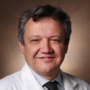 Peter Hedera合作论文数Department of Neurology, Vanderbilt University31
 C. Chris Kao合作论文数Department of Neurological Surgery, Vanderbilt University Medical Center28
 Srivatsan Pallavaram合作论文数Dept. of Electrical Eng. & Computer Science, Vanderbilt University25Chandler E Gill合作论文数24
 Dario J. Englot合作论文数Vanderbilt University Medical Center22
 E. Duco Jansen合作论文数Department of Biomedical Engineering, School of Engineering, Vanderbilt University18
 Hacker Mallory L合作论文数Department of Neurology, Vanderbilt University17Phibbs Fenna T合作论文数17
 Anita Mahadevan-Jansen合作论文数Biophotonics Center, Vanderbilt University17T. L. Davis合作论文数15Hong Yu合作论文数14
 David Bergqvist合作论文数Department of Surgical Sciences, Uppsala University11Fenna T. Phibbs合作论文数11Michael S. Remple合作论文数Department of Neurosurgery, Vanderbilt Medical Center11J. Spooner合作论文数10C. Kao合作论文数10H. Yu合作论文数10
 J. Michael Fitzpatrick合作论文数Vanderbilt University10John Spooner合作论文数10Camalier Corrie R合作论文数Department of Neurological Surgery, Vanderbilt University Medical Center9Jonathon Wells合作论文数9B Husberg合作论文数8
 Jonathan A Forbes合作论文数Department of Neurological Surgery, Vanderbilt University Medical Center8
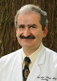 Bassel W. Abou Khalil合作论文数Department of Neurology, School of Medicine, Vanderbilt University8
 Jonathan M. Cayce合作论文数Departments of Biomedical Engineering and Neurosurgery, Vanderbilt University8T. Davis合作论文数7Maxim Turchan合作论文数7Amanda D. Currie合作论文数7
 Hernán F J González合作论文数5Vanderbilt University Institute of Imaging Science, Vanderbilt University Medical Center7
 Walter J Jermakowicz合作论文数Sch Med, Univ Miami7
 Victoria L Morgan合作论文数Department of Radiology, Vanderbilt University Medical Center;Vanderbilt University Institute of Imaging Science7Lauren Heusinkveld合作论文数Cleveland Clinic Lerner College of Medicine, Case Western Reserve University6T. Weaver合作论文数6Ebru Cetinkaya合作论文数6
 Saramati Narasimhan合作论文数Vanderbilt University Medical Center6Sarah K Bick合作论文数Department of Neurosurgery, Vanderbilt University Medical Center6
 Jason M. Schwalb合作论文数6Tolleson Christopher合作论文数6Rodriguez William合作论文数6Molinari Anna L合作论文数6
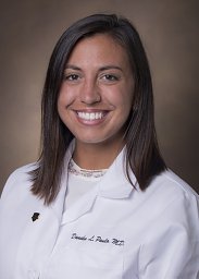 Paulo Danika合作论文数6Rui Li合作论文数6Michael G. Tramontana合作论文数6S. Falowski合作论文数5




