Manually managing the inventory of surgical tools during operations, preoperative and postoperative checks, and sterile processing department (SPD) workflows can be time-consuming and prone to human error. Errors may lead to instrument loss through the sterile processing workflow and missing instruments during surgery. Recent developments in deep-learning-based object detection models offer a promising solution by automatically identifying and counting surgical instruments on trays. This study demonstrates that modern object detection model architectures can achieve high accuracy on fine-grained detection of highly similar surgical instruments in real-time. We train and evaluate two CNN-based detectors of different sizes (YOLOv10-S and YOLOv10-L) and a transformer-based end-to-end detector (RT-DETR) on a dataset of 453 overhead images containing 20 tool classes, including multiple similar forceps, clamps, and retractor variants that are visually similar. YOLOv10-L, the large variant of YOLOv10, achieves the highest overall detection accuracy, with a mean Average Precision (mAP) of 96.5% across IoU thresholds from 0.5 to 0.95. Although the large variant of RT-DETR (RT-DETR-L) reaches a slightly lower mAP (94.8%), it achieves better fine-grained classification reliability within the detection task as it reduces similar-tool classification errors and decreases false background detections compared to YOLOv10-L. Latency is 11.0-19.1 ms for YOLOv10-L and RT-DETR-L models, meeting real-time requirements. These findings demonstrate the practical value of transformer-based object detectors for automated surgical tool detection and inventory systems. By reducing manual counting and enabling real-time verification, such systems have the potential to streamline sterile processing operations, decrease human error, and increase patient safety.
Abstract Meningiomas arising at the level of the internal auditory canal (IAC) and cerebellopontine angle (CPA) present several unique challenges for neurosurgeons and neurotologists. While observation currently plays a significant role, management with stereotactic radiosurgery or microsurgery is also common. The current chapter reviews the presentation, workup, classification, management, and outcomes surrounding lateral skull base meningiomas.
Gliomas are the most common primary malignant brain tumor in adults. Genome-wide association studies (GWAS) have identified 25 loci associated with glioma risk. Using transcriptome-wide association study (TWAS) methodology with joint-tissue imputation, our group investigated 3 genetic repositories to identify genes predictive of the glioma phenotype using the Glioma International Case Control Study (GICC), the Gliomascan GWAS, and the UK Biobank. We characterized the genetic basis of glioma by performing a GWAS using de-identified patient data from these 3 databases in a case-controlled design. For each GWAS, we performed a TWAS utilizing the PrediXcan framework in parallel to identify associations between genetically predicted gene expression in glioma. We applied 59 gene expression prediction models trained on data from GTEx and enriched via joint-tissue imputation (JTI) which borrows information across different tissues, leveraging shared genetic regulation, to improve prediction performance in a tissue-dependent manner. We used a r2 threshold of 0.1 for expression prediction. Bonferroni correction was used for multiple testing adjustment. 7094 cases of glioma and 427,814 controls were included in our meta-analysis. Notably, 12 genes were found to be significantly associated with the glioma phenotype in the Gliomscan database and 17 genes in the GICC database. From the JTI analysis, TERT upregulation in the nucleus accumbens and substantia nigra were found to be significantly associated with the glioma phenotype. Furthermore, ARFRP1 in the amygdala was highly predictive of the glioma phenotype. Glioma is a genetically complex diagnosis with generally poor prognosis. Several novel genes were identified as being predictive of the glioma phenotype. JTI identified several genes including TERT, ARFRP1, and ZGPAT within the nucleus accumbens, substantia nigra, and amygdala predicting glioma. These genes provide further avenues for precision medicine in glioma and targets for therapy.
OBJECTIVE:Patient Reported Outcome Measures (PROMs) quantify the patient's quality of life and symptom burden. This study assesses the implementation of an automated PROM-capture system in an outpatient cranial neurosurgery clinic. METHODS:We conducted both quantitative and qualitative analyses of the implementation of a PROM tool in a single-center, dedicated brain tumor neurosurgery clinic. The primary measures were the PROMIS-10 and FACT-Br. The primary outcome of interest was PROM tool completion. For qualitative analysis, patients and providers were queried over a 6-month period to participate in a phone interview on their PROM tool experience. RESULTS:2,211 patients completed the PROM tool. The quantitative analysis found that white race (OR 1.42, 95% CI 1.21 - 1.68), being of non-Hispanic ethnicity (OR 1.20, 95% CI 1.06 - 1.37), or having private insurance (OR 1.28, 95% CI 1.08 - 1.53) were all associated with higher odds of PROM tool completion. Having an inactive patient portal - My Health At Vanderbilt (MHAV [OR 0.62, 95% CI 0.42 - 0.90]), or appointment providers were associated with decreased odds of PROM tool completion. In-person visits for new patients or for follow-up care were less likely to complete PROM tools as compared to telehealth. Fifteen patients and two providers participated in the phone interview, and the demographics of this group matched the overall cohort. Both patients and providers felt the PROM tool was pertinent, beneficial to patient care, and focused communication between the patient and provider. CONCLUSIONS:This implementation study identified several barriers to developing an automated PROM-collection system in an outpatient cranial neurosurgery practice. These findings have been used to refine this system at our institution and may inform similar implementations elsewhere.
Introduction: An understanding of the trajectory of patient-reported outcome measures (PROMs) based on different management strategies is an important element of patient counseling. In this study, we retrospectively assess prospectively collected PROM data to assess the impact of different management approaches for VS on patients' quality of life (QOL).
Introduction The middle fossa craniotomy (MFCs) is commonly utilized for spontaneous cerebrospinal fluid (CSF) leaks, encephaloceles, and superior semicircular canal dehiscence (SSCD). This study compares postoperative outcomes of MFCs with and without LD use. Methods A retrospective cohort study of adults over the age of 18 years presenting for the repair of nonneoplastic CSF leak, encephalocele, or SSCD via MFC from 2009 to 2021 was conducted. The main exposure of interest was the placement of an LD. The primary outcome was the presence of postoperative complications (acute/delayed neurologic deficit, meningitis, intracranial hemorrhage, and stroke). Secondary outcomes included operating room (OR) time, length of stay, recurrence, and need for reoperation. Results In total, 172 patients were included, 96 of whom received an LD and 76 who did not. Patients not receiving an LDweremore likely to receive intraoperativemannitol ( n = 24, 31.6% vs. n = 16, 16.7%, p = 0.02). On univariate logistic regression, LD placement did not influence overall postoperative complications (OR: 0.38, 95% confidence interval [CI]: 0.05- 2.02, p = 0.28), CSF leak recurrence (OR: 0.75, 95% CI: 0.25-2.29, p = 0.61), or need for reoperation (OR: 1.47, 95% CI: 0.48-4.96, p = 0.51). While OR time was shorter for patients not receiving LD (349 +/- 71 vs. 372 = 85 minutes), this difference was not statistically significant (p = 0.07). Conclusion No difference in postoperative outcomes was observed in patients who had an intraoperative LD placed compared to those without LD. Operative times were increased in the LD cohort, but this difference was not statistically significant. Given the similar outcomes, we conclude that LD is not necessary to facilitate safe MCF for nonneoplastic skull base pathologies.
Introduction: Vestibular schwannoma (VS) is currently managed with observation, radiation therapy, and microsurgery. While a genetic association with neurofibromatosis type II has been described, little is known about the causal genetic underpinnings of VS. A transcriptome-wide association study (TWAS), which tests the contribution of genetically determined gene expression to disease development, may shed light on biological mechanisms associated with VS.
INTRODUCTION:Meningiomas have varying degrees of aggressive behavior. Some systemic hematologic makers are associated with malignancy, but their value in predicting aggressive meningioma behavior is not fully understood. OBJECTIVE:To evaluate the association between preoperative markers such as neutrophil-lymphocyte ratio (NLR), neutrophil-monocyte ratio (NMR), monocyte-lymphocyte ratio (MLR), platelet-lymphocyte ratio (PLR), and prognostic nutritional index (PNI), and diagnostic and prognostic factors including WHO grade, proliferation index, presence of edema on preoperative MRI, and tumor recurrence. METHODS:A retrospective review of patients treated between 2000 and 2019 with a preoperative complete blood count (CBC) differential lab draw before intracranial meningioma resection was conducted. All preoperative steroid dosages were converted to dexamethasone equivalents. Primary outcomes included presence/absence of perilesional edema, WHO grade, Ki-67/MIB-index, and recurrence. Univariate and multivariable regression analyses were conducted. RESULTS:A total of 209 meningioma patients were included. Of these, 143 (68 %) were WHO grade I, 61 (29 %) grade II and 5 (2 %) were grade III. Recurrence was reported in 19 (9.1 %) tumors. No hematologic markers were associated with recurrence. In separate multivariable logistic analyses, no biomarkers were associated with perilesional edema or WHO grade. MLR was associated with higher MIB-index (p = 0.018, OR 6.57, 95 % CI 1.37-30.91). CONCLUSION:Most hematologic markers were not associated with meningioma invasiveness, grade, proliferative index, or aggressiveness. Preoperative MLR was associated with high proliferation index in patients undergoing surgery for intracranial meningioma. Higher MLR could be a surrogate for meningioma proliferation and has potential to be used as an adjunct for risk-stratifying meningiomas.
OBJECTIVE:Excision of intracranial meningiomas often requires resection or coagulation of the dura mater. The choice of dural closure technique is individualized and based on surgeon preference. The objective of this study was to determine outcomes following various dural closure techniques for supratentorial meningiomas.METHODS:A retrospective, single-center cohort study was performed for patients who underwent excision of supratentorial meningiomas from 2000 to 2019. Outcomes including operative time, postoperative in-hospital complications, readmission, causes of readmission including surgical site infection, pseudomeningocele, need for shunt surgery, and imaging appearance of pseudomeningocele on long-term follow-up imaging were compared. Univariate and multivariable analyses were conducted.RESULTS:A total of 353 patients who had complete clinical and operative data available for review were included. Of these patients, 227 (64.3%) had nonsutured dural graft reconstruction and 126 (35.7%) had sutured dural repair, including primary closure, artificial dura, or pericranial graft. There was significant variability in using nonsutured dural reconstruction compared with sutured dural repair technique among surgeons (p < 0.001). Tumors with sagittal sinus involvement were more likely to undergo nonsutured closure (n = 79, 34.8%) than dural repair (n = 26, 20.6%) (p = 0.003). There were no other differences in preoperative imaging findings or WHO grade. Frequency of surgical site infection and pseudomeningocele, need for shunt surgery, and recurrence were similar between those undergoing nonsutured and those undergoing sutured dural repair. The mean operative time for the study cohort was 234.9 (SD 106.6) minutes. The nonsutured dural reconstruction group had a significantly shorter mean operative time (223.9 [SD 99.7] minutes) than the sutured dural repair group (254.5 [SD 115.8] minutes) (p = 0.015). In a multivariable linear regression analysis, after controlling for tumor size and sinus involvement, nonsutured dural graft reconstruction was associated with a 36.8-minute reduction (95% CI -60.3 to -13.2 minutes; p = 0.002) in operative time.CONCLUSIONS:Dural reconstruction using a nonsutured graft and sutured dural repair exhibit similar postoperative outcomes for patients undergoing resection for supratentorial meningiomas. Although sutured grafts may sometimes be necessary, nonsutured graft reconstruction for most supratentorial meningioma resections may suffice. The decreased operative time associated with nonsutured grafts may ultimately result in cost savings. These findings should be taken into consideration when selecting a dural reconstruction technique for supratentorial meningioma.
Introduction: Current instruments used for neurosurgical procedures are rigid and have limited range of motion. This is especially problematic in the endoscopic endonasal approach (EEA) where there the operative corridors are narrow. Due to these limitations, neurosurgeons are often forced to expand the approach increasing tissue damage, or in extreme cases, leave tumor behind when the instruments cannot safely navigate the operative anatomy. Thus, there is a need for steerable instruments that have increased range of motion.
BACKGROUND:Patients' comorbidities might affect the immediate postoperative morbidity and discharge disposition after surgical resection of intracranial meningioma.OBJECTIVE:To study the impact of comorbidities on outcomes and provide a web-based application to predict time to favorable discharge.METHODS:A retrospective review of the prospectively collected national inpatient sample (NIS) database was conducted for the years 2009-2013. Time to favorable discharge was defined as hospital length of stay (LOS). A favorable discharge was defined as a discharge to home and a non-home discharge destination was defined as an unfavorable discharge. Cox proportional hazards model was built. Full model for time to discharge and separate reduced models were built.RESULTS:Of 10,757 patients who underwent surgery for meningioma, 6554 (60%) had a favorable discharge. The median hospital LOS was 3 days (interquartile range [IQR] 2-5). In the full model, several clinical and socioeconomic factors were associated with a higher likelihood of unfavorable discharge. In the reduced model, 13 modifiable comorbidities were negatively associated with a favorable discharge except for drug abuse and obesity, which are not associated with discharge. Both models accurately predicted time to favorable discharge (c-index:0.68-0.71).CONCLUSION:We developed a web application using robust prognostic model that accurately predicts time to favorable discharge after surgery for meningioma. Using this tool will allow physicians to calculate individual patient discharge probabilities based on their individual comorbidities and provide an opportunity to timely risk stratify and address some of the modifiable factors prior to surgery.
Introduction: The use of lumbar drain (LD) in non-tumor middle fossa craniotomies (MFCs) is commonly utilized for spontaneous cerebrospinal fluid (CSF) leaks, encephaloceles, and semicircular canal dehiscence (SSCD). Debate exists regarding the utility of lumbar drain use in MFC cases in providing brain relaxation and gentle temporal lobe retraction. This study compares postoperative outcomes of MFCs with and without LD use.
INTRODUCTION: Understanding the role of social determinants of health (SDoH) is critical in informing preoperative care and postoperative follow-up for meningioma patients undergoing surgery. METHODS: A retrospective cohort study of adults presenting for meningioma resection from 1999-2020 was conducted. Primary outcomes included recurrence, readmission, and LTF (6-month and total). The main exposure of interest was Area Deprivation Index (ADI), a composite score of socioeconomic disadvantages. Additional SDoH factors analyzed included drive time to the hospital, insurance, and race. Multivariable logistic regression analyses for LTF and Cox regression for recurrence- and readmission-free survival were conducted. Covariates included age, WHO grade, and extent of resection. RESULTS: In total, 736 patients were included. Recurrence was observed in 90 patients, and 99 patients were readmitted following discharge. 121 patients missed initial and 6-month follow-up, but 107 of those eventually followed-up after six months. Fourteen patients were entirely LTF. ADI (OR = 1.11 per 10-point increase, 95% CI 1.02-1.21, p = 0.017) and drive time (OR = 1.03 per 30-minute increase, 95% CI 1-1.06, p = 0.022) predicted 6-month LTF. Drive time predicted complete LTF (OR = 1.04 per 30-minute increase, 95% CI 1-1.07, p = 0.017). Medicare/Military insurance predicted lower recurrence risk (HR = 0.43, 95% CI 0.23-0.81, p = 0.009) and higher readmission risk (HR = 2.00, 95% CI 1.17-3.41, p = 0.011). Other SDoH factors were not associated with recurrence or readmission. Lower WHO grade predicted lower recurrence risk (HR = 0.5, 95% CI 0.33-0.78, p = 0.002), while age (HR = 0.75 per 10-year increase, 95% CI 0.62-0.89, p < 0.001) and gross total resection (HR = 0.56, 95% CI 0.35-0.9, p = 0.016) predicted lower readmission risk. CONCLUSIONS: Socioeconomically disadvantaged neighborhoods and further distance from the hospital were barriers to follow-up after meningioma resection. Other SDoH associated with readmission and recurrence are reported. Individualized follow-up plans should consider these SDoH to ensure care continuity and improve outcomes.
BACKGROUND:Recurrence after meningioma resection warrants serial surveillance imaging, but little evidence guides the optimal time interval between imaging studies/surveillance duration.OBJECTIVE:To describe recurrence-free survival (RFS) after meningioma resection, conditioned to short-term RFS.METHODS:A retrospective cohort study for adults presenting for meningioma resection from 2000 to 2018 was conducted. The primary outcome was postoperative follow-up RFS. Conditional RFS Kaplan-Meier analysis was performed at 1, 2, 3, 5, and 10 years, conditioned to 6-month and 12-month RFS. RFS probabilities conditioned to 6-month RFS were estimated in subgroups, stratified by World Health Organization grade, extent of resection, and need for postoperative radiation.RESULTS:In total, 723 patients were included. Median age at surgery was 57.4 years (IQR = 47.2-67.2). Median follow-up was 23.5 months (IQR = 12.3-47.8). Recurrence was observed in 90 patients (12%), with median time to recurrence of 14.4 months (IQR = 10.3-37.1). Conditioned to 6-month postoperative RFS, patients had 90.3% probability of remaining recurrence-free at 2 years and 69.4% at 10 years. Subgroup analysis conditioned to 6-month RFS demonstrated grade 1 meningiomas undergoing gross total resection (GTR) had 96.0% probability of RFS at 1 year and 82.8% at 5 years, whereas those undergoing non-GTR had 94.5% and 79.9% probability, respectively. RFS probability was 78.8% at 5 years for non-grade 1 meningiomas undergoing GTR, compared with 69.7% for non-grade 1 meningiomas undergoing non-GTR. Patients with non-grade 1 meningiomas undergoing upfront radiation had a 1-year RFS of 90.1% and 5-year RFS of 51.7%.CONCLUSION:Recurrence risk after meningioma resection after an initial recurrence-free period is reported, with high-risk subgroups identified. These results can inform objective shared decision-making for optimal follow-up.
Objectives: Cranioplasty is a commonly performed neurosurgical procedure that restores cranial anatomy. While plastic surgeons are commonly involved with cranioplasties, the cost of performing a cranioplasty with neuro-surgery alone (N) vs. neurosurgery and plastic surgery (N + P) is unknown.Methods: A single-center, multi-surgeon, retrospective cohort study was undertaken on all cranioplasties per-formed from 2012 to 22. The primary exposure variable of interest was operating team, comparing N vs. N + P. Cost data was inflation-adjusted to January 2022 using Healthcare Producer Price Index as calculated by the US Bureau of Labor Statistics.Results: 186 patients (105 N vs. 81 N + P) underwent cranioplasties. The N + P group has a significantly longer length-of-stay (LOS) 4.5 +/- 1.6days, vs. 6.0 +/- 1.3days (p < 0.001), but no significant difference in reoperation, readmission, sepsis, or wound breakdown. N was significantly less expensive than N + P during both the initial cranioplasty cost ($36,739 +/- $4592 vs. $41,129 +/- $4374, p 0.014) and total cranioplasty costs including reoperations ($38,849 +/- $5017 vs. $53,134 +/- $6912, p < 0.001). Univariable analysis (threshold p = 0.20) was performed to justify inclusion into a multivariable regression model. Multivariable analysis for initial cranio-plasty cost showed that sepsis (p = 0.024) and LOS (p = 0.003) were the dominant cost contributors compared to surgeon type (p = 0.200). However, surgeon type (N vs. N + P) was the only significant factor (p = 0.011) for total cost including revisions.Conclusions: Higher costs to N + P involvement without obvious change in outcomes were found in patients undergoing cranioplasty. Although other factors are more significant for the initial cranioplasty cost (sepsis, LOS), surgeon type proved the independent dominant factor for total cranioplasty costs, including revisions.
BACKGROUND Although posterior myelotomy leaves patients with dorsal column deficits, few reports have explored the anterior cervical approach for cervical intramedullary tumors. The authors describe the resection of a cervical intramedullary ependymoma through an anterior approach with a two-level corpectomy and fusion. OBSERVATIONS A 49-year-old male presented with a C3–5 ventral intramedullary mass with polar cysts. Because of the ventral location of the tumor and the added benefit of avoiding a posterior myelotomy and dorsal column deficits, an anterior C4–5 corpectomy offered a direct route and excellent visualization of the ventrally located tumor. After a C4–5 corpectomy, microsurgical resection, and C3–6 anterior fusion with a fibular allograft filled with autograft, the patient remained neurologically intact. Magnetic resonance imaging (MRI) on postoperative day (POD) 1 confirmed gross-total resection. The patient was extubated on POD 2 and was discharged home on POD 4 with a stable examination. At 9 months, the patient developed mechanical neck pain refractory to conservative treatment and underwent a posterior fusion to address pseudarthrosis. MRI at 15 months showed no evidence of tumor recurrence with the resolution of neck pain. LESSONS An anterior cervical corpectomy provides a safe corridor to access ventral cervical intramedullary tumors and avoids posterior myelotomy. Although the patient required a three-level fusion, we believe the tradeoff of decreased motion compared to dorsal column deficits is preferred.
-BACKGROUND: Acute subdural hematoma is a neuro-surgical emergency. Thrombocytopenia poses a manage-ment challenge for these patients. We aimed to determine the impact of thrombocytopenia on preoperative hemor-rhage expansion and postoperative outcomes. -METHODS: This retrospective study evaluated patients presenting at our institution with acute subdural hematoma between 2009 and 2019. Patients who underwent surgery, had thrombocytopenia (platelets <150,000/mL), and had multiple preoperative computed tomography scans were included. Case control 1:1 matching was performed to generate a matched cohort with no thrombocytopenia. Univariate analyses were conducted to determine changes in subdural thickness and midline shift, postoperative Glasgow Coma Scale score, mortality, length of stay, and readmission rates. -RESULTS: We identified 19 patients with both throm-bocytopenia and multiple preoperative computed tomog-raphy scans. Median platelet count was 112,000/mL (Q1 69,000, Q3 127,000). Comparing the thrombocytopenia cohort with the control group, there was a statistically significant difference in change in subdural thickness (median 5 mm [Q1 2, Q3 7.4] vs. 0 mm [Q1 0, Q3 1.5]; P = 0.001) and change in midline shift (median 3 mm [Q1 0, Q3 9.5] vs. median 0.5 mm [Q1 0, Q3 1.5]; P = 0.018). The thrombocytopenia cohort had higher in-hospital mortality (10 [52.6%] vs. 2 [10.5%]; P = 0.003). No significant differences were found in postoperative Glasgow Coma Scale score, length of stay, number of readmissions, and n umber of reoperations. -CONCLUSIONS: Thrombocytopenia is significantly associated with expansion of hematoma preoperatively in patients with acute subdural hematoma. While the benefit of early platelet correction cannot be determined from this study, patients who present with thrombocytopenia will benefit from close monitoring, a low threshold to obtain repeat imaging, and anticipating early surgical evacuation after platelet optimization.
Introduction: Outpatient patient visit no-shows result in clinic workflow inefficiencies, delayed access to care, and opportunity costs for providers and staff. Early identification of patients likely to no-show could allow for targeted patient outreach or strategic clinic scheduling/overbooking. Predictors of clinic no-show among the skull base/brain tumor patient population have not been previously characterized.
 Reid C. Thompson合作论文数25
 Lola B. Chambless合作论文数24
 Zuckerman Scott L合作论文数Spinal Column Surgical Outcomes and Quality Research Laboratory, Vanderbilt University23
 Dewan Michael合作论文数Dept Neurol Surg, Vanderbilt Univ20
 Silky Chotai合作论文数17
 Alan Tang合作论文数16
 Patrick D Kelly合作论文数15Bradley S. Guidry合作论文数12
 Yengo-Kahn Aaron M合作论文数Vanderbilt Sports Concussion Center, Vanderbilt University School of Medicine12Mocco J合作论文数10
 Robert Dambrino合作论文数Department of Neurological Surgery, Vanderbilt University Medical Center;Vanderilt Policy and Costs in Surgery (VPaCS) Research Center, Vanderbilt University Medical Center10B. Root合作论文数7Jake R. McDermott合作论文数6
 Candace J Grisham合作论文数Sch Med, Vanderbilt Univ6Singer Robert J合作论文数6John C. Wellons合作论文数Pediatric Neurosurgery Children’s Hospital6Matthews Lan合作论文数4Katherine Kelly合作论文数4
 Eric R Gamazon合作论文数Division of Genetic Medicine, Department of Medicine, School Of Medicine, Vanderbilt University Medical Center;University of Cambridge4Anthony J Sermarini合作论文数3Diana G. Douleh合作论文数3Gunther Wong合作论文数3
 Hani Chanbour合作论文数Department of Neurological Surgery, Vanderbilt University Medical Center3
 Harrison C Thomas合作论文数Sch Med, Vanderbilt Univ3
 Bekelis Kimon合作论文数Neurosurg Sect, Dartmouth Hitchcock Med Ctr3Chitale Rohan合作论文数3Mistry Akshitkumar M合作论文数Department of Medicine, Vanderbilt University Medical Center3
 Aaron A. Cohen-Gadol合作论文数Goodman Campbell Brain and Spine, Indiana University3
 Nishant Kumar合作论文数3
 Allen K Sills合作论文数3Joseph S. Cheng合作论文数3Fatima Gauhar合作论文数2Justiss A Kallos合作论文数2Whitney E Muhlestein合作论文数2Brian M. Rapp合作论文数2
 Robert J Singer合作论文数2Brad S. Guidry合作论文数2Rut Thakkar合作论文数2
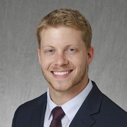 Chase H. Foster合作论文数Department of Neurological Surgery, The GW School of Medicine & Health Sciences2
 Nathan R Lindquist合作论文数Dept Otolaryngol Head & Neck Surg, Vanderbilt Univ2
 Jeffrey Chen合作论文数Department of Anesthesiology, School of Medicine, University of California, San Diego2Chi Le合作论文数2
 Jacob Jo合作论文数2
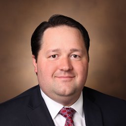 Thomas G Stewart合作论文数School of Data Science, University of Virginia2Le He合作论文数2Cheung-Flynn Joyce合作论文数2
 Bret Mobley合作论文数2
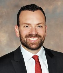 Jonathan A Forbes合作论文数Department of Neurological Surgery, Vanderbilt University Medical Center2
 Liles Campbell合作论文数2




