Functional magnetic resonance imaging (fMRI) allows to display functional activities of certain brain areas. In combination with a three dimensional anatomical dataset, acquired with a standard magnetic resonance (NIR) scanner, it can be used tu identify eloquent brain areas, resulting in so-called functional neuronavigation, supporting the neurosurgeon while planning and performing the operation. But during the operation brain shift leads to an increasing inaccuracy of the navigation system. Intraoperative NIR imaging is used to update the neuronavigation system with a new anatomical dataset. To preserve the advantages of functional neuronavigation, it is necessary to save the functional information. Since fMRI cannot be repeated intraoperatively with the unconscious patient easily we tried to solve this problem by means of image processing and pattern recognition algorithms. In this paper we present an automatic approach for transfering preoperative markers into an intraoperative 3-D dataset. In the first step the brains are segmented in both image sets which are then registered and aligned. Next, corresponding points are determined. These points are then used to determine the position of the markers by estimating the local influence of brain shift.
Mittels funktioneller Magnetresonanztomographie können neuro-funktionelle Bereiche im Gehirn bestimmt werden, welche dem Chirurgen während der Operation zur Identifikation von Gehirnbereichen dienen. Verschiebungen des Gehirns führen jedoch zu Ungenauigkeiten, die nur durch eine intraoperative Aufnahme eines Volumenbildes kompensiert werden können. Funktionelle Informationen (fMRI-Marker) gehen dabei verloren. Zur Übertragung dieser Informationen wird ein automatisches Verfahren vorgestellt, welches prä- und intraoperativen Datensätze registriert und dann nach korrespondierenden Gehirnarealen sucht, umso die Markerposition schätzen zu können.
In this paper we present the image processing system STROKE-CT for quantitatively evaluating CT images of stroke patients during treatment and rehabilitation [3,2]. It was designed to supersede the fully manual method proposed by Blunk [1]. STROKE-CT was developed to allow systematic outcome studies within a framework community based study of stroke, where systematically all stroke patients from the population of a 100.000 inhabitant city in Germany are monitored[4].
Based on a sample of 8000 manually segmented video images acquired in a subway station, we examine if heads of pedestrians can be detected by use of wavelet features and parameter free statistical classifiers. In particular we address the question of which wavelet filter to use for feature generation and how to model class conditional densities of features. We show that a markov chain based approach in combination with HAAR-wavelets is a good compromise between high recognition rates and computational efficiency. The latter is required since developed algorithms are supposed to run on a dedicated low cost hardware.
In this paper we describe the architecture of a vision-based workspace monitoring system for different kinds of nonautonomous service robots. Based on a hardware system with a movable color CCD-camera, mounted on a linear sledge above the workspace, we are developing a hierarchical, modular software system with a flexible control which is not restricted to a single application. During the initialization a 3D map of the workspace is generated by tracking prominent features like edges and corners while the camera is moved. In the following, regions of interest are determined by means of motion detection and/or color classification. These regions are used for the search of known objects, e.g. humans, the robot itself, etc. After an object is identified, its posture and location is estimated by matching a geometric 3D model of the object with the detected regions. The feasibility of this approach will be proved with two applications.
In diesem Beitrag untersuchen wir, in wie weit Waveletmerkmale in Verbindimg mit parameterfreien statistischen Klassifikatoren geeignet sind, Personengruppen in Bildern einer Videosequenz vom Szenenhintergrund zu unterscheiden. Anhand einer manuell segmentierten Stichprobe, die 2850 Bilder umfaßt, werden parameterfreie statistische Klassifikatoren überwacht trainiert. Die Klassifikatoren basieren auf Bin-Histogrammen und Markovketten und zeichnen sich durch eine sehr geringe algorithmische Komplexität und hohe Geschwindigkeit aus. Es wurden mittlere Erkennungsraten von über 80% im Test erreicht.
In this paper we propose a new system of using external markers for the determination of camera parameters for multiple DSA images. A calibration frame was defined containing just five different simple geometrical shapes which were multiply located over the frame. A software system was developed which allows the fully automated segmentation and identification of the markers. The overall marker size could be reduced by 60 percent in comparision to a former model and the interactive effort for physicians could be minimized.
Automatic tooth restoration systems produce dental restorations for individual given teeth with a prepared cavity. Whereas the shape of the inlay inside the tooth is determined by the shape of the cavity the chewing surface yet has to be defined. We present RecOS, a method that makes use of an intact chewing surface of a model tooth to determine the chewing surface of an inlay, onlay or crown to be ground by an NC-machine e.g. from ceramics. The method uses the technique of image deformation to provide a congruence between range images of the model tooth and the prepared tooth such that the missing part is determined by the deformed model tooth. The image deformation is defined by a number of pairs of mutually corresponding feature points in both range images. Feature extraction techniques including active contours [4] are used to detect these points. A new approach for contour-matching is proposed to match corresponding feature points of the two different teeth. Our implementation was tested on a number of range images with manually marked cavities. The mean height difference between the restored surface and the original surface was between 0.2 mm and 1.0 mm. This is only half of the difference measured on machine-made inlays of a commercial system. The method can be extended to consider the chewing surface of antagonistic teeth as well.
In this paper a robust method for visual motion estimation under ego-motion is developed. The possible application of this method is image sequence analysis of road traffic or airport runway/taxiway scenes, where the camera is located in a moving vehicle. The method combines an application independent estimation of visual motion with specific methods for instantaneous detection of the vanishing point in the image plane and of the over-road location of the camera. The stationary background is separated from the obstacles while detecting the ego-motion corrected visual motion of on-road objects.
Unterschiedliche Faktoren beeinflußen die Neubildung von Gefäßen und somit auch das Wachsen und Metastasieren von Tumoren. Ihre Auswirkungen werden anhand des planaren Gefäßsystems von embryonalen Haushühnern analysiert. Durch eine Öffnung an der Eispitze wird der Embryo am 4. Bruttag mit einer auf einem Mikroskop fixierten CCD-Kamera aufgenommen.
This article investigates two approaches to the automatic detection of suspicious intervals in measurement sequences obtained from test cars. They ar vector quantization combined with hidden Markov models and (artificial) neural networks. The results obtained are discussed. Both approaches also yield a symbolic description of the test route.
In diesem Aufsatz werden zwei Ansätze zur automatischen Detektion von Fehlern bzw. verdächtigen Abschnitten in Meßreihen von PKW-Testfahrten vorgestellt, nämlich die Verwendung von Vektorquantisierung kombiniert mit Hidden-Markov-Modellen und von (künstlichen) neuronalen Netzen. Die Ergebnisse werden vergleichend gegenübergestellt. Es wird darauf hingewiesen, daß die verwendeten Ansätze auch eine symbolische Beschreibung der Fahrstrecke und der Fahrzustände liefern können.
We present a means of evaluating the relative recognition rate of unsupervised classifiers. Self organizing maps (so called Kohonen Maps) have some important features. They can classify data, reduce the dimension of input vectors and can be trained using unlabeled data. In our efforts to develop an automatic, context dependent classifier we highly relied on these features. Using unlabeled time series data, we had no way to compare different topologies with respect to “correct classifications”. We solved this problem by introducing a formal measure we called pseudo classes. This is an elegant, but heuristic method which can also be applied on other fields with context-dependent data
With MRI, esp. fast imaging sequences (FISP…), different soft tissue components can be analysed in detail by their different signal and contrast behaviour. Early stages of cartilage degeneneration of the knee are detectable [1][3]. However, evaluations have shown to be too inaccurate and time consuming without adequate hardware and software support.
 Heinrich Niemann合作论文数Department of Computer Science, Friedrich-Alexander-Universität Erlangen-Nürnberg17
 M Wolf合作论文数5
 Christopher Nimsky合作论文数Department of Neurosurgery, University Marburg3
 Matthias Wolf合作论文数3Dirk Wetzel合作论文数3Klaus Glückert合作论文数3T. Vogel合作论文数2
 Gerhard Sagerer合作论文数Bielefeld University2Dorothea Faulhaber合作论文数2
 Klaus Frank Gluckert合作论文数2H Niemann合作论文数1T Hanning合作论文数1R. Graumann合作论文数1Mossner, P.合作论文数1Wolf, M.合作论文数1M Vonrosenberg合作论文数1
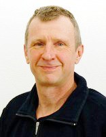 Dietrich Paulus合作论文数Institute for Computational Visualistics, Universität Koblenz-Landau1
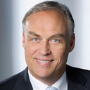 Bernd Kladny合作论文数Fachklinik Herzogenaurach In der Reuth 11Christian L. Knüvener合作论文数1
 Matthias Pelka合作论文数1Jens Höper合作论文数1Georg Pisinger合作论文数1Włodzimierz Kasprzak合作论文数Institute of Control and Computation Engineering
Warsaw University of Technology1Sebastian Meller合作论文数1
Matthias Pelka合作论文数1Jens Höper合作论文数1Georg Pisinger合作论文数1Włodzimierz Kasprzak合作论文数Institute of Control and Computation Engineering
Warsaw University of Technology1Sebastian Meller合作论文数1




